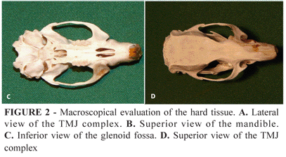
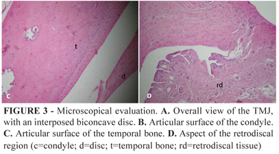
PURPOSE: To describe and evaluate normal rat temporomandibular joints from anatomic and histopathologic point of view and make a comparison between this joint in rats and humans. METHODS: Twelve male adult Wistar rats (12 same side joints) were used in this procedure. The following anatomical structures were histologically evaluated in a qualitative fashion: condyle, disc, temporal bone, retrodiscal tissue and synovia. The macroscopical and microscopic study of the human TMJ was based on the current literature. RESULTS: The TMJ is surrounded by a thin capsule, consisting of fibrous tissue, and a synovial lining. The mandibular angle has a prominent shape. The glenoid fossa is flat, with no eminences. Histologically, the TMJ is composed of different tissues that comprise the mandibular head, mandibular fossa and fibrocartilaginous disc. A layer of hyaline cartilage covers the articulating cortical condyle and temporal bone. CONCLUSION:Morphologically and histologically, the articular structure of rats is, on the whole, similar to that of humans. In these animals there is no articular eminence.
Temporomandibular Joint; Anatomy; Animal Experimentation; Rats


