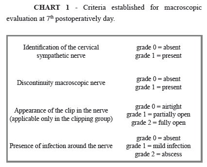
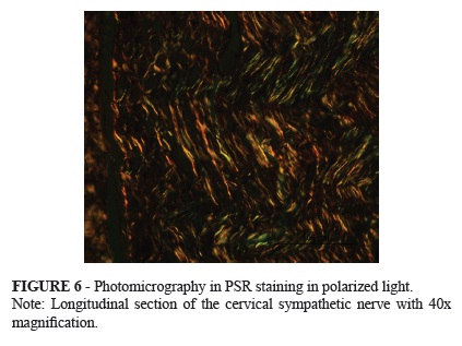
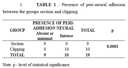
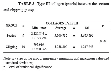
PURPOSE: To study the macro and microscopic evaluation of the damage caused by clamping or section of cervical sympathetic nerve in rabbits, quantifying the collagen in the lesions. METHODS: Twenty rabbits were divided into two groups of ten, doing in group 1 (section) section of the right cervical sympathetic nerve, while in group 2 (clipping) clipping of the nerve. All rabbits were induced to death on the seventh day after surgery. The macroscopic variables were: consequences of nerve lesion, clip appearance, presence of infection and adhesions around the nerve. Microscopy used hematoxylin-eosin staining to evaluate the stages and the degree of inflammation and necrosis, and F3BA Picrosirius red staining to quantify collagen. Mann-Whitney test was used for comparisons of collagen types I and III between groups. Fisher exact test analyzed the macroscopic variables, the degree of inflammation and necrosis. RESULTS: There was no discontinuity of nerve injury in the clipping group, as well as the clip was closed in all animals. The presence of severe adhesions was significantly higher in the clipping group (p<0.05). There was no significant difference on other variables macroscopically analyzed. There was no significant difference between groups regarding the type of inflammatory process and its intensity, as well as the presence of necrosis and collagen deposition in the nerves. CONCLUSIONS: In the macroscopic evaluation, the section caused discontinuity, which did not occur in the clamping group; there was no development of local infection; the clipping of the cervical sympathetic nerve was linked to the presence of a greater number of adhesions in comparison to the section group. Microscopically, no difference existed in relation to the type and intensity of inflammation reaction between the groups; occurred predominance of chronic and severe inflammation on the specimens; the necrosis was noticed equally in both groups; there was predominance of type I collagen deposition in relation to type III in both groups.
Sympathetic Nervous System; Surgical Stapling; Autonomic Nerve Block; Rabbits