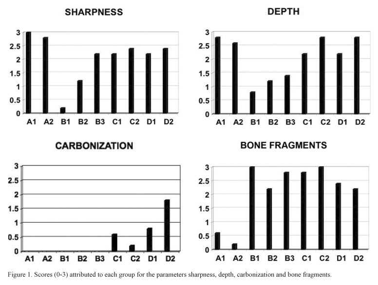
The aim of this study was to compare the peripheral bone damage induced by different cutting systems. Four devices were tested: Er:YAG laser (2.94 mm), Piezosurgery, high-speed drill and low-speed drill. Forty-five bone sections, divided into 9 groups according to different parameters, were taken from pig mandibles within 1 h post mortem. Specimens were fixed in 10% buffered formalin, decalcified and cut in thin sections. Four different parameters were analyzed: cut precision, depth of incision, peripheral carbonization and presence of bone fragments. For statistical analysis, the Kruskal-Wallis test was applied to assess equality of sample medians among groups. All sections obtained with the Er:YAG laser showed poor peripheral carbonization. The edges of the incisions were always well-shaped and regular, no melting was observed. Piezosurgery specimens revealed superficial incisions without thermal damage but with irregular edges. The sections obtained by traditional drilling showed poor peripheral carbonization, especially if obtained at lower speed. There was statistically significant differences (p<0.01) among the cutting systems for all analyzed parameters. Er:YAG laser, gave poor peripheral carbonization, and may be considered an effective method in oral bone biopsies and permits to obtain clear and readable tissue specimens.
laser; piezosurgery; bone biopsy; peripheral damage