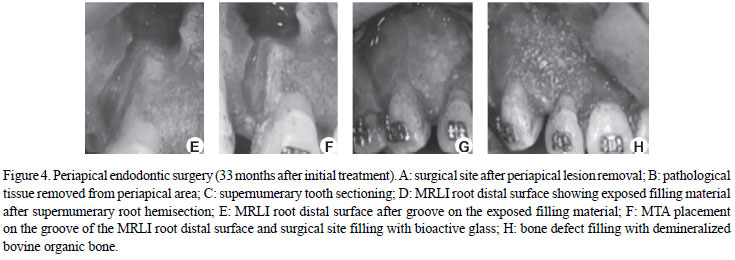
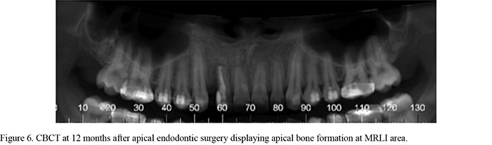
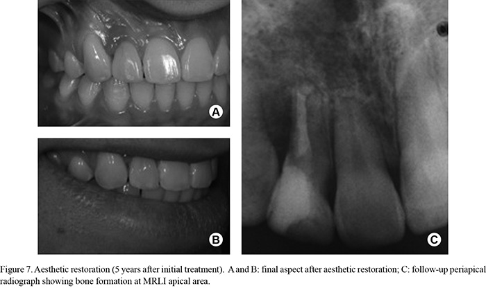
This paper describes and discusses the multidisciplinary treatment involving a permanent maxillary lateral incisor fused to a supernumerary tooth, both presenting pulp necrosis and periapical lesion. A 15-year-old male patient sought treatment complaining of pain, swelling and mobility on the maxillary right lateral incisor. After clinical and radiographic examination, root canal preparation was performed according to the crown-down technique and a calcium hydroxide dressing was placed for 15 days. The patient returned and the definitive endodontic filling was done with thermomechanical compaction of gutta-percha and sealer. After 18 months, clinical and radiographic examinations were carried out and no pain or swelling was reported. Two years after endodontic treatment, the patient returned for periodontal and cosmetic treatments. Nine months later, a cone-beam computed tomography (CBCT) revealed that the previously detected periodontal defect and periapical lesion were persistent. Apical endodontic surgery was indicated. The supernumerary tooth was removed, the communicating distal surface was filled and the surgical site received bioactive glass and demineralized bovine organic bone. The pathological tissue was submitted to histopathological examination and the diagnosis was periapical cyst. One year after the apical endodontic surgery, CBCT showed bone formation at maxillary lateral incisor apical area. Two years after the surgery, the restoration was replaced due to aesthetic reasons and periapical radiograph showed success after 5 years of treatment. A correct diagnosis and establishment of an adequate treatment plan resulted in a successful management of the case.
Fusion; apical surgery; cone-beam computed tomography; supernumerary tooth