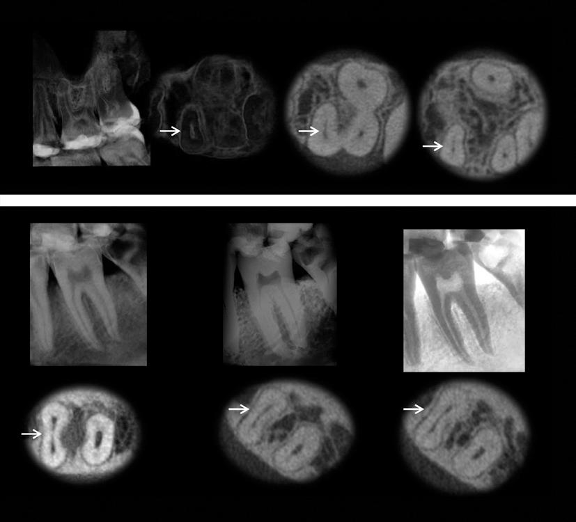
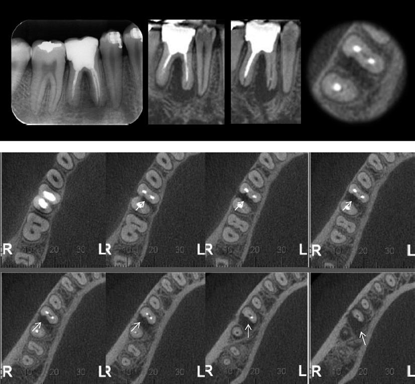
The aim of this study was to detect root isthmuses in maxillary and mandibular molars and evaluate their frequencies using map-reading dynamics in CBCT images. Two hundred extracted human maxillary and mandibular molars were used in ex vivo assay. A consecutive sample of two hundred maxillary and mandibular molars (first and second) was selected from CBCT exams. The isthmuses were detected from the pulp orifice to the apex and were recorded according to their beginning and their end, into categories: 1. begin and end in cervical third; 2. beginning in cervical third and end in middle third; 3. beginning in cervical third and end in apical third; 4. beginning and end in middle third; 5. begin in middle third and end in apical third; 6. beginning and end in apical third; 7. no isthmus. The scans were obtained in different planes with map-reading in axial slices of 0.5 mm/0.5 mm involved the coronal to apical direction. The frequencies of isthmus were analyzed according to the level of root and evaluated by Chi-square test. The level of significance was set at α=0.05. The presence of isthmus detected in maxillary molars was 86% in ex vivo assay and 62% in vivo assay, whereas in mandibular molars was observed 70% in ex vivo assay and 72% in vivo assay. The frequency of isthmus was high in both study models. The map-reading dynamics in CBCT images was found to be precise to detect the localization of isthmus.
anatomy; isthmus; endodontic failure; success; apical periodontitis; healing