Abstract
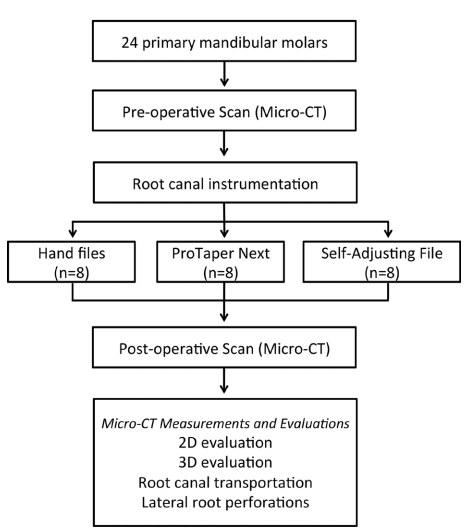
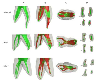
The aim of the study was to evaluate canal preparation in primary molars with hand files, ProTaper Next and Self-Adjusting File (SAF) by 2D and 3D micro-computed tomography (micro-CT) analysis. Canals of 24 primary molars were prepared with hand files (HF), ProTaper Next (PTN) and SAF (n=8/group). The teeth were scanned before and after root canal preparation and the pre- and postoperative micro-CT images were reconstructed. Changes in 2D (area, perimeter, roundness, minor and major diameter) and 3D [volume, surface area, structure model index (SMI)] morphological parameters, as well as canal transportation and lateral perforations were evaluated (Kruskal-Wallis and ANOVA; a=0.05). SAF presented smaller changes in minor diameter, volume and surface area compared with HF and PTN (p<0.05). PTN presented more circular canals after preparation. 3D analysis revealed greater transportation in HF. PTN and SAF presented more centered canal preparation, especially in curved areas. SAF and HF presented, respectively, the lowest (0.05±0.02 and 0.07±0.04) and highest (0.14±0.11 and 0.29±0.17) apical transportation. There were fewer lateral perforations in SAF (4.2%) and PTN (7.7%) than in HF (47.8%) (p<0.05). In primary molars, mechanical preparation showed better shaping ability than hand files, promoting more centered preparations and lower occurrence of lateral perforations and canal transportation. Clinical Relevance: Manual instrumentation is still reported as the main choice in the primary teeth preparation; however, studies have shown limitations in its use. The morphological characteristics of primary teeth and the limited knowledge of shaping procedures in these teeth using mechanical preparation become a challenge for clinical practice and might impair the predictability of endodontic treatment.
Key Words:
primary teeth; root canal preparation; micro-computed tomography; self-adjusting file; rotary system.
 Thumbnail
 Thumbnail
 Thumbnail
 Thumbnail