BACKGROUND:
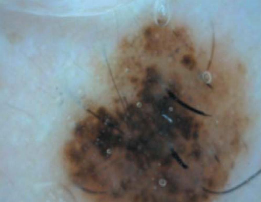
The incidence of cutaneous melanoma is increasing worldwide. Since it is an aggressive neoplasm, it is difficult to treat in advanced stages; early diagnosis is important to heal the patient. Melanocytic nevi are benign pigmented skin lesions while atypical nevi are associated with the risk of developing melanoma because they have a different histological pattern than common nevi. Thus, the clinical diagnosis of pigmented lesions is of great importance to differentiate benign, atypical and malignant lesions. Dermoscopy appeared as an auxiliary test in vivo, playing an important role in the diagnosis of pigmented lesions, because it allows the visualization of structures located below the stratum corneum. It shows a new morphological dimension of these lesions to the dermatologist and allows greater diagnostic accuracy. However, histopathology is considered the gold standard for the diagnosis.
OBJECTIVES:
To establish the sensitivity and specificity of dermoscopy in the diagnosis of pigmented lesions suspected of malignancy (atypical nevi), comparing both the dermatoscopic with the histopathological diagnosis, at the Dermatology Service of the outpatient clinic of Hospital de Base, São José do Rio Preto, SP.
METHODS:
Analysis of melanocytic nevi by dermoscopy and subsequent biopsy on suspicion of atypia or if the patient so desires, for subsequent histopathological diagnosis.
RESULTS:
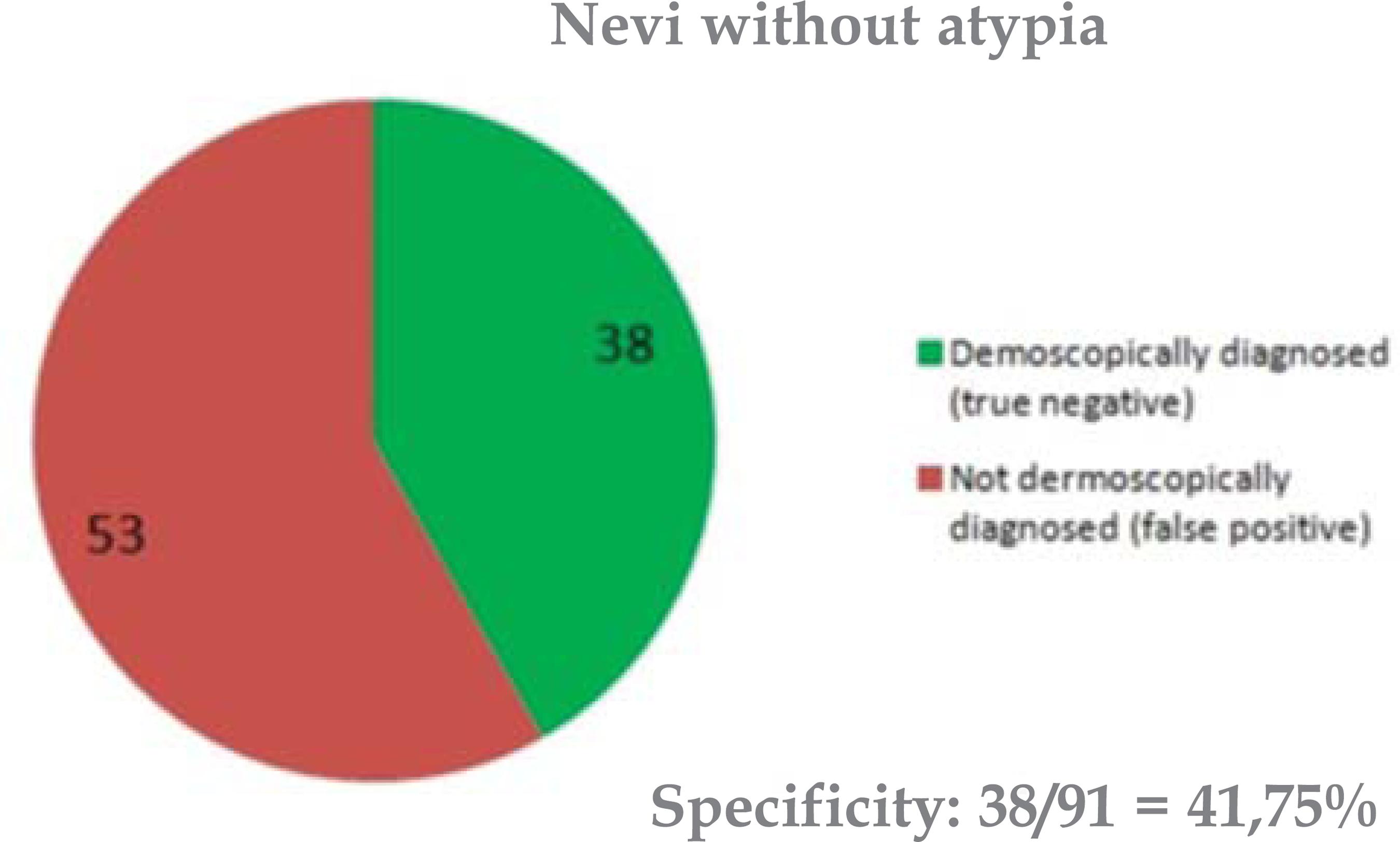
Sensitivity: 93%. Specificity: 42%.
CONCLUSIONS:
Dermoscopy is a highly sensitive method for the diagnosis of atypical melanocytic nevi. Despite the low specificity with many false positive diagnoses, the method is effective for scanning lesions with suspected features of malignancy.
Dermoscopy; Diagnosis; Nevus, pigmented