Abstract
BACKGROUND:
Basal cell carcinoma is the most frequent cancer in fair-skinned populations and dermoscopy is an important, non-invasive technique that aids in the diagnosis of Basal cell carcinoma.
OBJECTIVES:
The aim of this study was to evaluate the relationship between histopathological subtypes and dermoscopic features of Basal cell carcinoma.
METHODS:
This study included 98 patients with clinically and histopathologically confirmed Basal cell carcinomas. The dermoscopic features of the lesions from each patient were analyzed before the histopathological findings were evaluated.
RESULTS:
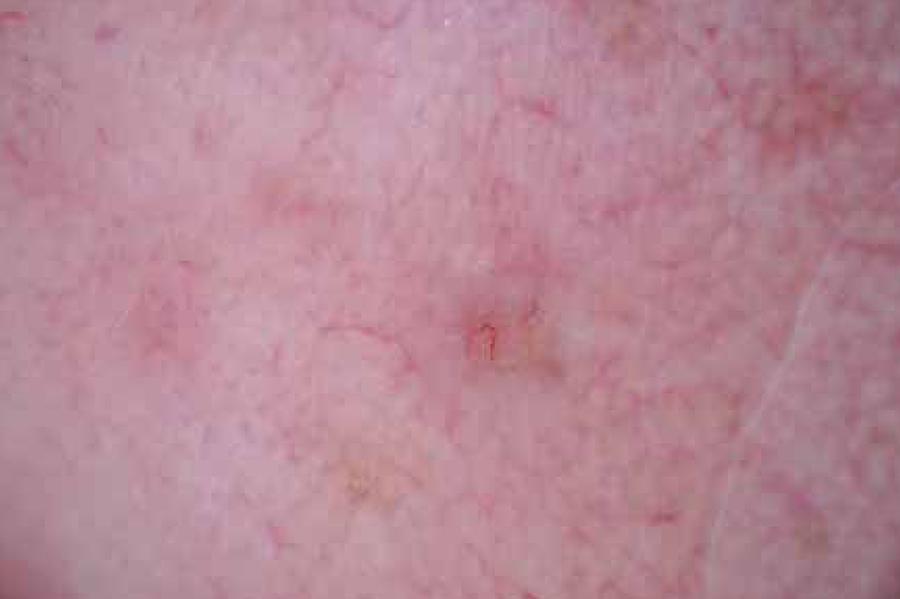
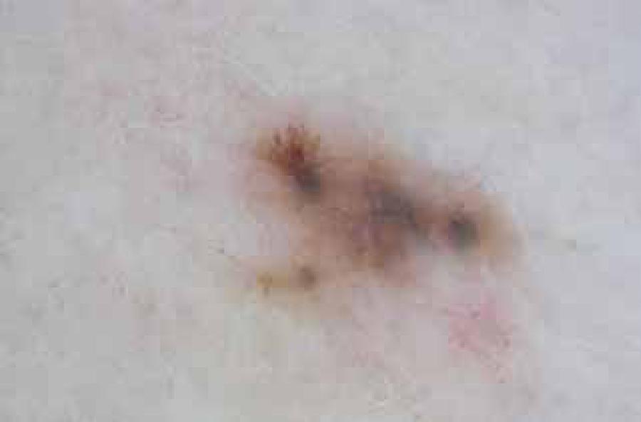
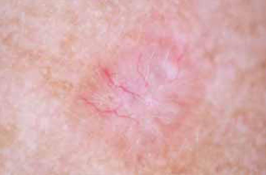
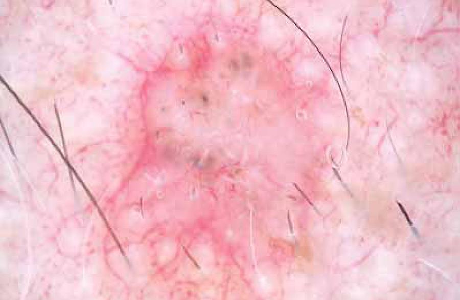
Dermoscopic structures were observed in all 98 patients and irregular vascularity was identified in 78 patients (79.6%). The most common vascular pattern was the presence of arborizing vessels (42 patients, 42.9%) followed by arborizing microvessels (21 patients, 21.4%) and short fine telangiectasias (SFTs; 15 patients, 15.3%). White streaks (38 patients, 38.8%), translucency (31 patients, 31.6%), a milky-pink to red background (42 patients, 42.9%), and erosion/ulceration (29 patients, 29.6%) were also observed. Pigmented islands were seen as blue-gray globules (7 patients, 7.1%) and blue-gray ovoid nests (42 patients, 42.9%). The pigment distribution pattern was maple leaf-like areas in 9 patients (9.2 %) and spoke wheel-like areas in 6 patients (6.1%).
CONCLUSIONS:
Basal cell carcinomas show a wide spectrum of dermoscopic features. Arborizing vessels were the most common dermoscopic findings in Basal cell carcinomas, while superficial Basal cell carcinomas displayed mainly milky-pink to red areas, and arborizing microvessels. The most common dermoscopic features of pigmented types were islands of pigment (blue-gray globules, blue-gray ovoid nests). In conclusion, dermoscopy can be used as a valuable tool for the diagnosis of Basal cell carcinomas and prediction of their histopathological subtypes.
Keywords:
Carcinoma, basal cell; Dermoscopy; Neoplasms, basal cell