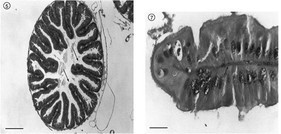
The present research describes the histology of the midgut, gastric caeca, and pyloric valve of Tropidacris collaris (Stoll, 1813), (Orthopetera: Romaleidae). We used light microscopy, staining (Gomori's trichrome and periodic acid-Schiff (PAS)), and a routine histological analysis method (hematoxilin-eosin). The insects were obtained from, and also bred in, the Laboratory of Entomology, Department of Biology, of the Rural Federal University of Pernambuco (UFRPE). The collected material was fixed in alcoholic Boüin and embedded in paraplast. The results demonstrated that the midgut wall is composed of an inner epithelial layer and two outer layers of striate muscles: one internal (circular) and the other external (longitudinal), with connective tissue between the muscle fibers. The epithelium is single-layered, with two cell types: regenerative and elongated columnar. The gastric caeca presents muscle layers similar to those of the midgut. Simple columnar epithelium lines the gastric caeca, which presents villi and projects towards the lumen. The pyloric valve is of striate muscle tissue, covered by a single epithelial-cell layer.
histology; midgut; gastric caeca; pyloric valve; Tropidacris collaris