Abstract
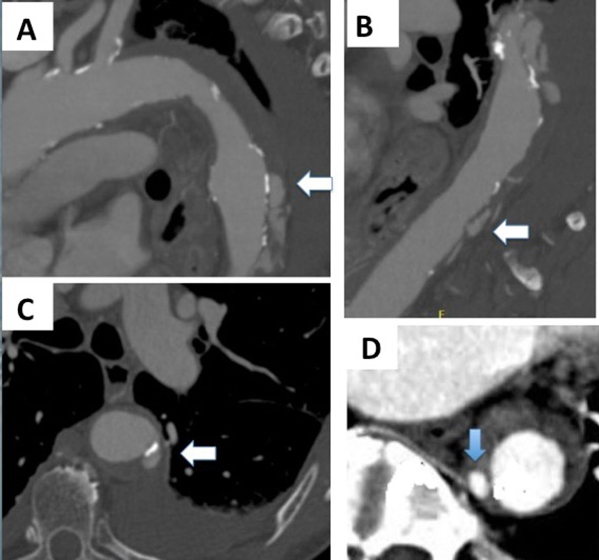
The natural histories of intramural hematoma (IMH) and penetrating atherosclerotic ulcer (PAU) are highly variable as they may progress to aneurysm formation, rupture, or dissection, or even resolve, in the specific case of IMH. Imaging plays an increasingly important role in clinical and surgical management of IMH and PAU. In contrast to ulcer-like projections, images of intramural blood pools have not been widely reported in CT studies of patients with IMH. Understanding the imaging characteristics and the natural course of each of these entities would help clinicians and surgeons to identify patients at greatest risk for bad prognosis and may improve outcomes. This paper discusses the pathophysiology of these entities, the controversies regarding their natural history, and the prognostic factors that should be identified in CT scans.
Keywords:
acute aortic syndrome; intramural aortic hematoma; aortic hematoma; penetrating ulcers of aorta
 Thumbnail
 Thumbnail
 Thumbnail