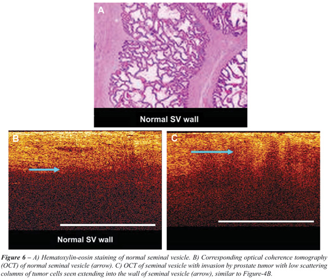
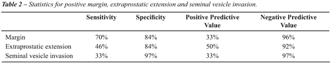
OBJECTIVE: Optical coherence tomography (OCT) is a unique technology, developed to provide high resolution, cross sectional images of human tissue. The objective of this study was to explore the feasibility of OCT for the evaluation of positive surgical margins and extra capsular extension in robotic prostatectomy specimens and compare it to histopathology. MATERIAL AND METHODS: Radical prostatectomy was performed in 100 patients. Twenty OCT images of each specimen were taken from the base of the seminal vesicles (SV), apical and vesicle margins, peripheral and posterolateral area and any palpable nodule. Predictions were made regarding positive surgical margin, SV involvement, capsular invasion and compared with the final histopathology. RESULTS: A total of 2000 OCT images were taken and analyzed. Out of 100 specimens, 85 had T2 disease, 15 had T3 disease with a median Gleason's score of 7 (range 6 to 9) and 10 had positive surgical margins. We predicted 21 specimens to have positive margins based on OCT images out of which 7 were truly positive and 14 were falsely positive. Based on OCT images, 79 specimens were predicted to have negative margins out of which 76 were truly negative and 3 were falsely negative. We found the sensitivity, specificity, positive predictive value and negative predictive value to be 70%, 84%, 33% and 96% respectively. CONCLUSION: Our initial feasibility study established the template for the visual OCT characteristics of the prostate, SV and cancerous tissue. The negative predictive value of evaluating surgical margins was high.
prostate cancer; prostatectomy; laparoscopy; robotic assisted; tomography; optical coherence