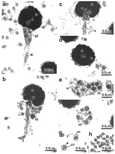
Fig. 1.
Spermiogenesis process of the Alestidae. Figure subunits are longitudinal sections of spermatids corresponding to (a, b) Brycinus lateralis, (c) Brycinus imberi, (d) Brachypetersius altus, (e) Alestopetersius compressus, (f) Micralestes acutidens. The flagellum is initially lateral to the nucleus. The nuclear rotation towards the flagellar axis is present. The nuclear rotation can be complete reaching 90 degrees as represented by the late spermatid of Alestopetersius compressus (e), or incomplete reaching about 20 degrees as occurs in the late spermatid of Micralestes acutidens (f). Legends: c: cytoplasm, d: distal centriole, f: flagellum, m: mitochondria, n: nucleus, p: proximal centriole, pi: midpiece, v: vesicle, asterisk: cytoplasmic canal, arrow: nuclear fossa.
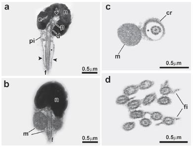
Fig. 2.
Spermatozoon of Bryconalestes longipinnis. a, b, c: longitudinal sections. d, e, f, g, h: transverse sections from top to posterior region. The nucleus (n) is spherical and lies medial to the flagellum (f). The centriolar complex (p, d) and nuclear fossa (arrow) is medial. The proximal centriole (p) is in a right angle relative to the distal centriole (d). The cytoplasmic canal is present (asterisk). The midpiece (pi) is asymmetrical, and contains the spherical mitochondria (m), vesicles (v) and a cytoplasmic sleeve (arrowhead). The flagellum has lateral fins (fi). Legends: cr: concentric membranous rings, d: distal centriole, f: flagellum, fi: lateral fins, m: mitochondria, n: nucleus, p: proximal centriole, v: vesicle, asterisk: cytoplasmic canal, arrow: nuclear fossa, arrowhead: cytoplasmic sleeve.
Fig. 3.
Spermatozoon of Alestopetersius compressus. a, b, c: longitudinal sections. d, e, f: transverse sections from top to posterior region. The nucleus (n) is spherical and lies eccentric to the flagellum (f). The centriolar complex (p, d) and nuclear fossa (arrow) is eccentric. The proximal centriole (p) is oblique relative to the distal centriole (d). The cytoplasmic canal is present (asterisk). The midpiece (pi) is strongly asymmetrical, and contains the spherical mitochondria (m) and vesicles (v). The flagellar membrane has some vesicles and the flagellum. Legends: d: distal centriole, f: flagellum, m: mitochondria, n: nucleus, p: proximal centriole, v: vesicle, asterisk: cytoplasmic canal, arrow: nuclear fossa.
Fig. 4.
Spermatozoon of Brachypetersius altus. a, b, c: longitudinal sections. d, e, f, g, h, i: transverse sections from top to posterior region. The nucleus (n) is spherical and lies medial to the flagellum (f). The centriolar complex (p, d) and nuclear fossa (arrow) is medial. The proximal centriole (p) is in a right angle relative to the distal centriole (d). The cytoplasmic canal is present (asterisk). The midpiece (pi) is asymmetrical, and contains the branched or "c" shape mitochondria (m), vesicles (v) and a cytoplasmic sleeve (arrowhead). Legends: a: axoneme, cr: concentric membranous rings, d: distal centriole, f: flagellum, m: mitochondria, n: nucleus, p: proximal centriole, v: vesicle, asterisk: cytoplasmic canal, arrow: nuclear fossa, arrowhead: cytoplasmic sleeve
Fig. 5.
Spermatozoon of Brycinus imberi. a, b, c, d: longitudinal sections. e, f, g, h, i, j, k: transverse sections from top to posterior region. The nucleus (n) is spherical and lies medial to the flagellum (f). The centriolar complex (p, d) and nuclear fossa (arrow) is medial. The proximal centriole (p) is in a right angle relative to the distal centriole (d). The cytoplasmic canal is present (asterisk). The midpiece (pi) is asymmetrical, and contains the spherical mitochondria (m), vesicles (v) and a cytoplasmic sleeve (arrowhead). Legends: a: axoneme, cr: concentric membranous rings, d: distal centriole, e: electron-lucent areas, f: flagellum, m: mitochondria, n: nucleus, p: proximal centriole, v: vesicle, asterisk: cytoplasmic canal, arrow: nuclear fossa, arrowhead: cytoplasmic sleeve.
Fig. 6.
Spermatozoon of Brycinus lateralis. a, b, c: longitudinal sections. d, e, f, g, h, i, j: transverse sections from top to posterior region. The nucleus (n) is spherical and lies medial to the flagellum (f). The centriolar complex (p, d) and nuclear fossa (arrow) is medial. The proximal centriole (p) is in a right angle relative to the distal centriole (d). The cytoplasmic canal is present (asterisk). The midpiece (pi) is asymmetrical, and contains the spherical mitochondria (m), vesicles (v) and a cytoplasmic sleeve (arrowhead). Legends: a: axoneme, cr: concentric membranous rings, d: distal centriole, e: electron-lucent areas, f: flagellum, m: mitochondria, n: nucleus, p: proximal centriole, v: vesicle, asterisk: cytoplasmic canal, arrow: nuclear fossa, arrowhead: cytoplasmic sleeve.
Fig. 7.
Spermatozoon of Micralestes acutidens. a, a-inset: longitudinal sections. b, c, d: transverse sections from top to posterior region. The nucleus (n) is spherical and lies strongly eccentric to the flagellum (f). The centriolar complex (p, d) and nuclear fossa (arrow) is strongly eccentric. The proximal centriole (p) is lateral and perpendicular relative to the distal centriole (d). The cytoplasmic canal is present (asterisk). The midpiece (pi) is strongly asymmetrical, and contains the spherical mitochondria (m) and vesicles (v). The membrane of the midpiece (me) has a striated aspect along its length (a, a- inset). Legends: cr: concentric membranous rings, d: distal centriole, f: flagellum, m: mitochondria, me: membrane of the midpiece, n: nucleus, p: proximal centriole, v: vesicle, asterisk: cytoplasmic canal, arrow: nuclear fossa.
Fig. 8.
Spermatozoon of Rhabdalestes rhodesiensis. a, d: longitudinal sections. b, c: transverse sections from top to posterior region. The nucleus (n) is spherical and lies strongly eccentric to the flagellum (f). The centriolar complex (p, d) and nuclear fossa (arrow) is strongly eccentric. The proximal centriole (p) is anterior and oblique relative to the distal centriole (d). The cytoplasmic canal is present (asterisk). The midpiece (pi) is strongly asymmetrical, and contains the spherical mitochondria (m) and vesicles (v). The membrane of the midpiece (me) has a striated aspect along its length (c, d). Legends: d: distal centriole, f: flagellum, m: mitochondria, me: membrane of the midpiece, n: nucleus, p: proximal centriole, v: vesicle, asterisk: cytoplasmic canal, arrow: nuclear fossa.
Fig. 9.
Spermatozoon of Chalceus epakros. a, b: longitudinal sections. c, d: transverse sections from top to posterior region. The nucleus (n) is spherical and lies medial to the flagellum (f). The centriolar complex (p, d) and nuclear fossa (arrow) is medial. The proximal centriole (p) is in a right or oblique angle relative to the distal centriole (d). The cytoplasmic canal is present (asterisk). The midpiece (pi) is asymmetrical, and contains only one "c" shape mitochondria (m), vesicles (v) and a cytoplasmic sleeve (arrowhead). The flagellum has lateral fins (fi). Legends: cr: concentric membranous rings, d: distal centriole, e: electron-lucent areas, f: flagellum, fi: flagellar fins, m: mitochondria, n: nucleus, p: proximal centriole, v: vesicle, asterisk: cytoplasmic canal, arrow: nuclear fossa, arrowhead: cytoplasmic sleeve.