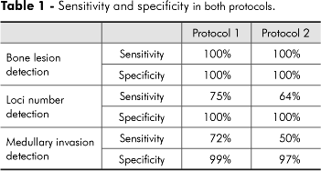
Computed tomography is the choice technique to assess oral and maxillofacial osseous lesions because it provides hard and soft tissues visualization in one examination without superimposition of surrounding structures. This examination offers a significant advance in maxillofacial lesions detection with an excellent anatomic resolution. The aim of this research was to evaluate the validity of two protocols, in axial sections, in simulated mandibular lesions. Two CT protocols were obtained in dry mandibles in which perforations were done simulating lesions. Two observers, previously calibrated, evaluated the images according to different parameters. The results indicated that the sensitivity and specificity in lesion detection were 100% for both protocols, but the detection of loci number of multilocular lesions and the location and detection of medullar invasion obtained reduced validity values, which were influenced by the acquisition protocol. We concluded that thinner axial slices and reconstructions were more effective in detecting early medullar invasion and loci number. Thicker protocols were not considered appropriate to detect multilocular lesions and early stages of medullar invasion.
Tomography; X-ray computed; Bone diseases; Mandible