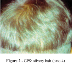
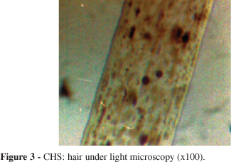
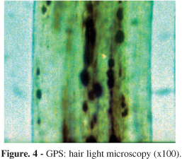
PURPOSE: To study and compare the appearance of hairs from patients with Chédiak-Higashi and Griscelli-Prunieras syndromes under light and polarized light microscopy. METHOD: Hairs from 2 Chédiak-Higashi and 2 Griscelli-Prunieras patients were obtained and examined under normal and polarized light microscopy. RESULTS: Under light microscopy, hairs from Chédiak-Higashi patients presented evenly distributed, regular melanin granules, larger than those seen in normal hairs. Under polarized light microscopy, shafts exhibited a bright and polychromatic refringence appearance. In contrast, hair from Griscelli-Prunieras patients, under light microscopy, exhibited bigger and irregular melanin granules, distributed mainly near the medulla. Under polarized light microscopy, shafts appeared monotonously white. CONCLUSION: Light microscopic examination of hair shafts of patients with Chédiak-Higashi or Griscelli-Prunieras syndrome reveals subtle differences that are useful in identifying both disorders, but not in distinguishing between them. We provide evidence that polarized light microscopy of hair shafts, an approach that has not been previously described, aids in differentiating between these syndromes. We propose hair study by polarized light microscopy as a helpful complementary diagnostic method for differential diagnosis between CHS and GPS, especially when the more sophisticated molecular studies are not available.
Chédiak-Higashi; Griscelli-Prunieras; Silvery hair; Polarized light microscopy