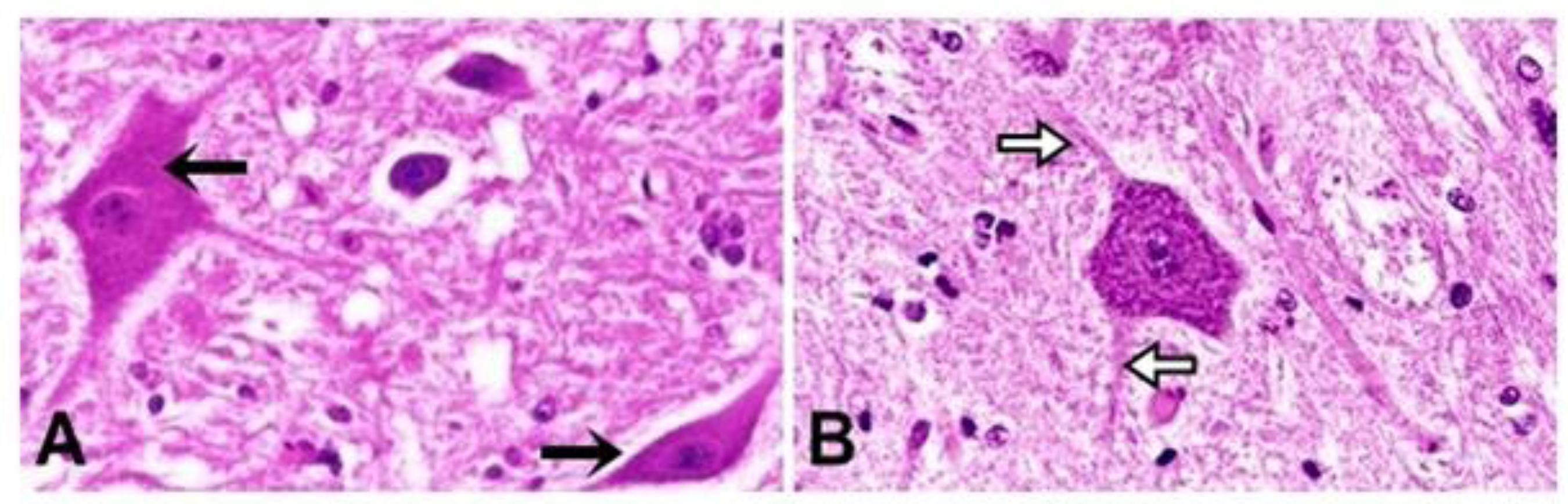
The aim of this work was to analyze the neuron morphology and morphometry of cervical, thoracic and lumbar areas of nonsymptomatic seropositive dogs’ spinal cord for toxoplasmosis. Twenty indefinite-breed adult dogs were used; ten dogs were healthy, with negative serology for toxoplasmosis, and were used as the control group (group 1), and ten dogs were nonsymptomatic but seropositive for toxoplasmosis (group 2). After the microtomy, with interval of 100 micrometers (µm), the histological 5-µm-thick cuts were dyed by hematoxylin-eosin and Masson's trichrome techniques. The glass slides were analyzed under light microscope to examine the neuron morphology. The parameters considered for the morphometric analysis were area, perimeter, maximum diameter, minimum diameter and shape factor of cytoplasm and nucleus of neuron. The results were statistically analyzed by Student’s t test at 5% probability level. The morphological characteristics between the two groups were similar and according to literature. The morphometric results showed that there were changes in neurons size and structure, and increase and loss of star shape were noticed in seropositive animals. The results suggest that the neurons of these dogs, yet nonsymptomatic, can have lost their conductor function.
canine; central nervous system; histology; Toxoplasma gondii