
Abstract
Aim: to evaluate the acute effects of expiratory positive airway pressure on cardiac autonomic modulation in chronic obstructive pulmonary disease patients during spontaneous breathing and slow deep breathing. Methods: 17 patients were evaluated. The R-R intervals were collected (Polar® S810i) during spontaneous breathing (10 minutes) and slow deep breathing (4 minutes), with and without 5 cmH2O expiratory positive airway pressure. Stable signals were analyzed by Kubios®. Heart rate variability indices were computed in time domain and in frequency domain. Results: Expiratory positive airway pressure application affected low frequency (spontaneous breathing: 62.5±4.1 vs slow deep breathing: 28.2±4.2, p<0.001) and high frequency (spontaneous breathing: 37.4±17.3 vs slow deep breathing: 58.9±18.1, p<0.001). Interactions were observed between expiratory positive airway pressure effect and slow deep breathing effect for low frequency (p<0.001), high frequency (p<0.001) and low frequency/high frequency ratio (p<0.001). When patients were stratified by disease’s severity, we identified a significant low frequency reduction (p<0.001) and high frequency increase (p<0.001) for all stages when slow deep breathing was associated with expiratory positive airway pressure. Conclusion: A 5 cmH2O expiratory positive airway pressure during spontaneous and slow deep breathing can elicit an acute response, resulting in a cardiac autonomic control improvement in moderate-to-very severe patients.
Key words
autonomic nervous system; COPD; HRV; positive-pressure respiration; sinus arrhythmia
INTRODUCTION
Sympathetic and parasympathetic control, peripheral oxygen saturation (SpO2), pulmonary stretch baroreceptors and cardiac and pulmonary reflexes control are altered in chronic obstructive pulmonary disease (COPD) patients (Pinsky 2005PINSKY MR. 2005. Cardiovascular issues in respiratory care. Chest 128(Suppl 2): 592S-597S., Reis et al. 2010aREIS MS, ANA P, RODRIGO P, ANICETO IAV, CATAI AM & BORGHI-SILVA A. 2010b. Autonomic control of heart rate in patients with chronic cardiorespiratory disease and in healthy participants at rest and during a respiratory sinus arrhythmia maneuver. Rev Bras Fisioter 14(2): 106-113., Mazzuco et al. 2015MAZZUCO A, MEDEIROS WM, SPERLING MPR, DE SOUZA AS, ALENCAR MCN, ARBEX FF, NEDER JA, ARENA R & BORGHI-SILVA A. 2015. Relationship between linear and nonlinear dynamics of heart rate and impairment of lung function in COPD patients. Int J Chron Obstruct Pulmon Dis 10: 1651-1661.). The bronchoconstriction mechanism, hypoxia, hypercapnia and systemic inflammation may explain the potential sympatho-vagal imbalance in COPD patients, leading to higher risk of morbimortality (Highcock et al. 2003HIGHCOCK MP, SHNEERSON JM & SMITH IE. 2003. Increased ventilation with NiIPPV does not necessarily improve exercise capacity in COPD. Eur Respir J 22(1): 100-105., Borghi-Silva et al. 2009BORGHI-SILVA A, ARENA R, CASTELLO V, SIMÕES RP, MARTINS LEB, CATAI AM & COSTA D. 2009. Aerobic exercise training improves autonomic nervous control in patients with COPD. Respir Med 103(10): 1503-1510., Roque et al. 2014ROQUE AL ET AL. 2014. Chronic obstructive pulmonary disease and heart rate variability: a literature update. Int Arch of Med 7: 43., Nicolino et al. 2015NICOLINO J, RAMOS D, LEITE MR, RODRIGUES FM, DE ALENCAR SILVA BS, TACAO GY, DE TOLEDO AC, VANDERLEI LC & RAMOS EM. 2015. Analysis of autonomic modulation after an acute session of resistance exercise at different intensities in chronic obstructive pulmonary disease patients. Int J Chron Obstruct Pulmon Dis 10: 223-229., Chhabra et al. 2015CHHABRA SK, GUPTA M, RAMASWAMY S, DASH DJ, BANSAL V & DEEPAK KK. 2015. Cardiac sympathetic dominance and systemic inflammation in COPD. COPD 12(5): 552-559.).
Scientific evidence suggests that non-invasive ventilation methods can positively modulate the autonomic control of the heart rate (HR) (Borghi-Silva et al. 2008BORGHI-SILVA A, REIS MS, MENDES RG, PANTONI CBF, SIMÕES RP, MARTINS LEB & CATAI AM. 2008. Noninvasive ventilation acutely modifies heart rate variability in chronic obstructive pulmonary disease patients. Respir Med 102(8): 1117-1123.). Different NIV modes are able to increase ventilation and SpO2 and decrease pulmonary complications and dyspnea symptoms, reducing respiratory muscle load and intrathoracic pressure during rest and physical exercise and improving physical and functional performance (Pinsky 2005PINSKY MR. 2005. Cardiovascular issues in respiratory care. Chest 128(Suppl 2): 592S-597S., Reis et al. 2010aREIS MS, ANA P, RODRIGO P, ANICETO IAV, CATAI AM & BORGHI-SILVA A. 2010b. Autonomic control of heart rate in patients with chronic cardiorespiratory disease and in healthy participants at rest and during a respiratory sinus arrhythmia maneuver. Rev Bras Fisioter 14(2): 106-113., Chhabra et al. 2015CHHABRA SK, GUPTA M, RAMASWAMY S, DASH DJ, BANSAL V & DEEPAK KK. 2015. Cardiac sympathetic dominance and systemic inflammation in COPD. COPD 12(5): 552-559.). Recent studies investigating bilevel positive airway pressure in COPD patients have shown that this method is effective to improve pulmonary ventilation, neural control of HR and sympatho-vagal balance, reducing blood pressure and illness exacerbation frequency (Skyba et al. 2007SKYBA P, JOPPA P, OROLIN M & TKACOVA R. 2007. Blood pressure and heart rate variability response to noninvasive ventilation in patients with exacerbations of chronic obstructive pulmonary disease. Physiol Res 56(5): 527-533., Borghi-Silva et al. 2008BORGHI-SILVA A, REIS MS, MENDES RG, PANTONI CBF, SIMÕES RP, MARTINS LEB & CATAI AM. 2008. Noninvasive ventilation acutely modifies heart rate variability in chronic obstructive pulmonary disease patients. Respir Med 102(8): 1117-1123.). The major effect of the application of an expiratory positive airway pressure (EPAP) device consists in increasing the expiratory flow and decreasing pulmonary hyperinflation; moreover, it maintains upper airways patency, prevents atelectasis and improves mucociliary clearance (Nicolini et al. 2013NICOLINI A, MERLIAK F & BARLASCINI C. 2013. Use of positive expiratory pressure during six minute walk test: results in patients with moderate to severe chronic obstructive pulmonary disease. Multidiscip Respir Med 8(1): 19.).
However, there is still lack of information about the effects of EPAP on cardiac autonomic modulation in COPD patients during spontaneous breathing (SB) or during slow deep breathing (DB) induced by respiratory sinus arrhythmia maneuver (RSA-M). The analysis of heart rate variability (HRV) has been widely used to investigate cardiac autonomic nervous activity and cardiorespiratory disease severity (Skyba et al. 2007SKYBA P, JOPPA P, OROLIN M & TKACOVA R. 2007. Blood pressure and heart rate variability response to noninvasive ventilation in patients with exacerbations of chronic obstructive pulmonary disease. Physiol Res 56(5): 527-533., Borghi-Silva et al. 2008BORGHI-SILVA A, REIS MS, MENDES RG, PANTONI CBF, SIMÕES RP, MARTINS LEB & CATAI AM. 2008. Noninvasive ventilation acutely modifies heart rate variability in chronic obstructive pulmonary disease patients. Respir Med 102(8): 1117-1123.). Therefore, the aim of the present study was to evaluate, by HRV analysis, the acute effects of EPAP on cardiac autonomic modulation in COPD patients during SB and DB. We hypothesized that EPAP would have a positive acute effect, with predominance of parasympathetic autonomic modulation during DB and SB in moderate – very severe COPD patients.
MATERIALS AND METHODS
Study design
The study was approved by the Comitê de Ética em Pesquisa da Universidade de Santa Cruz do Sul, protocol n. 1.100.926, and all volunteers signed an informed consent statement prior to participation. Cross-sectional case performed on a convenience non-probability sample in accordance to STROBE statement. Our research was conducted within Programa de Reabilitação Pulmonar do Hospital Santa Cruz (Santa Cruz do Sul, RS, Brazil).
Subjects
After acceptance of the patients, 20 patients with a clinical diagnosis of COPD confirmed by pulmonary function test, with good cognition and no illness exacerbation in the last 15 days were included in the study. Patients with musculoskeletal disorders or neurological sequelae that affected the locomotor system in such a way that would preclude participation in the research protocol, with a clinical diagnosis of lung cancer, current alcoholism, complex arrhythmias, uncontrolled metabolic disease or electrocardiogram (ECG) alterations were excluded from the study (n=3) (Figure 1).
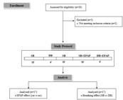
Study protocol phases. SB: spontaneous breathing; DB: slow deep breathing; EPAP: Expiratory Positive Airway Pressure; 10’: 10 minutes; 4’: 4 minutes; 15’: 15 minutes.
Measurements
Patients were evaluated in a laboratory at a temperature of 22°C and relative humidity between 50% and 60%. They were instructed to avoid stimulants and alcoholic drinks and not to perform exhausting physical exercise the day before the test; they were also instructed not to smoke or use bronchodilators for 6 hours before the test and heart rate stimulants. Patients underwent a clinical assessment for surveying sex, age and COPD stage. Afterwards, vital signs were evaluated SpO2 with a portable oximeter (Nonin, Onyx, 9500) and for systolic blood pressure (SBP) and diastolic blood pressure (DBP) an aneroid sphygmomanometer (Welchallyn Durashock DS44, Tycos) and stethoscope (Littman classic II).
Pulmonary function
A digital spirometer (Microloop®, MK8, Care Fusion, Hoechberg, Germany) was used to assess pulmonary function. Spirometry was performed following the American Thoracic Society recommendations, and the results were analyzed according to the values predicted by Pereira et al. 2007PEREIRA CAC, SATO T & RODRIGUES SC. 2007. New reference values for forced spirometry in white adults in Brazil. J Bras Pneumol 33(4): 397-406.. The exam provided measures of the slow vital capacity, the forced vital capacity (FVC), the forced expiratory volume in 1 second (FEV1) and the FEV1/FVC ratio. The severity of airflow limitation in COPD was classified according to the Global Initiative for Chronic Obstructive Lung Disease (GOLD) recommendations and patients were classified as moderate (GOLD II), severe (GOLD III), or very severe (GOLD IV) (GOLD 2017GOLD - GLOBAL INITIATIVE FOR CHRONIC OBSTRUCTIVE LUNG DISEASE. Pocket Guide to COPD Diagnosis, Management and Prevention. 2017. Global Initiative for Chronic Obstructive Lung Disease. Available at: http://www.goldcopd.it/materiale/2017/GOLD_Pocket_2017.pdf. Accessed on 12 dec 2017.
http://www.goldcopd.it/materiale/2017/GO...
).
Respiratory muscle strength
A digital manometer (MDI®, MVD300, Porto Alegre, Brazil) provided measures of the maximum inspiratory pressure (MIP) and the maximum expiratory pressure (MEP). MIP and MEP were obtained from residual volume and total lung capacity, respectively. During the maneuver, the subjects remained seated, wearing nose clips and with a rigid, plastic flanged mouthpiece in place, according to the recommendations for the Brazilian population (Neder et al. 1999NEDER JA, ANDREONI S, LERARIO MC & NERY LE. 1999. Reference values for lung function tests. II. Maximal respiratory pressures and voluntary ventilation. Braz J Med Biol Res 32(6): 719-727.). The values were analyzed as described in literature and expressed as percentage of predicted values (Neder et al. 1999NEDER JA, ANDREONI S, LERARIO MC & NERY LE. 1999. Reference values for lung function tests. II. Maximal respiratory pressures and voluntary ventilation. Braz J Med Biol Res 32(6): 719-727.). Respiratory muscle weakness was determined for a MIP of <60 cmH2O (Beaumont et al. 2015BEAUMONT M ET AL. 2015. Inspiratory muscle training during pulmonary rehabilitation in chronic obstructive pulmonary disease: A randomized trial. Chron Respir Dis 12(4): 305-312.).
Expiratory positive airway pressure
A facial mask connected to a Spring Loaded® (EPAP kit) valve was adapted to the patient’s face and regulated with a cephalic fixator to avoid air escaping. Positive expiratory pressure (PEP) was adjusted to 5 cmH2O, based on previous findings which demonstrated that a 5 cmH2O PEP was safe and determined lower autonomic imbalance in COPD patients (Reis et al. 2010bREIS MS, ARENA R, DEUS AP, SIMÕES RP, CATAI AM & BORGHI-SILVA A. 2010c. Deep breathing heart rate variability is associated with respiratory muscle weakness in patients with chronic obstructive pulmonary disease. Clinics 65(4): 369-375.). Patients remained seated in a comfortable chair with adjustable height and backrest, with their legs at 90 degrees, their feet resting on the ground and their spine erect, for 10 minutes of SB and 4 minutes of DB.
Heart rate variability
The most stable signal RR intervals (iR-R) sections containing 256 points were selected for HRV analysis from the signals recorded during SB and DB, with and without EPAP (Figure 1), DB (4 minutes) and SB were the initial and final minutes were excluded (in order to obtain a stable signal portion). The HR and iR-R were recorded using a telemetric cardiac monitor (Polar® S810i, Kempele, Finland); an elastic band (Polar T31 transmitter) was placed around the patient’s thorax at the level of the lower third of the sternum while the patient was in sitting position, and signals were continuously transmitted to the receiving unit by an electromagnetic field. Recorded data were then transferred to Kubios HRV® analysis software (version 2.2, Matlab Kuopio, Finland) for subsequent analysis.
The time domain analysis: mean HR, HR standard deviation (STD) and RR tri-index. The Frequency domain analysis: low-frequency (LF) and high-frequency (HF) indexes indicates sympathetic and parasympathetic activity respectively and the LF/HF ratio, expressed in normalized units (nu).
Controlled deep breathing
Subjects were instructed to perform a series of deep and slow inspirations and expirations, with a pulmonary volume that varied from the total lung capacity (maximal inspiration) to the residual volume (maximal expiration); each respiratory cycle was performed in 10 seconds, with a 5-second inspiration and a 5-second expiration, resulting in five to six respiratory cycles per minute (Reis et al. 2010bREIS MS, ARENA R, DEUS AP, SIMÕES RP, CATAI AM & BORGHI-SILVA A. 2010c. Deep breathing heart rate variability is associated with respiratory muscle weakness in patients with chronic obstructive pulmonary disease. Clinics 65(4): 369-375.). According to a previous study Hayano et al. 1994HAYANO J, MUKAI S, SAKAKIBARA M, OKADA A, TAKATA K & FUJINAMI T. 1994. Effects of respiratory interval on vagal modulation of heart rate. Am J Physiol 267(1): H33-H40., this controlled breathing technique provides maximal respiratory sinus arrhythmia response that is able to induce maximal parasympathetic modulation on HR control. In order to control the respiratory rhythm, the participants’ breathing rate was monitored and analyzed with a pointer clock and, at the same time, verbal feedback was provided by the researcher, based on their ECG signals and HR plotted on a computer monitor, which confirmed whether or not the respiratory cycle had been performed correctly.
Statistical analysis
Data were analyzed using the Sigmaplot® statistical package (version 11.0, Systat Software Inc., San Jose, CA, USA). Data were checked for normality using the Shapiro–Wilk test and presented descriptively as mean and standard deviation (parametric) or as median and minimum and maximum interval (non-parametric). The results were compared using two-way analysis of variance on ranks, with post-hoc Dunn’s, in order to identify statistically significant differences in: (i) clinical parameters; (ii) HRV time domain parameters; and (iii) HRV frequency domain parameters. For these analyses, subjects were categorized according to EPAP effect (yes vs no) and breathing effect (SB vs DB). Residuals were evaluated under the assumptions of normality, constant variance, and independence (p<0.05).
RESULTS
Sample characteristics
17 patients meeting the inclusion criteria for this study were selected, in which we can observe a higher frequency of male patients, it is possible to observe similar distribution between COPD stages and inspiratory muscle strength. The clinical characteristics of the sample are listed in Table I.
Cardiovascular response parameters during SB and DB, with and without EPAP
The aim of the present study was to evaluate by HRV analysis, the acute effects of EPAP on cardiac autonomic modulation in COPD patients during SB and DB. Our results showed that the EPAP application significantly affected LF (p<0.001) and HF (p<0.001). COPD patients presented a positive response to the EPAP application both during SB, with a sympathetic modulation predominance, and during DB, with a parasympathetic modulation predominance (Table II).
Interaction of EPAP effect and DB effect in COPD patients
When comparing EPAP effect and DB effect, it was observed that an interaction between EPAP effect and DB effect in COPD patients for STD HR (p=0.01), RR tri index (p=0.01), LF (p<0.001), HF (p<0.001) and LF/HF ratio (p<0.001).
HRV frequency domain parameters stratified according to disease staging
When stratifying patients by disease severity, we identified a significant LF reduction and a significant HF increase when DB was associated with EPAP, for all COPD stages, indicating increased parasympathetic modulation and reduced sympathetic activation (Figure 2).

HRV frequency domain parameters stratified according to disease staging. (a) LF- normalized low frequency power; (b) HF- normalized high frequency power.
DISCUSSION
The present study was the first to evaluate the EPAP effect during SB and DB on cardiac autonomic modulation in COPD patients. We observed that an EPAP of 5cmH2O was enough to determine a better autonomic response in COPD patients. In addition, when DB was associated with EPAP, we observed an adequate autonomic response with increase of parasympathetic modulation. Positive effects were observed independently of disease severity.
In the present study, we demonstrated that EPAP was able to correct these autonomic control imbalances during controlled breathing (DB) with parasympathetic accentuation. The use of ventilatory patterns as a therapeutic tool (i.e slow deep breathing) is frequently used for these patients in clinical setting. In this sense, our study brings out the importance of EPAP use as adjunctive therapy to ventilatory patterns, however this should be scientifically improved (Macklem 2010MACKLEM PT. 2010. Therapeutic implications of the pathophysiology of COPD. Eur Respir J 35(3): 676-680.). It is well known that EPAP promotes reduction of respiratory work, improves expiration time, avoids airway collapse, improves pulmonary ventilation and gas exchange and decreases carbon dioxide (CO2) retention (Borghi-Silva et al. 2008BORGHI-SILVA A, REIS MS, MENDES RG, PANTONI CBF, SIMÕES RP, MARTINS LEB & CATAI AM. 2008. Noninvasive ventilation acutely modifies heart rate variability in chronic obstructive pulmonary disease patients. Respir Med 102(8): 1117-1123., Reis et al. 2010cREIS MS, SAMPAIO LM, LACERDA D, DE OLIVEIRA LV, PEREIRA GB, PANTONI CB, THOMMAZO LD, CATAI AM & BORGHI-SILVA A. 2010a. Acute effects of different levels of continuous positive airway pressure on cardiac autonomic modulation in chronic heart failure and chronic obstructive pulmonary disease. Arch Med Sci 6(5): b719-b727.). Reis et al. 2010aREIS MS, ANA P, RODRIGO P, ANICETO IAV, CATAI AM & BORGHI-SILVA A. 2010b. Autonomic control of heart rate in patients with chronic cardiorespiratory disease and in healthy participants at rest and during a respiratory sinus arrhythmia maneuver. Rev Bras Fisioter 14(2): 106-113. also demonstrated that a 5 cmH2O continuous positive airway pressure treatment in COPD patients was safer and produced better effects on their ventilatory parameters and autonomic balance than when a pressure of 10 cmH2O was applied.
DB naturally affects the autonomic nervous system, inducing an increased parasympathetic response (Roque et al. 2014ROQUE AL ET AL. 2014. Chronic obstructive pulmonary disease and heart rate variability: a literature update. Int Arch of Med 7: 43.). In our study, DB without the use of EPAP induced increased sympathetic response and reduced parasympathetic response in COPD patients, which would characterize an adverse response to DB. We can emphasize that this cardiac autonomic modulation abnormality in COPD patients has been previously explained by reduced FEV1 and increased bronchoconstriction, hypoxemia and respiratory muscle weakness, which are pathophysiological characteristics of these patients (Goulart et al. 2016GOULART CL, SIMON JC, SCHNEIDERS PB, SAN MARTIN EA, CABIDDU R, BORGHI-SILVA A, TRIMER R & GONÇALVES DA SILVA AL. 2016. Respiratory muscle strength effect on linear and nonlinear heart rate variability parameters in COPD patients. Int J Chron Obstruct Pulmon Dis 11: 1671-1677.). COPD patients can present a fast and short breathing pattern due to reduced diaphragmatic incursion, which is limited by pulmonary hyperinflation. Thus, the ergoreceptor may be activated early and determine a fast-central response in cardiac autonomic control (Reissmann et al. 2000REISSMANN HK, RANIERI VM, GOLDBERG P & GOTTFRIED SB. 2000. Continuous positive airway pressure facilitates spontaneous breathing in weaning chronic obstructive pulmonary disease patients by improving breathing pattern and gas exchange. Intensive Care Med 26(12): 1764-1772.).
During SB with EPAP COPD patients showed an increase in sympathetic modulation, we believe that this adverse behavior may be due to the disease’s pathophysiology, primarily due to pulmonary hyperinflation. In the Reis et al. 2010aREIS MS, ANA P, RODRIGO P, ANICETO IAV, CATAI AM & BORGHI-SILVA A. 2010b. Autonomic control of heart rate in patients with chronic cardiorespiratory disease and in healthy participants at rest and during a respiratory sinus arrhythmia maneuver. Rev Bras Fisioter 14(2): 106-113., in which it applied CPAP of 10cmH2O in COPD patients, they highlighted altered autonomic regulation, leading to an increase in sympathetic activity and a reduction in parasympathetic activity, which is not favorable, as it has been related to diseases and an increased risk of mortality.
In our study COPD patients in distinct stages of disease severity benefited from the use of 5 cmH2O EPAP, presenting a parasympathetic response during DB; this finding has great clinical relevance since several studies report an inadequate response during DB without EPAP in patients with COPD (Reis et al. 2010aREIS MS, ANA P, RODRIGO P, ANICETO IAV, CATAI AM & BORGHI-SILVA A. 2010b. Autonomic control of heart rate in patients with chronic cardiorespiratory disease and in healthy participants at rest and during a respiratory sinus arrhythmia maneuver. Rev Bras Fisioter 14(2): 106-113., c, Goulart et al. 2016GOULART CL, SIMON JC, SCHNEIDERS PB, SAN MARTIN EA, CABIDDU R, BORGHI-SILVA A, TRIMER R & GONÇALVES DA SILVA AL. 2016. Respiratory muscle strength effect on linear and nonlinear heart rate variability parameters in COPD patients. Int J Chron Obstruct Pulmon Dis 11: 1671-1677.). In COPD, the respiratory system is primarily hampered by the additional elastic load associated with dynamic hyperinflation. These factors predispose patients to respiratory failure by increasing the load on the respiratory muscles, while decreasing their mechanical efficiency and capacity for generating maximal pressure (Reissmann et al. 2000REISSMANN HK, RANIERI VM, GOLDBERG P & GOTTFRIED SB. 2000. Continuous positive airway pressure facilitates spontaneous breathing in weaning chronic obstructive pulmonary disease patients by improving breathing pattern and gas exchange. Intensive Care Med 26(12): 1764-1772.). Müller et al. 2017MÜLLER PT, CHRISTOFOLETTI G, KOCH R, ZARDETTI NOGUEIRA JH, PEREIRA PATUSCO LA & CHIAPPA GR. 2017. CPAP and EPAP elicit similar lung deflation in a non-equivalent mode in GOLD 3-4 COPD patients. Clin Respir J 12(4): 1598-1606. highlighted that CPAP and EPAP would reduce static hyperinflation in COPD patients, and that EPAP would demonstrate equivalent effects to CPAP on static hyperinflation indirectly measured by inspiratory capacity.
Considering that there was no significant change in peripheral oxygen saturation, that respiratory rate was controlled during DB and that in all stages of the disease the HRV response to the EPAP application was beneficial and significant, we hypothesize that the decrease of respiratory muscle overload and the delay of early airway collapse, caused by the pneumatic splint acquired through the use of 5 cmH2O EPAP, may be the causal factors of the alterations observed in this study.
Some limitations need to be highlighted, including our difficulty in screening patients with 95% pure sinus beats; moreover, our sample may not be representative of the entire COPD patients, since patients are known to present several types of arrhythmias that compromise the collection and analysis of signals. This study has great significance and importance for the clinical management of patients with COPD; the hereby presented novel results about the EPAP application may contribute to the development of new research tools in the clinical environment as well as to the improvement of the effects of the interventions implemented in pulmonary rehabilitation programs, since the EPAP application could improve ventilation, promote airway stability, reduce air trapping and promote cardiac autonomic balance. In addition, EPAP devices are easy to apply and accessible for patients with COPD.
Our results suggest that the application of 5 cmH2O EPAP during SB and DB is able to elicit an acute response, which results in a cardiac autonomic control improvement, in moderate-to-very severe COPD patients. Future studies are needed to assess the long-term effects of the EPAP application on HRV, in order to consider its inclusion in pulmonary rehabilitation programs as an adjunct treatment tool for these subjects.
ACKNOWLEGMENTS
We would like to thank our volunteers patients for participating and making possible this research. Financial support: Universidade de Santa Cruz do Sul- UNISC, Hospital Santa Cruz e Conselho Nacional de Desenvolvimento Científico e Tecnológico (CNPq); Fundação de Amparo à Pesquisa do Estado de São Paulo (FAPESP). (n. 2013/23013-0 and 2015/04101-1); CAPES -Programa Atração de Jovens Talentos (n. 88881.062123/2014-01 // 88887.067710/2014-00).
REFERENCES
- BEAUMONT M ET AL. 2015. Inspiratory muscle training during pulmonary rehabilitation in chronic obstructive pulmonary disease: A randomized trial. Chron Respir Dis 12(4): 305-312.
- BORGHI-SILVA A, ARENA R, CASTELLO V, SIMÕES RP, MARTINS LEB, CATAI AM & COSTA D. 2009. Aerobic exercise training improves autonomic nervous control in patients with COPD. Respir Med 103(10): 1503-1510.
- BORGHI-SILVA A, REIS MS, MENDES RG, PANTONI CBF, SIMÕES RP, MARTINS LEB & CATAI AM. 2008. Noninvasive ventilation acutely modifies heart rate variability in chronic obstructive pulmonary disease patients. Respir Med 102(8): 1117-1123.
- CHHABRA SK, GUPTA M, RAMASWAMY S, DASH DJ, BANSAL V & DEEPAK KK. 2015. Cardiac sympathetic dominance and systemic inflammation in COPD. COPD 12(5): 552-559.
- GOLD - GLOBAL INITIATIVE FOR CHRONIC OBSTRUCTIVE LUNG DISEASE. Pocket Guide to COPD Diagnosis, Management and Prevention. 2017. Global Initiative for Chronic Obstructive Lung Disease. Available at: http://www.goldcopd.it/materiale/2017/GOLD_Pocket_2017.pdf Accessed on 12 dec 2017.
» http://www.goldcopd.it/materiale/2017/GOLD_Pocket_2017.pdf - GOULART CL, SIMON JC, SCHNEIDERS PB, SAN MARTIN EA, CABIDDU R, BORGHI-SILVA A, TRIMER R & GONÇALVES DA SILVA AL. 2016. Respiratory muscle strength effect on linear and nonlinear heart rate variability parameters in COPD patients. Int J Chron Obstruct Pulmon Dis 11: 1671-1677.
- HAYANO J, MUKAI S, SAKAKIBARA M, OKADA A, TAKATA K & FUJINAMI T. 1994. Effects of respiratory interval on vagal modulation of heart rate. Am J Physiol 267(1): H33-H40.
- HIGHCOCK MP, SHNEERSON JM & SMITH IE. 2003. Increased ventilation with NiIPPV does not necessarily improve exercise capacity in COPD. Eur Respir J 22(1): 100-105.
- MACKLEM PT. 2010. Therapeutic implications of the pathophysiology of COPD. Eur Respir J 35(3): 676-680.
- MAZZUCO A, MEDEIROS WM, SPERLING MPR, DE SOUZA AS, ALENCAR MCN, ARBEX FF, NEDER JA, ARENA R & BORGHI-SILVA A. 2015. Relationship between linear and nonlinear dynamics of heart rate and impairment of lung function in COPD patients. Int J Chron Obstruct Pulmon Dis 10: 1651-1661.
- MÜLLER PT, CHRISTOFOLETTI G, KOCH R, ZARDETTI NOGUEIRA JH, PEREIRA PATUSCO LA & CHIAPPA GR. 2017. CPAP and EPAP elicit similar lung deflation in a non-equivalent mode in GOLD 3-4 COPD patients. Clin Respir J 12(4): 1598-1606.
- NEDER JA, ANDREONI S, LERARIO MC & NERY LE. 1999. Reference values for lung function tests. II. Maximal respiratory pressures and voluntary ventilation. Braz J Med Biol Res 32(6): 719-727.
- NICOLINI A, MERLIAK F & BARLASCINI C. 2013. Use of positive expiratory pressure during six minute walk test: results in patients with moderate to severe chronic obstructive pulmonary disease. Multidiscip Respir Med 8(1): 19.
- NICOLINO J, RAMOS D, LEITE MR, RODRIGUES FM, DE ALENCAR SILVA BS, TACAO GY, DE TOLEDO AC, VANDERLEI LC & RAMOS EM. 2015. Analysis of autonomic modulation after an acute session of resistance exercise at different intensities in chronic obstructive pulmonary disease patients. Int J Chron Obstruct Pulmon Dis 10: 223-229.
- PEREIRA CAC, SATO T & RODRIGUES SC. 2007. New reference values for forced spirometry in white adults in Brazil. J Bras Pneumol 33(4): 397-406.
- PINSKY MR. 2005. Cardiovascular issues in respiratory care. Chest 128(Suppl 2): 592S-597S.
- REIS MS, ANA P, RODRIGO P, ANICETO IAV, CATAI AM & BORGHI-SILVA A. 2010b. Autonomic control of heart rate in patients with chronic cardiorespiratory disease and in healthy participants at rest and during a respiratory sinus arrhythmia maneuver. Rev Bras Fisioter 14(2): 106-113.
- REIS MS, ARENA R, DEUS AP, SIMÕES RP, CATAI AM & BORGHI-SILVA A. 2010c. Deep breathing heart rate variability is associated with respiratory muscle weakness in patients with chronic obstructive pulmonary disease. Clinics 65(4): 369-375.
- REIS MS, SAMPAIO LM, LACERDA D, DE OLIVEIRA LV, PEREIRA GB, PANTONI CB, THOMMAZO LD, CATAI AM & BORGHI-SILVA A. 2010a. Acute effects of different levels of continuous positive airway pressure on cardiac autonomic modulation in chronic heart failure and chronic obstructive pulmonary disease. Arch Med Sci 6(5): b719-b727.
- REISSMANN HK, RANIERI VM, GOLDBERG P & GOTTFRIED SB. 2000. Continuous positive airway pressure facilitates spontaneous breathing in weaning chronic obstructive pulmonary disease patients by improving breathing pattern and gas exchange. Intensive Care Med 26(12): 1764-1772.
- ROQUE AL ET AL. 2014. Chronic obstructive pulmonary disease and heart rate variability: a literature update. Int Arch of Med 7: 43.
- SKYBA P, JOPPA P, OROLIN M & TKACOVA R. 2007. Blood pressure and heart rate variability response to noninvasive ventilation in patients with exacerbations of chronic obstructive pulmonary disease. Physiol Res 56(5): 527-533.
Publication Dates
-
Publication in this collection
20 July 2020 -
Date of issue
2020
History
-
Received
20 Apr 2018 -
Accepted
12 Feb 2019


