
Abstract
Solidago microglossa is used as an anti-inflammatory agent in traditional Brazilian medicine, and this work evaluated the anti-inflammatory potential of the crude ethanolic extract of the flowers of S. microglossa in vivo, as assayed by paw edema models induced by carrageenan, prostaglandin E2, bradykinin and compound 48/80. In the chemical profile, we identified compounds by electrospray ionization mass spectrometry and quantified them by HPLC-DAD. Additionally, this study analyzed the potential to activate the in vitro transcriptional activity of PPARγ, which is a nuclear receptor linked to the anti-inflammatory response. It was possible to identify five compounds: quinic acid, quercetin, chlorogenic acid, hyperoside, and rutin. In the paw edema evaluation, it was possible to show the potential of reducing edema during the inflammatory process. The crude ethanolic extract of the flowers of S. microglossa activated PPARγ compared to the full agonist rosiglitazone and in a dose-response manner. It is possible to conclude that the extract of the flowers of S. microglossa showed anti-inflammatory activity, and the phenolic compounds present in this species might be responsible for this activity.
Key words
natural products; popular use; Asteraceae; inflammation; chemical profile
INTRODUCTION
Solidago microglossa is a plant native to South America, and its botanical synonym is Solidago chilensis, which is used in traditional Brazilian medicine and appears in the list of Relação Nacional de Plantas Medicinais de Interesse ao Sistema Único de Saúde (Renisus). Infusions and macerates of the rhizomes, leaves or flowers of S. microglossa are used for the treatment of gastric disorders, intestinal ulcers, healing, rheumatism, and injuries as an anti-inflammatory drug, popularly known as Brazilian Arnica (Goulart et al. 2007, Bieski et al. 2015BIESKI GC ET AL. 2015. Ethnobotanical study of medicinal plants by population of Valley of Juruena Region, Legal Amazon, Mato Grosso, Brazil. J Ethnopharmacol 173: 383-423.). Solidago is the largest representative of the Asteraceae family, containing approximately 120 species distributed throughout the world. This genus is widely used in traditional medicine by many countries, particularly for the treatment of inflammatory and diuretic problems (Blumenthal et al. 1998BLUMENTHAL M, BUSSE WR & GOLDBERG A. 1998. The complete German commission e monographs. Boston: American Botanical Council, Austin and Integrative Medicine Communications 1st ed., 685 p.). Phytochemical studies have identified the presence of monoterpenes, sesquiterpenes, sesquiterpene lactones, triterpenes, steroids, saponins, flavonoids, and cinnamic acid derivatives (Souza et al. 2017SOUZA DMF, SA RD, ARAUJO EL & RANDAU K. 2017. Anatomical, phytochemical and histochemical study of Solidago chilensis Meyen. An Acad Bras Cienc 90: 2107-2120.).
In addition to being used in traditional medicine, especially in developing countries, natural products may be the basis for the study and development of new drugs. Between 1981 and 2014, 4% of newly released drugs were isolated from natural products, while 27% were synthetic compounds. Observing the source of other products, 59.1% were semisynthetic molecules, which are synthetic drugs with pharmacophoric groups based on the structures of natural products or structures that mimic natural products or botanical products (Newman & Cragg 2016NEWMAN DJ & CRAGG GM. 2016. Natural Products as Sources of New Drugs from 1981 to 2014. J Nat Prod 79(3): 629-661.). These data demonstrate the importance of natural product studies for the safety of communities that use these plants as well as for the development of new medicines.
Bearing this in mind, we evaluated the anti-inflammatory potential of the crude ethanolic extract of S. microglossa flowers in vivo using paw edema models induced by carrageenan, prostaglandin E2 and compound 48/80 and identified the compounds present by electrospray ionization mass spectrometry (ESI-MS/MS). To understand the molecular mechanism of the anti-inflammatory effects of the S. microglossa flower extract, we also investigated its effects on the transcriptional activity of PPARγ, a nuclear receptor linked to the anti-inflammatory response.
MATERIALS AND METHODS
Plant material and preparation of the crude extract
S. microglossa (SM) was collected in the city of Nova Friburgo (22°19’54” S; 42°32’28” W) in January 2015. The specimen was identified and deposited in the herbarium of the
Instituto de Biologia da Universidade Estadual de Campinas, deposit number: UEC 177791. This study was registered in the Sistema Nacional de Gestão do Patrimônio Genético e do Conhecimento Tradicional Associado – SISGEN (no A3B47E6). The plant was stabilized and dried in a circulating air oven at a temperature of 40 °C, and the leaves were separated from the flowers. The flowers were ground and subjected to maceration in 99% ethanol with a powder/solvent ratio of 1:5 (mass/volume), and the solvent was changed every 24 h. The extract was dried in a rotary evaporator and stored in an amber bottle at a temperature between 2 and 8 °C.
Analysis of the chemical profile of the extract by ESI-MS/MS
Preliminary analysis of the extract of the plant species was performed by direct infusion in a TQD Acquity mass spectrometer (Micromass-Waters Manchester, England) using an ESI source in negative ionization mode. Samples (1 mg/ml) were dissolved in a solution containing 50/50 (v/v) chromatographic grade methanol (Merck, Darmstadt, Germany) and 0.5% ammonium hydroxide in water. The instrument parameters were as follows: source temperature of 100 °C, capillary voltage of 3.0 kV and a 30 V cone voltage, and a collision energy of 30 V. The constituents were identified by comparison of their ESI-MS/MS fragmentation spectra with commercial standards (quercetin (Sigma-Aldrich® ≥95%), quinic acid (Sigma-Aldrich®) and chlorogenic acid (Sigma-Aldrich® ≥95%)). All compounds were compared with data from the database www.massbank.eu.
HPLC-DAD quantification
Quantification of the main components of the extract were performed on an Agilent HPLC Infinity 1260 equipped with a quaternary gradient pump, automatic injector, temperature control column oven and diode array detector (DAD). For HPLC analysis, an Inertsil® ODS-3 column, 15 cm in length with a 4.6 mm internal diameter and 5 μm particles was used. Analyses were performed at 30 °C, a flow rate of 0.8 mL/min, and an injection volume of 20 µL, and chromatograms were obtained at 326, 354, and 372 nm. The solvents used were 1% acetic acid in water (solvent A) and acetonitrile (solvent B) with the following gradient program: 0-5 min (20% B), 5-15 min (20-40% B), 15-25 min (40-80% B), 25-26 min (80-100% B), 26-30 min (100% B) 30-31 min (100-20% B) and 31-35 min (20% B). The extract was dissolved in methanol and water 80:20 v/v to obtain a 10 mg/mL dilution. This solution was cleaned with a C-18 solid phase extraction cartridge (SPE) previously activated with methanol and conditioned with methanol and water 80:20 (v/v). This sample was eluted and dissolved to obtain a 4 mg/mL dilution for quercetin quantification and a 0.4 mg/mL dilution for chlorogenic acid and rutin quantification. Samples were prepared in 6 replicates. Chlorogenic acid (≥ 98%, Sigma-Aldrich), quercetin (≥ 98%, Sigma-Aldrich) and rutin (≥ 98%, Sigma-Aldrich) standards were prepared at concentrations of 100 µg/mL in methanol. Then, chlorogenic acid and quercetin dilutions of 2, 4, 8, 12 and 16 µg/mL in methanol were obtained, while rutin dilutions in methanol were 5, 10, 15, 20 and 25 µg/mL. The patterns were analyzed in triplicate by HPLC, and from the peak area results, the respective analytical curves were obtained.
Animals
Adult female Swiss mice, 25-30 g, from the Núcleo de Animais de Laboratório of the Universidade Federal Fluminense (NAL-UFF) were used and kept in the Laboratório Multiusário de Pesquisa Biomédica de Nova Friburgo. The study was approved by the Committee of Ethics in Animal Experimentation of Universidade Federal Fluminense (UFF) (no 925), according to the protocols described below.
Paw edema induced by carrageenan
Anti-inflammatory activity was determined using the carrageenan-induced hind paw edema method. Swiss mice were divided into four groups (n = 7 per group) and treated per os with vehicle (saline solution 0.9% + 1% Tween 80, negative control), indomethacin (10 mg/kg) or the extract of S. microglossa at doses of 125 and 250 mg/kg. After 30 min, 300 µg of carrageenan solution (a mixture of κ and λ carrageenans) (30 μL) was injected into the hind paw of each animal. Paw volumes were measured with digital caliper 0, 1, 2, 3 and 4 h after carrageenan administration. The results are expressed as the percent of paw thickness (Winter et al. 1962).
Paw edema induced by prostaglandin E2, bradykinin and compound 48/80
Swiss mice were divided into four groups (n = 8 per group) and treated per os with vehicle (saline solution 0.9% + 1% Tween 80, negative control), reference drug or extract of S. microglossa at doses of 125 and 250 mg/kg. Thirty minutes later, edema was induced by intraplantar injection (right hind paw, 30 μL) of the inflammatory compounds, and the volume of the paws was measured before and 15, 30, 60 and 90 min after inflammation induction. Inflammatory inducers and reference drugs, when used, were as follows: PGE2 (20 μg/paw, indomethacin 10 mg/kg), bradykinin (50 μg/paw, dexamethasone 1 mg/kg), compound 48/80 (10 μg/paw, positive control cyproheptadine, 4 mg/kg) (Vendramini-Costa et al. 2015VENDRAMINI-COSTA DB, SPINDOLA HM, DE MELLO GC, ANTUNES E, PILLI RA & CARVALHO JE. 2015. Anti-inflammatory and antinociceptive effects of racemic goniothalamin, a styryl lactone. Life Sci 139: 83-90.). The paw thickness was measured with a digital caliper, and the results are expressed as the percent of paw thickness.
Transactivation and Reporter Gene Assay
Human cervical cancer HeLa cells (4 × 10⁴ cells/well, in a 48-well plate), from the Cell Bank of Rio de Janeiro, Brazil, were cotransfected with expression vectors containing cDNA for the human PPARγ receptor (pCMV-SPORT6-PPARγ) and its responsive element fused to the luciferase reporter gene, which was kindly provided by Dr. Paul Webb from the Methodist Research Institute, TX, United States. Transfections were performed with Lipofectamine (Lipofectamine 2000 Transfection Reagent) according to the manufacturer’s instructions. Cells were exposed for 24 h to vehicle (DMSO) or to the S. microglossa extract at concentrations ranging from 0.05 to 0.20 mg/ml. Luciferase activity was measured in a luminometer with a luciferase reporter assay kit (Promega) according to the manufacturer’s instructions. The results were reported as luciferase activity induced by the extract of S. microglossa relative to the vehicle (DMSO). The experiment was performed in triplicate and repeated at least three times (Milton et al. 2017MILTON FA ET AL. 2017. Dibutyltin Compounds Effects on PPARγ/RXRα Activity, Adipogenesis, and Inflammation in Mammalians Cells. Front Pharmacol 2(8): 1-11.).
Statistical analyses
GraphPad Prism 5® software and R statistical program were used for the statistical analyses. The results are shown as the mean ± SEM. In the experiments performed for the evaluation of paw edema induction, each group was analyzed by columns for each time point, and the analyses were performed by two-way ANOVA followed by Tukey’s multiple comparisons of means 95% familywise confidence level. In the experiments performed for the evaluation of PPARγ activation, the analyses were performed at a fixed time of treatment using one-way ANOVA followed by Tukey’s post hoc test. Values of p less than 0.05 were considered significant.
RESULTS
Analysis of the chemical profile of the extract and fractions by ESI-MS/MS and HPLC-DAD
The extract of S. microglossa was analyzed by ESI-MS/MS in negative ion mode. Thus, by comparing its MS/MS fragments with data from the literature and a database, it was possible to identify five compounds: quinic acid, quercetin, chlorogenic acid, hyperoside, and rutin (Figures 1, 2 and 3). The spectra and fragmentation profiles of the identified molecules are described in Table I. In the HPLC-DAD analyses, it was possible to detect and quantify chlorogenic acid, quercetin, and rutin. The signals at 4.14 min, 7.50 min and 17.23 min showed appropriate resolution and were identified as chlorogenic acid, quercetin, and rutin, respectively (Figure 4). The results show that the methodology developed for HPLC-DAD quantification presented selectivity for the analyzed components.

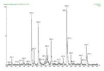
Enlargement of the ESI(-)-MS spectrum of S. microglossa flowers in the mass range of 300 to 500 m/z.

ESI(-)-MS/MS spectra of the compounds quinic acid (a), quercetin (b), chlorogenic acid (c), hyperoside (d) and rutin (e).

HPLC-DAD chromatogram of S. microglossa extract. Temperature 30 °C; flow rate 0.8 mL/min; 20 μL injection volume; and detection at 354 nm. Gradient program of 1% acetic acid in water (solvent A) and acetonitrile (solvent B): 0-5 min (20% B), 5-15 min (20-40% B), 15-25 min (40-80% B), 25-26 min (80-100% B), 26-30 min (100% B) 30-31 min (100-20% B) and 31-35 min (20% B).
Identified substances in the ethanolic extract of S. microglossa flowers by comparison of the ESI(-)-MS/MS spectrum with the database www.massbank.eu.
The analytical curves (Figure 5) obtained from the results of 3 replicates for each point showed linearity, with determination coefficients (R2) greater than 0.99 for the three substances. The relative standard deviations for each point on each curve were all below 2%. Area data (mAU) for curve preparation were obtained from chromatograms with detection at the most sensitive wavelengths for each substance, namely 326, 354 and 372 nm for chlorogenic acid, quercetin, and rutin, respectively. The results for the linearity analyses are summarized in Table II.

The quantification of the investigated components showed a relative standard deviation of 5.8% for chlorogenic acid, 5.7% for quercetin and 4.2% for rutin. These values can be considered suitable for the analysis of chemical markers in complex matrices. The concentration found for each substance was 7.3 mg chlorogenic acid per g extract, 3.4 mg quercetin per g stratum and 47.8 mg rutin per g extract (Table III).
Data for the quantification of chlorogenic acid, quercetin and rutin in the S. microglossa extract: average area, relative standard deviation (DPR) and concentration of the substance in the extract.
Carrageenan-induced paw edema
For data analysis of the experiments with animals, the R statistical program was used. The results of the paw edema measurements were submitted to a two-way analysis of variance (ANOVA) (compound and time). Tukey’s post hoc test was used to identify specific differences in the variables for which the p values found were higher than the established statistical significance criterion (p <0.05).
According to Figure 6, the paw thickness percentiles increased with carrageenan treatment, ranging from 26.98% at the first hour to 40.86% at the fourth hour, while the negative control varied from 38.42% to 80.56% after 4 h of the experiment. The group treated with the reference drug ranged from 28.75 to 36.40% after 4 h of the experiment. Two-way ANOVA (compound and time) showed significant effects from time factors [F = 4,769; p <0.01] and compound factors [F = 27,736; p <0.001], in addition to the interaction of compound and time factors [F = 2,606; p <0.01] on the formation of carrageenan-induced paw edema.

Variation in paw thickness percent 1 to 4 h after carrageenan administration. Animals were pretreated with vehicle (saline solution + 1% Tween 80, 10 ml/kg), indomethacin 10 mg/kg, S. microglossa extract 125 mg/kg, or S. microglossa extract 250 mg/kg 30 min before intraplantar injection of carrageenan (300 μg/paw). Edema was measured with a digital caliper at each time point marked. N=7 for all groups. Statistical analyses were performed between the treated groups and the group treated only with the vehicle at certain times (1 h, 2 h, 3 h, and 4 h). Analysis of variance (two-way ANOVA) followed by Tukey’s multiple comparison of the means at the 95% familywise confidence level.
Post hoc analysis showed that all compounds were more efficient in reducing paw edema relative to saline (p <0.001). Indomethacin was more effective in reducing paw edema compared to S. microglossa 125 mg (p <0.01). S. microglossa 250 mg was more effective than S. microglossa 125 mg (p <0.01). There was no significant difference between S. microglossa 250 mg and indomethacin in reducing carrageenan-induced paw edema (p> 0.1).
Prostaglandin E2-, bradykinin- and compound 48/80-induced paw inflammation
In the analysis of Prostaglandin E2 (PGE 2)-induced paw inflammation, the increase in the thickness of the paw by the vehicle ranged from 53.84% to 7.14%, between 15 and 90 min. Indomethacin-treated paws ranged between a 34.47% and 3.95% increase. Two-way ANOVA (compound and time) showed significant effects from time factors [F = 131,164; p <0.001] and compound factors [F = 24,391; p <0.001], in addition to the interaction of compound and time factors [F = 3,602; p <0.001] on the formation of prostaglandin-induced paw edema. Post hoc analysis showed that only indomethacin was more efficient in reducing paw edema than saline (p <0.001). There was no significant difference between animals treated with S. microglossa and saline in reducing prostaglandin-induced paw edema (p> 0.1) (Figure 7).

Variation in paw thickness percent 15 to 90 min after PGE2 administration. Animals were pretreated with vehicle (saline solution + 1% Tween 80, 10 ml/kg), indomethacin 10 mg/kg, S. microglossa extract 125 mg/kg or S. microglossa extract 250 mg/kg 30 min before intraplantar injection of PGE2 (20 μg/paw). Edema was measured with a digital caliper at each time point marked. N=8 for all groups. Statistical analyses were performed between the treated groups and the group treated only with the vehicle at certain times (15 min, 30 min, 60 min, and 90 min). Analysis of variance (two-way ANOVA) followed by Tukey’s multiple comparison of the means at the 95% familywise confidence level.
When the inflammatory process was induced by injecting bradykinin, only the group treated with dexamethasone presented a significant difference when compared with the vehicle (Figure 8).
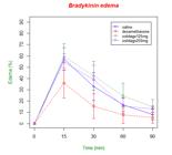
Variation in paw thickness percent 15 to 90 min after bradykinin administration. Animals were pretreated with vehicle (saline solution + 1% Tween 80, 10 ml/kg), dexamethasone 1 mg/kg, S. microglossa 125 mg/kg or S. microglossa 250 mg/kg 30 min before intraplantar injection of bradykinin (10 μg/paw). Edema was measured with a digital caliper at each time point marked. N=8 for all groups. Statistical analyses were performed between the treated groups and the group treated only with the vehicle at certain times (15 min, 30 min, 60 min, and 90 min). Analysis of variance (two-way ANOVA) followed by the Tukey’s multiple comparison of the means at the 95% familywise confidence level.
In the assay where compound 48/80 was used, 4 mg/kg cyproheptadine was used as a reference drug. Two-way ANOVA (compound and time) showed significant effects from time factors [F = 51.12; p <0.001] and compound factors [F = 75.39; p <0.001], in addition to the interaction of compound and time factors [F = 35.71; p <0.001] on the formation of compound 48/80-induced paw edema. Post hoc analysis showed that all compounds were effective in reducing paw edema relative to saline (p <0.001). S. microglossa at 250 mg/kg was more effective than S. microglossa at 125 mg (p <0.01). Cyproheptadine was shown to have a more effective initial effect than S. microglossa 250 mg at time points 1 and 2 (p <0.001), however S. microglossa 250 mg was shown to have a more efficient late effect than cyproheptadine at time point 4 (p <0.05) (Figure 9).

Variation in paw thickness percent 15 to 90 min after compound 48/80 administration. Animals were pretreated with vehicle (saline solution + 1% tween 80, 10 ml/kg), cyproheptadine 4 mg/kg, S. microglossa 125 mg/kg or S. microglossa 250 mg/kg 30 min before intraplantar injection of compound 48/80 (50 μg/paw). Edema was measured with a digital caliper at each time point marked. N=8 for all groups. Statistical analyses were performed between the treated groups and the group treated only with the vehicle at certain times (15 min, 30 min, 60 min, and 90 min). Analysis of variance (two-way ANOVA) followed by Tukey’s multiple comparison of the means at the 95% familywise confidence level.
Effects of S. microglossa on PPARγ transcriptional activity
The extract of S. microglossa was able to increase the transcriptional activity of PPARγ in a dose-dependent manner, indicating that it can have one or more compounds with agonist activity. The activation promoted by S. microglossa was 1.6 ± 0.25-fold at 0.05 mg/ml, 2.9 ± 1.6-fold at 0.075 mg/ml, 3.2 ± 1.2-fold at 0.10 mg/ml, 4.3 ± 1.6-fold at 0.15 mg/ml, and 4.5 ± 1.9-fold at 0.2 mg/ml. The EC50 value was 0.075 mg/mL (Figure 10a). The highest activation promoted by S. microglossa was approximately 50% of that induced by rosiglitazone, a known agonist of PPARγ, which increased the transcriptional activity 9.2 ± 1.4-fold at 10-5 M (Figure 10b).

S. microglossa activates PPARγ. HeLa cells were cotransfected with plasmids containing the expression vector pCMV-SPORT6-PPARγ and the PPRE luciferase reporter and treated with vehicle (DMSO), rosiglitazone (Rosi) or increasing concentrations of S. Microglossa. S. microglossa activated PPARγ in a dose-dependent manner with an EC50 value of 0.75 mg/mL (a). The highest concentration of S. Microglossa (2.0 mg/mL) induced approximately 50% of the PPARγ activation promoted by rosiglitazone at 10-5 M (b). The luciferase activity was measured after 24 h. *p ≤ 0.05, **p ≤ 0.01, ***p ≤ 0.001 (compared to vehicle). Data are presented as the mean (SD) of three independent experiments conducted in triplicate. The statistical significance of the difference between the groups was assessed by one-way ANOVA followed by Tukey’s post hoc test. Values of p less than 0.05 were considered significant.
DISCUSSION
Natural products have been recognized as remedies since ancient times and have long been used as folk medicines for inflammatory conditions such as fevers, pain, migraines, and arthritis (Yuan et al. 2006). S. microglossa is present in the first edition of the Brazilian Pharmacopeia (Brazilian Pharmacopea 1929, apud Liz et al. 2008LIZ R, VIGIL SVG, GOULART S, MORITZ MIG, SCHENKEL EP & FRÖDE TS. 2008. The anti-inflammatory modulatory role of Solidago chilensis Meyen in the murine model of the air pouch. J Pharm Pharmacol 60(4): 515-521.) and is popularly used by the Brazilian population in substitution with Arnica montana as a diuretic, analgesic and anti-inflammatory agent to treat burns and rheumatic disease among other ailments (Mors et al. 2000MORS WB, RIZZINI CT & PEREIRA NA. 2000. Medicinal plants of Brazil. Algonac: Reference Publications, 1st ed., 549 p., Lorenzi & Matos 2002LORENZI H & MATOS FJA. 2002. Plantas medicinais no Brasil – nativas e exóticas: Solidago chilensis Meyen. São Paulo: Instituto Plantarum de Estudos da Flora, 1a ed., 450 p.).
The Ministério da Saúde Brasileiro initiated a Política Nacional de Práticas Integrativas e Complementares no SUS (MS 2018) to guarantee the safe access and rational use of medicinal and phytotherapeutic plants for the Brazilian population, as well as promoting the sustainable use of biodiversity and developing the productive chain and the national industry. This program encourages the study of several medicinal plants, including S. microglossa. Some studies have been conducted in recent years to confirm the popular use of Brazilian Arnica, but these works have focused on the leaves of the species. Our study was conducted with the flowers of S. microglossa, showing that the macerate containing the flowers can be effective in treating inflammation.
The analysis of the chemical profile with electrospray ionization of the samples in electrospray ionization mass spectrometry (ESI-MS/MS) is known as its fingerprint and has been used in the analysis of complex matrices, as well as in extracts of plant origin. By this method, it is possible to identify previously described compounds (Moller et al. 2007, Roesler et al. 2007). Flavonoids (quercetrin, quercetin, rutin) and terpenes (pumiloxide, solidagenone, limonene, germacrene D, and γ-cadinene) have been described in this plant (Gastaldi et al. 2018GASTALDI B, CATALÁN CAN, SILVA-SOFRÁS FM & GONZÁLEZ SB. 2018. Solidago chilensis Meyen (Asteraceae), a medicinal plant from South America. A comprehensive review: ethnomedicinal uses, phytochemistry and bioactivity. Bol Latinoam Caribe Plantas Med Aromát 17(1): 17-29.). Some of these compounds have been found in this extract by ESI-MS/MS, corroborating data from the literature.
Baggio and colleagues conducted a study with S. microglossa grown in South Brazil and found the presence of the classes of saponins, flavonoids, tannins and alkaloids (Baggio et al. 2012BAGGIO AL, BUSKIEVICZ A, FORNARI A, FRANKE TA & LUCCA P. 2012. Pesquisa fitoquímica dos princípios ativos presentes na droga vegetal Solidago Microglossa, D.C. (Arnica Brasileira). Them et Scie 2(1): 149-152). Another study on the leaves of S. microglossa from South Brazil demonstrated the capacity to inhibit reactive oxygen species and lipid peroxidation, and it was possible to identify the phenolic compounds rutin, quercetin, quercitrin and gallic acid by HPLC-DAD (Sabir et al. 2012SABIR SM, AHMAD SD, HAMIDC A, KHAN MQ, ATHAYDE ML, SANTOS DB, BOLIGON AA & ROCHA JBT. 2012. Antioxidant and hepatoprotective activity of ethanolic extract of leaves of Solidago microglossa containing polyphenolic compounds. Food Chem 131(3): 741-747.). These compounds coincide with those described in the S. microglossa flowers collected from this study. However, studies on the toxicity and safety of these species are still scarce (Smolarek et al. 2009SMOLAREK FSF, NUNES PMP, CANSIAN FC, MERCALI CA, CARVALHO JLS, DIAS JFG & MIGUEL OG. 2009. Abordagem Fitoquímica e das Atividades Biológicas da Espécie vegetal Solidago microglossa D.C. Visão Acadêmica 10(1): 77-82.).
Some studies have shown the anti-inflammatory potential of the compounds that were identified in S. microglossa. Chlorogenic acid has anti-inflammatory effects in lipopolysaccharide-stimulated RAW 264.7 cells. This compound significantly inhibited NO production and the expression of COX-2 and iNOS without any cytotoxicity. Chlorogenic acid also attenuated proinflammatory cytokines (including IL-1β and TNF-α) and other inflammation-related markers, such as IL-6, in a dose-dependent manner. Additionally, endotoxin-induced adhesion of macrophages and the expression level of ninjurin1 (Ninj1) decreased after chlorogenic acid treatment. Finally, chlorogenic acid inhibited the nuclear translocation of NF-κB (Hwang et al. 2014HWANG SJ, KIM YW, PARK Y, LEE HJ & KIM KW. 2014. Anti-inflammatory effects of chlorogenic acid in lipopolysaccharide-stimulated RAW 264.7 cells. J Inflamm Res 63(1): 81-90.).
Quercetin, one of the most studied flavonoids in recent years for different pathologies, is found in many fruits and vegetables. In a recent review, the action of quercetin on inflammation was described using in vitro cell line assays, which showed that quercetin inhibits lipopolysaccharide (LPS)-induced tumor necrosis factor α (TNF-α) production in macrophages and LPS-induced IL-8 production in lung A549 cells. In glial cells, it was shown that quercetin could inhibit the LPS-induced mRNA level changes of TNF-α and interleukin (IL)-1α. The effects of quercetin resulted in diminished apoptotic neuronal cell death induced by microglial activation. Quercetin inhibits the production of inflammation-producing enzymes (cyclooxygenase (COX) and lipoxygenase (LOX)) (Li et al. 2016LI Y, YAO J, HAN C, YANG J, CHAUDHRY MT, WANG S, LIU H & YIN Y. 2016. Quercetin, Inflammation and Immunity. Nutrients 8(3): 167-181.).
Rutin and quercetin inhibited the acute and chronic phases of an experimental model of inflammation in rats (Guardia et al. 2001GUARDIA T, ROTELLI AE, JUAREZ AO & PELZER LE. 2001. Anti-inflammatory properties of plant flavonoids. Effects of rutin, quercetin and hesperidin on adjuvant arthritis in rat. Farmaco 56(9): 683-687.). Thus, evaluating the data described in the literature, we can recognize that phenolic compounds have been intensely studied for their pharmacological potential, so the compounds identified in the extract of the flowers of S. microglossa may be responsible for its anti-inflammatory activity (Shen et al. 2002SHEN SC, LEE WR, LIN HY, HUANG HC, KO CH, YANG LL & CHEN YC. 2002. In vitro and in vivo inhibitory activities of rutin, wogonin, or quercetin on lipopolysaccharide-induced nitric oxide and prostaglandin E (2) production. Eur J Pharmacol 446(1-3): 187-194., Kim et al. 2002KIM DW, CHI YS, SON KH, CHANG HW, KIM JS, KANG SS & KIM HP. 2002. Effects of sophoraflavanone G, a prenylated flavonoid from Sophora flavescens, on cyclooxygenase-2 and in vivo inflammatory response. Arch Pharm Res 25(3): 329-335., Morikawa et al. 2003MORIKAWA K, NONAKA M, NARAHARA M, TORII I, KAWAGUCHI K, YOSHIKAWA T, KUMAZAWA Y & MORIKAWA S. 2003. Inhibitory effect of quercetin on carrageenan-induced inflammation in rats. Life Sci 74(6): 709-721.).
In this work, the carrageenan-induced inflammation model was used for an initial screening in the evaluation of nonsteroidal anti-inflammatory drugs (NSAIDs), which is useful for evaluating the contribution of mediators involved in cellular changes, such as histamine, serotonin and kinins over the 4 h that it was evaluated (Winter et al. 1962, Kale et al. 2007KALE M, MISAR AV, DAVE V, JOSHI M & MUJUMDAR AM. 2007. Anti-inflammatory activity of Dalbergia lanceolaria bark etanol extract in mice and rats. J Ethnopharmacol 112(2): 300-304.). A bradykinin assay was performed to observe whether S. microglossa extract can reduce vasodilation and vascular permeability as well as release NO and regulate the production of prostaglandins and histamines. When the prostaglandin E2 assay was performed, the S. microglossa extract was able to reduce paw edema, which during the inflammatory process induces the vasodilation, chemotaxis and chemokinesis of leukocytes, the release of mediators of inflammation and the sensitization of nerve endings, causing hyperalgesia during the inflammatory response (Koeberle & Werz 2015KOEBERLE A & WERZ O. 2015. Perspective of microsomal prostaglandin E2 synthase-1 as drug target in inflammation-related disorders. Biochem Pharmacol 98(1): 1-15., Robbins & Cotran 2005ROBBINS SS & COTRAN RS. 2005. Pathologic basis of diseases. 7a ed., Philadelphia: Sauders, 1524 p.).
S. microglossa extract was not able to inhibit the inflammatory process induced by bradykinin within the evaluated doses. Bradykinin is a peptide with vasodilatory action formed by the activity of enzymes on kininogens. In the inflammatory process, bradykinin is formed due to the increase in vascular permeability, which causes the extravasation of plasma as well as in the release and modulation of the production of prostaglandins and histamines (Brown & Robert 2001BROWN NJ & ROBERTS LJ. 2001. Histamine, bradykinin, and their antagonists. In: Hardman JG & Limbird LE (Eds). Goodman and Gilaman’s the pharmacological basis of therapeutics. 10th ed., New York: McGraw-Hill Companies, p. 218-315., Costa et al. 2006COSTA R, FERNANDES ES, MENEZES-DE-LIMA JR. O, CAMPOS MM & CALIXTO JB. 2006. Effect of novel selective non-peptide kinin B1 receptor antagonists on mouse pleurisy induced by carrageenan. Peptides 27(11): 2967-2975., Oliveira et al. 2010OLIVEIRA C ET AL. 2010. Effect of plant neutrophil elastase inhibitor on leucocyte migration, adhesion and cytokine release in inflammatory conditions. Br J Pharmacol 161(4): 899-910., Bender et al. 2017BENDER L, WEIDMANN H, ROSE-JOHN S, RENNÉ T & LONG AT. 2017. Factor XII-Driven Inflammatory Reactions with Implications for Anaphylaxis. Front Immunol 8(1): 1115.). Bradykinin can stimulate prostaglandins during an inflammatory process, whereas glucocorticoids inhibit prostaglandin production in endothelial cells by interacting with specific steroid receptors (Lewis et al. 1986LEWIS GD, CAMPBELL WB & JOHNSON AR. 1986. Inhibition of Prostaglandin Synthesis by Glucocorticoids in Human Endothelial Cells. Endocrinology 119(1): 62-69.). The S. microglossa extract was not able to act at this point of the inflammation cascade and showed that its anti-inflammatory activity is not the same as that of glucocorticoids.
In another experiment, compound 48/80 was used to identify the potential of the extract to inhibit the release of histamine and serotonin during the inflammatory process. According to previous work, when injected into the paw, serotonin at doses of 0.2-20 µg produced dose-related edema (Cirino et al. 1989). Compound 48/80 is a polybasic mixture of low molecular weight polymers that induces an intense release of serotonin and histamine by tissue mast cells (Vendramini-Costa et al. 2015VENDRAMINI-COSTA DB, SPINDOLA HM, DE MELLO GC, ANTUNES E, PILLI RA & CARVALHO JE. 2015. Anti-inflammatory and antinociceptive effects of racemic goniothalamin, a styryl lactone. Life Sci 139: 83-90.). S. microglossa extract, at the doses tested, was able to decrease the edema caused by this agent only at 250 mg/kg for 30 min.
To elucidate the molecular mechanisms underlying the anti-inflammatory effects of the S. microglossa extract, we investigated its effects on PPARγ transcriptional activity. A luciferase gene reporter assay showed that S. microglossa was able to activate PPARγ in a dose-dependent manner. PPARγ is a transcription factor that belongs to the superfamily of nuclear receptors and plays a central role in metabolic homeostasis of lipid and glucose metabolism and inflammation (Tyagi et al. 2011TYAGI S, GUPTA P, SAINI AS, KAUSHAL C & SHARMA S. 2011. The peroxisome proliferator-activated receptor: A family of nuclear receptors role in various diseases. JAPTR 2(4): 236-240.). It is a target of several compounds that exhibit anti-inflammatory effects, such as nonsteroidal anti-inflammatory drugs (Puhl et al. 2015PUHL AC ET AL. 2015. Mechanisms of peroxisome proliferator activated receptor γ regulation by non-steroidal anti-inflammatory drugs. Nucl Recept Signal 13: e004.) and natural products, such as Amorpha fruticosa L. (Weidner et al. 2012WEIDNER C ET AL. 2012. Amorfrutins are potent antidiabetic dietary natural products. Proc Natl Acad Sci USA 109(19): 7257-7262.), Cymbopogon citratus (Katsukawa et al. 2010KATSUKAWA M, NAKATA R, TAKIZAWA Y, HORI K, TAKAHASHI S & INOUE H. 2010. Citral, a component of lemongrass oil, activates PPARα and γ and suppresses COX-2 expression. Biochim Biophys Acta 1801(11): 1214-1220.) and Rosmarinus officinalis (Rau et al. 2006RAU O, WURGLICS M, PAULKE A, ZITZKOWSKI J, MEINDL N & BOCK A. 2006. Carnosic acid and carnosol, phenolic diterpene compounds of the labiate herbs rosemary and sage, are activators of the human peroxisome proliferator-activated receptor gamma. Planta Med 72(10): 881-887.).
The inhibitory effects of activated PPARγ on prostaglandin E2 and other proinflammatory molecules, such as thromboxane A2 (TXA2), inducible nitric oxide synthase (iNOS) and cyclooxygenase (COX)–2, have been shown in both primary and secondary cell lines (Mendez & LaPointe 2003MENDEZ M & LAPOINTE MC. 2003. PPARδ Inhibition of Cyclooxygenase-2, PGE2 Synthase, and Inducible Nitric Oxide Synthase in Cardiac Myocytes. Hypertension 42(4): 844-850., Yamazaki et al. 2002YAMAZAKI T, YOKOO T, TOMIOKA Y, SUZUKI H, HISHINUMA T & MIZUGAKI M. 2002. Effect of peroxisome proliferator-activated receptor gamma on thromboxane A (2) and prostaglandin E (2) production in macrophage cell lines. Prostag Leukotr Ess 67(4): 245-251.). Other factors are likely to be involved in the anti-inflammatory effects of the S. microglossa extract observed in this study. However, the effects on PPARγ certainly contributed to understanding its mechanisms of action.
Thus, the ethanolic extract of S. microglossa flowers has anti-inflammatory potential, and the phenolic compounds identified and quantified should be responsible for this activity since previous works have demonstrated this anti-inflammatory potential of this plant. The compounds present in the extract may interact with PPARγ and possibly decrease the concentration of proinflammatory molecules, thereby decreasing the inflammatory process.
CONCLUSION
The extract of S. microglossa flowers showed anti-inflammatory activity. In a transactivation assay, the extract was able to increase PPARγ transcriptional activity. These data demonstrate the pharmacological potential of this species, corroborating its popular use.
ACKNOWLEGMENTS
We thank Fundação de Amparo à Pesquisa do Estado do Rio de Janeiro/Faperj for financial support (grant number E-26/110.110/2014) and Fundação de Amparo à Pesquisa do Estado de São Paulo/ FAPESP grant (2008/58035-6) for the use of the UHPLC–MS equipment.
REFERENCES
- BAGGIO AL, BUSKIEVICZ A, FORNARI A, FRANKE TA & LUCCA P. 2012. Pesquisa fitoquímica dos princípios ativos presentes na droga vegetal Solidago Microglossa, D.C. (Arnica Brasileira). Them et Scie 2(1): 149-152
- BENDER L, WEIDMANN H, ROSE-JOHN S, RENNÉ T & LONG AT. 2017. Factor XII-Driven Inflammatory Reactions with Implications for Anaphylaxis. Front Immunol 8(1): 1115.
- BIESKI GC ET AL. 2015. Ethnobotanical study of medicinal plants by population of Valley of Juruena Region, Legal Amazon, Mato Grosso, Brazil. J Ethnopharmacol 173: 383-423.
- BLUMENTHAL M, BUSSE WR & GOLDBERG A. 1998. The complete German commission e monographs. Boston: American Botanical Council, Austin and Integrative Medicine Communications 1st ed., 685 p.
- BROWN NJ & ROBERTS LJ. 2001. Histamine, bradykinin, and their antagonists. In: Hardman JG & Limbird LE (Eds). Goodman and Gilaman’s the pharmacological basis of therapeutics. 10th ed., New York: McGraw-Hill Companies, p. 218-315.
- COSTA R, FERNANDES ES, MENEZES-DE-LIMA JR. O, CAMPOS MM & CALIXTO JB. 2006. Effect of novel selective non-peptide kinin B1 receptor antagonists on mouse pleurisy induced by carrageenan. Peptides 27(11): 2967-2975.
- GASTALDI B, CATALÁN CAN, SILVA-SOFRÁS FM & GONZÁLEZ SB. 2018. Solidago chilensis Meyen (Asteraceae), a medicinal plant from South America. A comprehensive review: ethnomedicinal uses, phytochemistry and bioactivity. Bol Latinoam Caribe Plantas Med Aromát 17(1): 17-29.
- GOULAR S, MORITZ MI, LANG KL, LIZ R, SCHENKEL EP & FRÖDE TS. 2007. Anti-inflammatory evaluation of Solidago chilensis Meyen in a murine model of pleurisy. J Ethnopharmacol 113(5): 346-353.
- GUARDIA T, ROTELLI AE, JUAREZ AO & PELZER LE. 2001. Anti-inflammatory properties of plant flavonoids. Effects of rutin, quercetin and hesperidin on adjuvant arthritis in rat. Farmaco 56(9): 683-687.
- HWANG SJ, KIM YW, PARK Y, LEE HJ & KIM KW. 2014. Anti-inflammatory effects of chlorogenic acid in lipopolysaccharide-stimulated RAW 264.7 cells. J Inflamm Res 63(1): 81-90.
- KALE M, MISAR AV, DAVE V, JOSHI M & MUJUMDAR AM. 2007. Anti-inflammatory activity of Dalbergia lanceolaria bark etanol extract in mice and rats. J Ethnopharmacol 112(2): 300-304.
- KATSUKAWA M, NAKATA R, TAKIZAWA Y, HORI K, TAKAHASHI S & INOUE H. 2010. Citral, a component of lemongrass oil, activates PPARα and γ and suppresses COX-2 expression. Biochim Biophys Acta 1801(11): 1214-1220.
- KIM DW, CHI YS, SON KH, CHANG HW, KIM JS, KANG SS & KIM HP. 2002. Effects of sophoraflavanone G, a prenylated flavonoid from Sophora flavescens, on cyclooxygenase-2 and in vivo inflammatory response. Arch Pharm Res 25(3): 329-335.
- KOEBERLE A & WERZ O. 2015. Perspective of microsomal prostaglandin E2 synthase-1 as drug target in inflammation-related disorders. Biochem Pharmacol 98(1): 1-15.
- LEHRKE M & LAZAR MA. 2005. The many faces of PPARgamma. Cell 123(6): 993-999.
- LEWIS GD, CAMPBELL WB & JOHNSON AR. 1986. Inhibition of Prostaglandin Synthesis by Glucocorticoids in Human Endothelial Cells. Endocrinology 119(1): 62-69.
- LI Y, YAO J, HAN C, YANG J, CHAUDHRY MT, WANG S, LIU H & YIN Y. 2016. Quercetin, Inflammation and Immunity. Nutrients 8(3): 167-181.
- LIZ R, VIGIL SVG, GOULART S, MORITZ MIG, SCHENKEL EP & FRÖDE TS. 2008. The anti-inflammatory modulatory role of Solidago chilensis Meyen in the murine model of the air pouch. J Pharm Pharmacol 60(4): 515-521.
- LORENZI H & MATOS FJA. 2002. Plantas medicinais no Brasil – nativas e exóticas: Solidago chilensis Meyen. São Paulo: Instituto Plantarum de Estudos da Flora, 1a ed., 450 p.
- MENDEZ M & LAPOINTE MC. 2003. PPARδ Inhibition of Cyclooxygenase-2, PGE2 Synthase, and Inducible Nitric Oxide Synthase in Cardiac Myocytes. Hypertension 42(4): 844-850.
- MILTON FA ET AL. 2017. Dibutyltin Compounds Effects on PPARγ/RXRα Activity, Adipogenesis, and Inflammation in Mammalians Cells. Front Pharmacol 2(8): 1-11.
- MINISTÉRIO DA SAÚDE. PROGRAMA NACIONAL DE PLANTAS MEDICINAIS E FITOTERÁPICOS. Disponível em< http://portalms.saude.gov.br/acoes-e-programas/programa-nacional-de-plantas-medicinais-e-fitoterapicos-ppnpmf/plantas-medicinais-e-fitoterapicos-no-sus>. Accessado em 14 de janeiro de 2019.
» http://portalms.saude.gov.br/acoes-e-programas/programa-nacional-de-plantas-medicinais-e-fitoterapicos-ppnpmf/plantas-medicinais-e-fitoterapicos-no-sus - MORIKAWA K, NONAKA M, NARAHARA M, TORII I, KAWAGUCHI K, YOSHIKAWA T, KUMAZAWA Y & MORIKAWA S. 2003. Inhibitory effect of quercetin on carrageenan-induced inflammation in rats. Life Sci 74(6): 709-721.
- MORS WB, RIZZINI CT & PEREIRA NA. 2000. Medicinal plants of Brazil. Algonac: Reference Publications, 1st ed., 549 p.
- NEWMAN DJ & CRAGG GM. 2016. Natural Products as Sources of New Drugs from 1981 to 2014. J Nat Prod 79(3): 629-661.
- OLIVEIRA C ET AL. 2010. Effect of plant neutrophil elastase inhibitor on leucocyte migration, adhesion and cytokine release in inflammatory conditions. Br J Pharmacol 161(4): 899-910.
- PUHL AC ET AL. 2015. Mechanisms of peroxisome proliferator activated receptor γ regulation by non-steroidal anti-inflammatory drugs. Nucl Recept Signal 13: e004.
- RAU O, WURGLICS M, PAULKE A, ZITZKOWSKI J, MEINDL N & BOCK A. 2006. Carnosic acid and carnosol, phenolic diterpene compounds of the labiate herbs rosemary and sage, are activators of the human peroxisome proliferator-activated receptor gamma. Planta Med 72(10): 881-887.
- ROBBINS SS & COTRAN RS. 2005. Pathologic basis of diseases. 7a ed., Philadelphia: Sauders, 1524 p.
- SABIR SM, AHMAD SD, HAMIDC A, KHAN MQ, ATHAYDE ML, SANTOS DB, BOLIGON AA & ROCHA JBT. 2012. Antioxidant and hepatoprotective activity of ethanolic extract of leaves of Solidago microglossa containing polyphenolic compounds. Food Chem 131(3): 741-747.
- SHEN SC, LEE WR, LIN HY, HUANG HC, KO CH, YANG LL & CHEN YC. 2002. In vitro and in vivo inhibitory activities of rutin, wogonin, or quercetin on lipopolysaccharide-induced nitric oxide and prostaglandin E (2) production. Eur J Pharmacol 446(1-3): 187-194.
- SMOLAREK FSF, NUNES PMP, CANSIAN FC, MERCALI CA, CARVALHO JLS, DIAS JFG & MIGUEL OG. 2009. Abordagem Fitoquímica e das Atividades Biológicas da Espécie vegetal Solidago microglossa D.C. Visão Acadêmica 10(1): 77-82.
- SOUZA DMF, SA RD, ARAUJO EL & RANDAU K. 2017. Anatomical, phytochemical and histochemical study of Solidago chilensis Meyen. An Acad Bras Cienc 90: 2107-2120.
- TYAGI S, GUPTA P, SAINI AS, KAUSHAL C & SHARMA S. 2011. The peroxisome proliferator-activated receptor: A family of nuclear receptors role in various diseases. JAPTR 2(4): 236-240.
- VENDRAMINI-COSTA DB, SPINDOLA HM, DE MELLO GC, ANTUNES E, PILLI RA & CARVALHO JE. 2015. Anti-inflammatory and antinociceptive effects of racemic goniothalamin, a styryl lactone. Life Sci 139: 83-90.
- WEIDNER C ET AL. 2012. Amorfrutins are potent antidiabetic dietary natural products. Proc Natl Acad Sci USA 109(19): 7257-7262.
- YAMAZAKI T, YOKOO T, TOMIOKA Y, SUZUKI H, HISHINUMA T & MIZUGAKI M. 2002. Effect of peroxisome proliferator-activated receptor gamma on thromboxane A (2) and prostaglandin E (2) production in macrophage cell lines. Prostag Leukotr Ess 67(4): 245-251.
Publication Dates
-
Publication in this collection
12 Aug 2020 -
Date of issue
2020
History
-
Received
2 Oct 2019 -
Accepted
29 Nov 2019


