ABSTRACT
A 42-year-old woman was admitted to our clinic with a complaint of glare in both eyes. Biomicroscopic examination of both the eyes revealed iris and lens colobomas in the inferior quadrant. Fundus examination of the right eye revealed an oval and gray inferotemporal optic pit and two choroid colobomas in the inferior quadrant. In the left eye, two choroid colobomas were detected that were inferior to the optic nerve head. Furthermore, a 21-year-old man presented to our clinic for a routine ophthalmologic examination. Bilateral biomicroscopic examination was normal. Fundus examination of the left eye revealed an oval and gray inferotemporal optic pit and a choroid coloboma that was inferior to the optic nerve head. Here we describe optic pits co-occurring with iris, lens, and choroidal colobomas. On the basis of these cases, a defect in the closure of the embryonic fissure is the most plausible etiology of the optic pit.
Keywords:
Coloboma; Eye abnormalities; Optic nerve abnormalities; Iris abnormalities; Choroid abnormalities; Lens, crystalline abnormalities
RESUMO
Uma mulher de 42 anos de idade foi internada em nossa clínica com queixa de ofuscamento em ambos os olhos. O exame biomicroscópico revelou coloboma de íris e cristalino no quadrante inferior em ambos os olhos. O exame de fundo do olho direito revelou um fosseta óptica oval e acinzentada na região inferotemporal e dois colobomas coroide no quadrante inferior. No olho esquerdo, dois colobomas de coroide foram detectados inferiormente à da cabeça do nervo óptico. Outro homem de 21 anos apresentou-se em nossa clínica para um exame oftalmológico de rotina. O exame biomicroscópico foi normal, bilateralmente. O exame de fundo do olho esquerdo revelou uma fosseta oval e acinzentada de nervo óptico óptico inferotemporal e um coloboma coroide inferior à cabeça do nervo óptico. Nestes relatos nós descrevemos fossetas ópticas ocorrendo simultaneamente com colobomas de íris, cristalino, e coroide. Com base nestes casos, o defeito no fechamento da fissura embrionária é uma provável etiologia da fosseta óptica.
Descritores:
Coloboma; Anormalidades do olho; Nervo óptico/anormalidades; Íris/anormalidades; Coroide/anormalidades; Cristalino/anormalidades
INTRODUCTION
Optic pit is a congenital anomaly of the optic nerve head that may cause acquired or congenital vision loss and serous macular detachment(11 Corbett JJ, Savino PJ, Schatz NJ, Orr LS. Cavitary developmental defects of the optic disc. Visual loss associated with optic pits and colobomas. Arch Neurol. 1980;37(4): 210-3.). It usually occurs unilaterally and temporally on the optic disc, is round and gray, and may coexist with the cilioretinal artery. In the early phase, the optic pit is hypofluorescent on fundus fluorescein angiography (FFA); however, it is hyperfluorescent in the late phase(22 Brown GC, Shields JA, Goldberg RE. Congenital pits of the optic nerve head II. Clinical studies in humans. Ophthalmology. 1980;87(1):51-65.).
Here we report two cases of co-occurring optic pit and uveal coloboma, including the first published case of an optic pit accompanied by a unilateral iris, lens, and choroid coloboma.
CASE 1
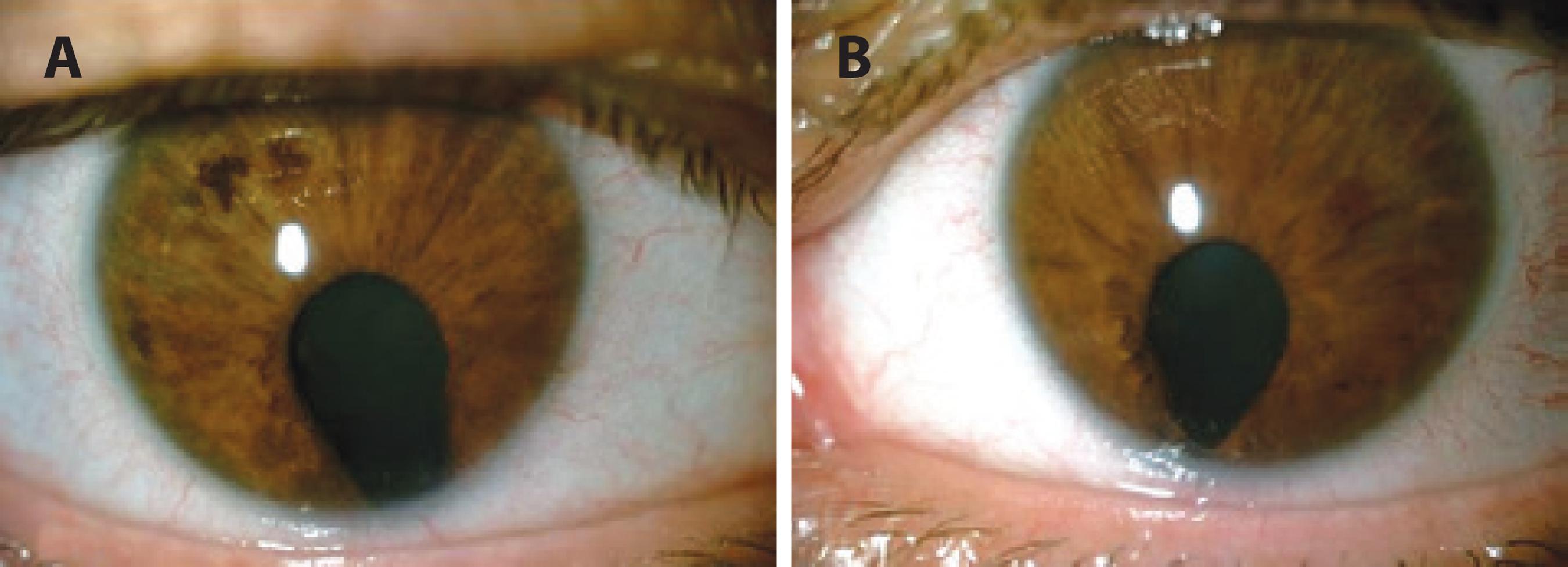
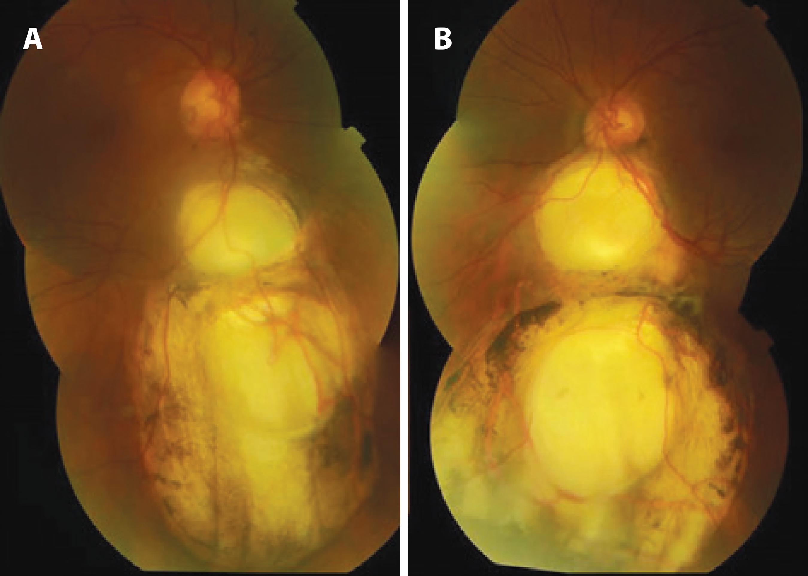
A 42-year-old woman was admitted with a complaint of glare in both the eyes. On ophthalmic examination, her best corrected visual acuity (BCVA) was 20/20 in both the eyes. Biomicroscopic examination of the right eye revealed iris and lens colobomas in the inferior quadrant, pigment precipitates on the lens, and posterior subcapsular cataract (Figure 1 A). In the left eye, there was an iris coloboma in the inferonasal quadrant and iris septa near the limbus that connects the iris tissue, pigment precipitates on the lens, and a lens coloboma in the inferonasal quadrant (Figure 1 B). Bilateral intraocular pressure was 18 mmHg. Fundus examination of the right eye revealed an oval and gray inferotemporal optic pit and two choroid colobomas (one was 2-disc size and an anteriorly located one was 8-disc size, extending to the equator) in the inferior quadrant (Figure 2 A). Under the left optic disc was a 4-disc-sized choroid coloboma that was covered with a membrane with a 1-optic disc-sized hole. A second 7-disc-sized coloboma was detected at the anterior of the coloboma, and pigment alterations were observed around both (Figure 2 B). No retinal detachment was found, and the bilateral maculae were normal. In the right eye, the optic pit revealed hypofluorescence in the early phase and relative hyperfluorescence in the late phase of FFA, as did the bilateral choroid colobomas (Figure 3). Results of optical coherence tomography (OCT) of the optic pit are shown in figures 3 C and 3 D.
A) Optic pit and choroidal colobomas of the right eye, case 1. B) Choroidal colobomas of the left eye inferior to the optic nerve, case 1.
A) Hypofluorescent optic pit and choroidal coloboma in the early phase of FFA of the right eye, case 1. B) Relatively hyperfluorescent optic pit and choroidal coloboma in the late phase of FFA of the right eye, case 1. C) Colorless image of the right fundus captured by OCT and a red arrow cross the optic pit, case 1. D) A deep optic pit is seen in the horizontal section of an OCT image of the right eye, case 1.
CASE 2
A 21-year-old man presented at our clinic for a routine bilateral ophthalmologic examination. His BCVA was 20/20 in the right eye and 20/50 in the left eye, with a refraction of -4.00. Bilateral biomicroscopic examination was normal. Intraocular pressure was 12 mmHg in the right eye and 9 mmHg in the left eye. Fundus examination of the left eye revealed an oval and gray inferotemporal optic pit and a choroid coloboma with half the size of an optic disc that was inferior to the optic nerve head. Additionally, two cilioretinal arteries were detected, one of which transited through the optic pit (Figure 4). The macula was normal, and there was no retinal detachment in the left eye. The fundus of the right eye was normal.
DISCUSSION
Two hypotheses have been proposed for optic pit development. The first proposes developmental anomaly of the primordial optic nerve(33 Gass JD. Serous detachment of the macula secondary to congenital pit of the optic nerve head. Am J Ophthalmol. 1969;67(6):821-41.), whereas the second attributes optic pit development to the incomplete closure of the embryonic fissure(44 Singerman LJ, Mittra RA. Hereditary optic pit and iris coloboma in three generations of a single family. Retina. 2001;21(3):273-5.
5 Brown GC, Augsberger JJ. Congenital pits of the optic nerve head and retinochoroidal colobomas. Can J Ophthalmol. 1980;15(3):144-6.
6 Gotsis S, Koutsandrea C, Apostopoulos M, Theodosiadis G. [Optic disk and choroidal coloboma]. J Fr Ophthalmol. 1989;12(10):683-5. French.-77 Saatci AO, Kocak N, Soylev FM. Unilateral coexistent optic pit and choroidal coloboma. Neuroophthalmology. 2002;28(1):41-3.). The coexistence of optic pits and uveal colobomas supports the latter hypothesis. Because of the defective closure of the embryonic fissure, colobomas may arise anywhere from the optic nerve head to the pupil. After invagination of the primary optic vesicle, the distal portion approaches the proximal part, thus creating the optic cup. Abnormal closure of the inner layer of the optic cup leads to chorioretinal coloboma, and the closure defect of the distal end of the embryonic fissure causes iris coloboma(88 Pagon RA. Ocular coloboma. Surv Ophthalmol. 1981;25(4):223-36.).
Cases of optic pit co-occurring with serous macular detachment, choroidal coloboma, or retinochoroidal coloboma have been reported, both unilaterally(66 Gotsis S, Koutsandrea C, Apostopoulos M, Theodosiadis G. [Optic disk and choroidal coloboma]. J Fr Ophthalmol. 1989;12(10):683-5. French.,77 Saatci AO, Kocak N, Soylev FM. Unilateral coexistent optic pit and choroidal coloboma. Neuroophthalmology. 2002;28(1):41-3.) and bilaterally(55 Brown GC, Augsberger JJ. Congenital pits of the optic nerve head and retinochoroidal colobomas. Can J Ophthalmol. 1980;15(3):144-6.). One report described a 2-year-old patient with aniridia and with an optic pit in one eye and a morning glory anomaly in the other(99 Traboulsi EI, Jurdi-Nuwayhid F, Torbey NS, Frangieh GT. Aniridia, atypical iris defects, optic pit and the morning glory disc anomaly in a family. Ophthalmic Paediatr Genet. 1986;7(2):131-5.). The optic pit and iris coloboma developing in three generations of the same family have also been reported(44 Singerman LJ, Mittra RA. Hereditary optic pit and iris coloboma in three generations of a single family. Retina. 2001;21(3):273-5.). Here we described a case of optic pit co-existing with iris, lens, and choroidal colobomas and a case of optic pit co-occurring with choroidal coloboma. Choroidal coloboma in the first patient had a hole in it, but retinal detachment was absent. Overall, a very small number of cases have been published regarding the co-occurrence of optic pits and choroidal colobomas(44 Singerman LJ, Mittra RA. Hereditary optic pit and iris coloboma in three generations of a single family. Retina. 2001;21(3):273-5.
5 Brown GC, Augsberger JJ. Congenital pits of the optic nerve head and retinochoroidal colobomas. Can J Ophthalmol. 1980;15(3):144-6.
6 Gotsis S, Koutsandrea C, Apostopoulos M, Theodosiadis G. [Optic disk and choroidal coloboma]. J Fr Ophthalmol. 1989;12(10):683-5. French.-77 Saatci AO, Kocak N, Soylev FM. Unilateral coexistent optic pit and choroidal coloboma. Neuroophthalmology. 2002;28(1):41-3.), and to the best of our knowledge, this is the first published description of optic pit accompanied by iris, lens, and choroid coloboma. On the basis of these cases, a defect in the closure of the embryonic fissure appears to be the most plausible etiology of the optic pit.
-
Funding: No specific financial support was available for this study.
REFERENCES
-
1Corbett JJ, Savino PJ, Schatz NJ, Orr LS. Cavitary developmental defects of the optic disc. Visual loss associated with optic pits and colobomas. Arch Neurol. 1980;37(4): 210-3.
-
2Brown GC, Shields JA, Goldberg RE. Congenital pits of the optic nerve head II. Clinical studies in humans. Ophthalmology. 1980;87(1):51-65.
-
3Gass JD. Serous detachment of the macula secondary to congenital pit of the optic nerve head. Am J Ophthalmol. 1969;67(6):821-41.
-
4Singerman LJ, Mittra RA. Hereditary optic pit and iris coloboma in three generations of a single family. Retina. 2001;21(3):273-5.
-
5Brown GC, Augsberger JJ. Congenital pits of the optic nerve head and retinochoroidal colobomas. Can J Ophthalmol. 1980;15(3):144-6.
-
6Gotsis S, Koutsandrea C, Apostopoulos M, Theodosiadis G. [Optic disk and choroidal coloboma]. J Fr Ophthalmol. 1989;12(10):683-5. French.
-
7Saatci AO, Kocak N, Soylev FM. Unilateral coexistent optic pit and choroidal coloboma. Neuroophthalmology. 2002;28(1):41-3.
-
8Pagon RA. Ocular coloboma. Surv Ophthalmol. 1981;25(4):223-36.
-
9Traboulsi EI, Jurdi-Nuwayhid F, Torbey NS, Frangieh GT. Aniridia, atypical iris defects, optic pit and the morning glory disc anomaly in a family. Ophthalmic Paediatr Genet. 1986;7(2):131-5.
Publication Dates
-
Publication in this collection
Sep-Oct 2016
History
-
Received
13 July 2015 -
Accepted
29 Oct 2015