Abstracts
BACKGROUND: Hepatocellular carcinoma is one of the most common malignant tumors worldwide. Imaging techniques, specially computed tomography and ultrasound, are among the most useful diagnostic tools, although the accuracy of these methods may have a significant variability. AIMS: To determine the prevalence of hepatocellular carcinoma in cirrhotic patients undergoing orthotopic liver transplantation at "Santa Casa de Misericórdia" of Porto Alegre, RS, Brazil; to estimate the sensitivity of computed tomography and ultrasound in pretransplantation detection of hepatocellular carcinoma in this population; to correlate the radiological characteristics with anatomopathological findings. MATERIALS AND METHODS: Retrospective prevalence study. Population: adult, cirrhotic patients undergoing orthotopic liver transplantation from January 1990 to July 2003. Among the 292 transplanted patients, 31 cases of hepatocellular carcinoma were diagnosed, of which 29 were included in the study. Tumor characteristics in both ultrasound and computed tomography were compared to those observed in anatomopathological examination. RESULTS: Prevalence of hepatitis C virus infection among patients with diagnosis of hepatocellular carcinoma was 93.5%, and the prevalence of hepatocellular carcinoma among transplanted patients was 10.6%. The overall sensitivity of the imaging techniques was 70.3% for computed tomography and 72% for ultrasound. CONCLUSION: The prevalence of hepatocellular carcinoma at our institution, as well as the sensitivity of both ultrasound and computed tomography to detect such tumors at pretransplantation screening were similar to those found by other authors, while the prevalence of hepatitis C virus infection, the most common etiological agent for liver disease in our patients, is one of the highest ever reported in literature. Factors influencing hepatocellular carcinoma detection rates were: time from examination to liver transplantation; acquisition of computed tomography images during arterial phase; lesion size. Arterial phase proved to be the most useful part of computed tomography examination in this study.
Hepatocellular carcinoma; Liver transplantation; Tomography; Ultrasonography
RACIONAL: O carcinoma hepatocelular é um dos tumores malignos mais comuns em todo o mundo. Exames de imagens, especialmente tomografia computadorizada e ultra-sonografia, estão entre as principais técnicas diagnósticas, embora a acurácia destes métodos possa apresentar significativa variabilidade. OBJETIVOS: Determinar a prevalência de carcinoma hepatocelular em pacientes cirróticos submetidos a transplante hepático na Santa Casa de Misericórdia de Porto Alegre, RS; estimar a sensibilidade da tomografia computadorizada e da ultra-sonografia na detecção pré-transplante de carcinoma hepatocelular nesse grupo de pacientes; correlacionar características radiológicas com achados anatomopatológicos. MATERIAIS E MÉTODOS: Estudo de prevalência retrospectivo. População: pacientes adultos, cirróticos, submetidos a transplante hepático de janeiro de 1990 a julho de 2003. Entre os 292 pacientes transplantados, foi diagnosticado 31 casos de carcinoma hepatocelular, dos quais 29 foram incluídos no estudo. As características tomográficas e ecográficas dos tumores diagnosticados pré-transplante foram comparadas com as observadas em exame anatomopatológico. RESULTADOS: A prevalência da infecção pelo vírus da hepatite C nos pacientes com diagnóstico de carcinoma hepatocelular foi de 93,5% e a prevalência deste entre os pacientes transplantados foi de 10,6%. A sensibilidade dos métodos de imagem na detecção de casos de carcinoma hepatocelular foi de 70,3% para tomografia computadorizada e de 72% para ecografia. CONCLUSÃO: A prevalência de carcinoma hepatocelular na instituição onde foi desenvolvido o estudo, bem como a sensibilidade da ultra-sonografia e da tomografia computadorizada para detecção dessa neoplasia na avaliação pré-transplante foi semelhante à relatada na literatura. Em contrapartida, a prevalência de infecção pelo vírus da hepatite C, fator etiológico de hepatopatia mais freqüente nos pacientes desta série, é das maiores já relatadas. Os fatores que influenciaram as taxas de detecção de carcinoma hepatocelular foram: tempo decorrido entre realização do exame e transplante; realização de tomografia computadorizada com fase arterial; tamanho da lesão. A fase arterial provou ser a mais importante no diagnóstico de carcinoma hepatocelular neste estudo.
Carcinoma hepatocelular; Transplante de fígado; Tomografia; Ultra-sonografia
ARTIGO ORIGINAL ORIGINAL ARTICLE
Hepatocellular carcinoma in patients undergoing orthotopic liver transplantation: radiological findings with anatomopathological correlation in Brazil
Carcinoma hepatocelular em pacientes submetidos a transplante hepático: achados radiológicos com correlação anatomopatológica no Brasil
Antônio Carlos MacielI; Carlos Thadeu CerskiII; Roger Klein MoreiraIII; Vinicius Labrea ResendeIV; Maria Lúcia ZanotelliV; Simone Berwig MatiottiVI
IDepartment of Radiology, "Santa Casa de Misericórdia de Porto Alegre"
IIDepartment of Pathology, Federal University of Rio Grande do Sul, Porto Alegre
IIIFederal University of Rio Grande do Sul, Porto Alegre
IVDepartment of Radiology, "Hospital de Clínicas de Porto Alegre"
VDivision of Transplantation, "Santa Casa de Misericórdia de Porto Alegre"
VIDepartment of Surgery, "Hospital Ernesto Dorneles", Porto Alegre, RS, Brazil
Address for correspondence Address for correspondence: Dr. Vinicius Labrea Resende Av. Protásio Alves, 3928/306 Petrópolis 91310-000 - Porto Alegre, RS, Brazil E-mail: vresende@terra.com.br
ABSTRACT
BACKGROUND: Hepatocellular carcinoma is one of the most common malignant tumors worldwide. Imaging techniques, specially computed tomography and ultrasound, are among the most useful diagnostic tools, although the accuracy of these methods may have a significant variability.
AIMS: To determine the prevalence of hepatocellular carcinoma in cirrhotic patients undergoing orthotopic liver transplantation at "Santa Casa de Misericórdia" of Porto Alegre, RS, Brazil; to estimate the sensitivity of computed tomography and ultrasound in pretransplantation detection of hepatocellular carcinoma in this population; to correlate the radiological characteristics with anatomopathological findings.
MATERIALS AND METHODS: Retrospective prevalence study. Population: adult, cirrhotic patients undergoing orthotopic liver transplantation from January 1990 to July 2003. Among the 292 transplanted patients, 31 cases of hepatocellular carcinoma were diagnosed, of which 29 were included in the study. Tumor characteristics in both ultrasound and computed tomography were compared to those observed in anatomopathological examination.
RESULTS: Prevalence of hepatitis C virus infection among patients with diagnosis of hepatocellular carcinoma was 93.5%, and the prevalence of hepatocellular carcinoma among transplanted patients was 10.6%. The overall sensitivity of the imaging techniques was 70.3% for computed tomography and 72% for ultrasound.
CONCLUSION: The prevalence of hepatocellular carcinoma at our institution, as well as the sensitivity of both ultrasound and computed tomography to detect such tumors at pretransplantation screening were similar to those found by other authors, while the prevalence of hepatitis C virus infection, the most common etiological agent for liver disease in our patients, is one of the highest ever reported in literature. Factors influencing hepatocellular carcinoma detection rates were: time from examination to liver transplantation; acquisition of computed tomography images during arterial phase; lesion size. Arterial phase proved to be the most useful part of computed tomography examination in this study.
Headings: Hepatocellular carcinoma. Liver transplantation. Tomography. Ultrasonography.
RESUMO
RACIONAL: O carcinoma hepatocelular é um dos tumores malignos mais comuns em todo o mundo. Exames de imagens, especialmente tomografia computadorizada e ultra-sonografia, estão entre as principais técnicas diagnósticas, embora a acurácia destes métodos possa apresentar significativa variabilidade.
OBJETIVOS: Determinar a prevalência de carcinoma hepatocelular em pacientes cirróticos submetidos a transplante hepático na Santa Casa de Misericórdia de Porto Alegre, RS; estimar a sensibilidade da tomografia computadorizada e da ultra-sonografia na detecção pré-transplante de carcinoma hepatocelular nesse grupo de pacientes; correlacionar características radiológicas com achados anatomopatológicos.
MATERIAIS E MÉTODOS: Estudo de prevalência retrospectivo. População: pacientes adultos, cirróticos, submetidos a transplante hepático de janeiro de 1990 a julho de 2003. Entre os 292 pacientes transplantados, foi diagnosticado 31 casos de carcinoma hepatocelular, dos quais 29 foram incluídos no estudo. As características tomográficas e ecográficas dos tumores diagnosticados pré-transplante foram comparadas com as observadas em exame anatomopatológico.
RESULTADOS: A prevalência da infecção pelo vírus da hepatite C nos pacientes com diagnóstico de carcinoma hepatocelular foi de 93,5% e a prevalência deste entre os pacientes transplantados foi de 10,6%. A sensibilidade dos métodos de imagem na detecção de casos de carcinoma hepatocelular foi de 70,3% para tomografia computadorizada e de 72% para ecografia.
CONCLUSÃO: A prevalência de carcinoma hepatocelular na instituição onde foi desenvolvido o estudo, bem como a sensibilidade da ultra-sonografia e da tomografia computadorizada para detecção dessa neoplasia na avaliação pré-transplante foi semelhante à relatada na literatura. Em contrapartida, a prevalência de infecção pelo vírus da hepatite C, fator etiológico de hepatopatia mais freqüente nos pacientes desta série, é das maiores já relatadas. Os fatores que influenciaram as taxas de detecção de carcinoma hepatocelular foram: tempo decorrido entre realização do exame e transplante; realização de tomografia computadorizada com fase arterial; tamanho da lesão. A fase arterial provou ser a mais importante no diagnóstico de carcinoma hepatocelular neste estudo.
Descritores: Carcinoma hepatocelular. Transplante de fígado. Tomografia. Ultra-sonografia.
INTRODUCTION
Hepatocellular carcinoma (HCC) is one of the most common malignant tumors worldwide, and is closely associated with chronic liver disease. Approximately 80% to 90% of the cases occur in patients with cirrhosis, which is believed to be the most important risk factor for this neoplasm. Cirrhotic patients have a 5-year risk of developing HCC of about 20%(13). Liver transplantation candidates, therefore, represent a very high risk population.
Imaging techniques, specially computed tomography (CT) and ultrasound (US), are among the most useful diagnostic tools for pretransplantation screening of HCC. However, the accuracy of these methods may vary due to both technical factors and tumor characteristics(6).
The objective of this study was to undertake a retrospective analysis of pretransplantation radiological findings of all cases of HCC diagnosed in adult patients who were submitted to liver transplantation at "Santa Casa de Misericórdia de Porto Alegre", Porto Alegre, RS, Brazil, from 1990 to 2003 correlating these with the results of anatomopathological examination of the explanted liver. The sensitivity of CT and US in detecting HCC in this population, using the present protocol for tumor screening at our institution, was estimated.
MATERIALS AND METHODS
Patients
We included in this study the adult cirrhotic patients who underwent orthotopic liver transplantation (OLT) at our institution from January of 1990 to July of 2003, and had post-transplantation anatomopathological diagnosis of HCC. Were performed 292 adult liver transplantations (which represents more than 80% of adult OLT carried out at the city of Porto Alegre during this period) and diagnosed 31 cases of HCC through pathological analysis of the explanted liver. Of these patients 22 (71%) were males, and 9 (29%) were females; mean age was 55.2 years (29-71 years). Regarding Child-Pugh functional class, 3 patients were categorized as class A (9.7%), 17 as class B (58.4%), and 11 as class C (35.5%). The etiology of chronic liver disease in the population of patients undergoing liver transplantation is described in Table 1. Two patients were excluded from the study because their imaging exams were not available.
Patients included in the transplantation waiting list were followed with medical examination, abdominal ultrasound and serum alpha-fetoprotein (AFP) at intervals of 3 to 6 months. In case of any abnormal findings, an abdominal CT was performed.
Pathological analysis
Following transplantation, the explanted liver was serially sectioned at 10-mm intervals for the anatomopathological examination. Nodules larger than 1.0 cm or with any differing characteristics, as compared to regenerative nodules, which would raise the suspicion of a malignant tumor, were examined microscopically. Malignant nodules were characterized as to their size, location and number. All cases were assessed by the same experienced pathologist blinded to the results of imaging.
Interpretation of imaging exams
All CT scans were retrospectively interpreted by one experienced radiologist who was unaware of any pathological characteristics of the tumor. The tumors seen on CT films were then characterized as to their size (diameter), total number of nodules, location (hepatic segment). Contrast-enhancement pattern was classified as hypodense, isodense, hyperdense or of mixed enhancement as compared to the surrounding hepatic parenchyma. CT scans showing one or more nodules, which were not suggestive of a benign process such as a cyst, hemangioma, or a regenerative nodule, were considered indicative of HCC and regarded as a positive exam. For the ultrasound exams, size, number, location, and ecogeniciy (hypoecoic, hyperecoic, or of mixed ecogenicity) of each lesion was registered. Examinations showing solid tumors, which were not suggestive of any benign process, were considered positive for HCC.
In all cases in which imaging exams were considered positive for HCC, we verified whether the lesions seen on pathological analysis corresponded to the ones observed on radiological exams. In the case of US, information about size and location of the nodules was used, whereas in the case of CT scans, films were compared to photographic images of liver cross-section specimens.
Medical records were carefully reviewed and any previous intervention that could alter radiological findings of tumors, such as percutaneous ethanol injection or arterial chemoembolization, was registered. Time from imaging exam to transplantation was also registered. CT exams used in this study were obtained in different centers. For this reason, specification of type of equipment used as well as description of technical details of imaging acquisition was not possible. Besides imaging exams, serum AFP measures were also obtained.
RESULTS
A total of 292 OLT carried out at our institution were analyzed. The most frequent etiological agent causing chronic liver disease in these patients was hepatitis C virus (HCV), followed by a combination of alcohol and HCV (as shown in Table 1). The prevalence of HCV infection in our population of patients was 93.5%. Among these patients, 31 cases of HCC were seen at anatomopathological examination, of which 29 were included in the study. A total of 73 individual nodules were detected at pathological analysis, with sizes that ranged from 0.3 to 8 cm.
CT, US, and serum AFP levels were available in 27, 25 and 28 of the 29 patients, respectively. Arterial phase images were obtained in 17 of the 27 CT scans. Most imaging exams were acquired less than 6 months before liver transplantation.
Of the 27 patients examined by CT, 19 had at least one nodule detected, resulting in a sensitivity of 70.3 % (CI 95%: 51.3-85.1). Considering individual nodules, only 37% of the 64 lesions (CI 95%: 26.3-49.7) were detected. This proportion was 6.9% for nodules less than 1 cm, 55.2% for nodules from 1 to 3 cm, and 100% for tumors larger than 3 cm.
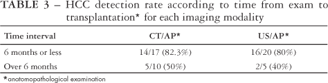
Sensitivity of CT was higher (82.3%) in the group of patients whose CT scans included arterial phase (Table 2). The contrast enhancement pattern during arterial, portal, and delayed phases is described in Table 3. In biphasic (arterial and venous phases) CT scans, the tumors most commonly appeared hyperdense during the arterial phase and isodense during de portal venous phase (35.3% of the cases), representing the most common enhancement pattern combination in our study (Figure 1). Considering individual phases separately, hyperdense lesions were the most common ones during arterial phase (41.1%), while hypodense nodules predominated during portal venous phase (56.5%). In biphasic CT scans, a significant proportion of tumors (41.2%) was visible only during arterial phase, while a smaller percentage of lesions (11.8%) was identified only during portal venous phase. Seven patients underwent CT scans after lipiodol injection. Of these, HCC were detected in six, resulting in a sensitivity of 85.7% for this technique.
In our study, ultrasound showed at least one nodule in 72% of patients with HCC (CI 95%: 52.3-86.8). A total of 68 individual malignant nodules were diagnosed in this group of patients. Of these, 39.7% (CI 95%: 28.6-51.6) were detected 12.1% of the lesions smaller than 1 cm, 37% of those with diameters from 1 to 3 cm, and 100% of nodules larger than 3 cm.
Both US and CT had their sensitivities affected by factors such as tumor size and time from examination to transplantation, as described in Tables 3 and 4. Of all nodules identified by these two imaging techniques, only two (one detected by US and one by CT) were not confirmed by histopathological examination to represent HCC.
Overall sensitivity of AFP was 32.1% (IC 95%: 16.9-50.8) and 10.7% (IC 95%: 2.8-26.4) when serum levels above 20 ng/mL and 100 ng/mL, respectively, were considered a positive result. Sensitivity for combination of screening exams was 83.3% for US and AFP, 82.6% for US and CT, and 86.9% for all three methods combined.
Of the 73 nodules diagnosed by anatomopathological examination, 21 received some kind of pretransplantantion treatment 18 were treated with percutaneous ethanol injection (Figure 2) and 3 with chemoembolization. Excluding satellite lesions (<0.5 cm and adjacent to a larger lesion), the average observed tumoral necrosis was 74.3% for the treated group and only 8% for the nodules which were not treated (P <0.0001). Tumor size in both groups did not differ significantly (Table 5).
DISCUSSION
Twenty-nine cirrhotic patients with diagnosis of HCC were included in this study. A HCV infection prevalence of 67.3% was observed in the population of transplanted patients, while an even higher prevalence (93.5%) was seen in the group of transplanted patients with HCC. The literature reports diverge significantly on this issue. HCV infection is thought to be associated with approximately 70% of cases of HCC in Japan, 77% in Spain, and 55% in the United States(11, 16), but these rates range from 7.6% to 81%(7, 21, 35). The prevalence of HCV infection associated with HCC, in our study, therefore, is among the highest reported in literature.
The risk of developing HCC in cirrhotic patients is approximately 3% to 5% per year(7, 20). Patients in transplantation waiting lists constitute a high-risk population(2, 11). Early diagnosis of HCC is of great importance, since several forms of therapy, such as percutaneous ethanol injection and arterial chemoembolization, are available and can alter the natural history of this tumor before transplantation can be carried out(4). OLT has been considered the treatment of choice for patients with tumors at early stages whose clinical conditions do not allow tumor resection(18). For these reasons, an adequate screening program is justified.
Patient assessment includes medical examination, laboratory workup, and imaging exams. Clinical assessment, however, have a very low sensitivity and specificity in detecting HCC. AFP, a glycoprotein which has its gene expression reactivated during hepatocarcinogenesis, is the most useful laboratory exam. Serum levels above 20 ng/mL are considered abnormal, although such level might be related to active hepatitis, cirrhosis and other non-malignant conditions. AFP, at this level, has a reported sensitivity of 55% to 80%(11, 34). Levels above 100 ng/mL are considered highly suggestive of HCC(34). In our study, AFP had a low sensitivity in detecting liver cancer.
The low sensitivity of AFP alone has led to the utilization of US as an additional screening technique. Overall US sensitivity reported in literature varies from 42% to 95%(5, 9). However, sensitivity for lesions smaller than 1 cm has been estimated to be about 37%(32), while for satellite nodules it may be as low as 16%(25). In this study, the observed sensitivity for US is similar to that found in most reports in which the entire explanted liver was examined at pathology. In studies in which surgical specimens were examined, however, reported sensitivity has usually been higher(8, 25, 26, 32).
For CT, HCC detection rates of 52% to 93.3% have been reported(8, 24, 25, 26), while sensitivity for individual nodules in patients undergoing OLT has ranged from 37% to 71%(14, 17, 24, 25, 26). In our study, despite the fact that many CT scans were not obtained using helical technique, the detection rate of HCC did not differ significantly from that reported by other authors.
Acquisition of images during arterial phase contributes significantly in increasing sensitivity for tumor detection by CT. The importance of arterial phase in CT is due to the fact that highly vascularized tumors, such as HCC, receive almost all their blood supply through the hepatic artery, resulting in early contrast enhancement. Normal hepatic parenchyma, on the other hand, receives only 25% of its blood supply through the hepatic artery, while the remaining 75% comes from the portal vein. As a result, tumor becomes conspicuous during arterial phase, since it becomes strongly impregnated by the contrast medium, as opposed to what happens to the surrounding liver parenchyma(19, 22). In cirrhotic livers, a greater proportion of blood supply comes from the hepatic artery, because it is less affected by the architectural changes secondary to fibrosis as compared to portal venous flow. For this reason, some authors believe that arterial phase assessment assumes an even greater importance in this group of patients(22). The use of arterial phase images can increase tumor detection rate in about 34% compared to CT scans with portal venous phase acquisitions alone. In 8% to 11% of patients, lesions can only be seen during arterial phase(1, 23). In our study, among patients with HCC at pathology whose CT scan included arterial phase images, the tumor detection rate was 82.3%, while the detection rate was only 50% for patients whose CT scans did not include arterial phase. In addition, 41.2% of the individual nodules assessed by biphasic CT were only seen during arterial phase.
The role of noncontrast CT in diagnosis of HCC remains controversial. Some authors report up to 25% of nodules being detected exclusively in noncontrast CT(3, 10). Other investigators question these findings, arguing that lesions which are visible in noncontrast CT can usually be seen during delayed images as well(31). In our sample, no nodules were seen only in noncontrast CT.
A small number of patients underwent CT evaluation after arterial injection of iodized oil. Sensitivity for this technique, as reported in recent studies with transplanted patients, is approximately 60%(27, 33).
Several factors may interfere with accuracy of imaging techniques in the diagnosis of HCC. Time from surveillance CT to transplantation, for instance, is an important variable to be taken into consideration when assessing the exams sensitivity. A prospective study carried out in China has estimated the mean duplication period for HCC to be approximately 117 days(30). In fact, some studies have already demonstrated a correlation between interval from CT examination to transplantation and reported sensitivity(24). As described in Table 3, our study confirms this finding. Sensitivity of both US and CT were higher in the group of patients who were assessed less than 6 months before transplantation.
Nodule size is also a factor that may greatly affect sensibility of imaging exams. In our sample, a low sensitivity for small lesions (<1 cm), and a high sensitivity for large tumors (>3 cm) were observed. We must emphasize, however, that a large number of satellite tumors were detected at anatomopathological examination, and all of them were considered individual lesions when assessing sensitivities of imaging techniques. In studies in which only surgical specimens are examined, as is the case of most series that include pathological correlation in literature, anatomopathological identification of undetected nodules distant from the main tumor is not possible. As a result, a falsely increased sensitivity may be reported.
Of the 29 patients included in our study, 12 underwent percutaneous ethanol injection and 3 were treated with chemoembolization prior to transplantation. Anatomopathological examination is the gold standard for assessment of tumor response after these forms of treatment(12), which is expressed by percentage of tumoral mass that has undergone necrosis. Several authors report complete tumoral necrosis in 50% to 73% of tumors treated with ethanol injection(15, 28, 29). Of patients submitted to ethanol injection, in our study, 14 out of 18 nodules (77%) underwent necrosis of at least 90% of total tumoral mass. Complete necrosis, however, was observed in only 27% of the cases.
CONCLUSION
The prevalence of HCC among population of adult, cirrhotic patients undergoing OLT at our institution was 10.6%, and HCV infection was the etiological agent in 93.5% of cases. Imaging exams used for screening of HCC in these patients had sensitivities which were similar to those reported in literature. Overall sensitivity of imaging techniques in detecting HCC in this population was 70.3% for CT and 72% for US. Sensitivity of both methods was low for lesions smaller than 1 cm and high for tumors larger than 3 cm. Factors which influenced sensibility of imaging methods were: interval from CT assessment to transplantation; availability of arterial phase images in CT scans; lesion size. Of nodules detected by biphasic CT, 41.2% were seen only during arterial phase, which was the most important phase in diagnosing HCC in our study. Finally, we observed significant tumoral necrosis in the group of patients treated with either percutaneous ethanol injection or chemoembolization prior to transplantation as compared to the group of patients who did not received any form of treatment.
Recebido em 22/12/2004.
Aprovado em 8/8/2005.
This work was performed at "Santa Casa de Misericórdia de Porto Alegre", RS, Brazil.
- 1. Baron RL, Oliver JH 3rd, Dodd GD 3rd, Nalesnik M, Holbert BL, Carr BI. Hepatocellular carcinoma: evaluation with biphasic, contrast-enhanced, helical CT. Radiology. 1996;199:505-11.
- 2. Bolondi L, Sofia S, Siringo S, Gaiani S, Casali A, Zironi G, Piscaglia F, Gramantieri L, Zanetti M, Sherman M. Surveillance programme of cirrhotic patients for early diagnosis and treatment of hepatocellular carcinoma: a cost effectiveness analysis. Gut. 2001;48:251-9.
- 3. Bressler EL, Alpern MB, Glazer GM, Francis IR, Ensminger WD. Hypervascular hepatic metastasis: CT evaluation. Radiology. 1987;162:49-51.
- 4. Bruix J. Treatment of hepatocellular carcinoma. Hepatology. 1997;25:259-62.
- 5. Chen DS, Sheu JC, Sung JL, Lai MY, Lee CS, Su CT, Tsang YM, How SW, Wang TH, Yu JY, Yang TH, Wang CY, Hsu CY. Small hepatocellular carcinoma: a clinicopathological study in thirteen patients. Gastroenterology. 1982;83:1109-19.
- 6. Collier J, Sherman M. Screening for hepatocellular carcinoma. Hepatology. 1998;27:273-8.
- 7. Colombo M, de Franchis R, Del Ninno E, Sangiovanni A, De Fazio C, Tommasini M, Donato MF, Piva A, Carlo V, Dioguardi N. Hepatocellular carcinoma in Italian patients with cirrhosis. N Engl J Med. 1991;325:675-80.
- 8. Cottone M, Marceno MP, Maringhini A, Rinaldi F, Russo G, Sciarrino E, Turri M, Pagliaro L. Ultrasound in the diagnosis of hepatocellular carcinoma associated with cirrhosis. Radiology. 1983;14:517-29.
- 9. Dodd GD3rd, Miller WJ, Baron RL, Skolnick ML, Campbell WL. Detection of malignant tumors in end-stage cirrhotic livers: efficacy of sonography as a screening technique. AJR Am J Roentgenol. 1992;159:727-33.
- 10. DuBrow RA, David CL, Libshitz HI, Lorigan JG. Detection of hepatic metastases in breast cancer: the role of nonenhanced and enhanced CT scanning. J Comput Assist Tomogr. 1990;14:366-9.
- 11. Everson GT. Increasing incidence and pretransplantation screening of hepatocellular carcinoma. Liver Transpl. 2000;6(6 suppl 2):s2-s10.
- 12. Fujimoto T. The experimental and clinical studies of percutaneous ethanol injection therapy (PEIT) under ultrasonography for small hepatocellular carcinoma. Acta Hepatol Jpn. 1988;61:619-26.
- 13. Ikeda K, Saitoh S, Koida I, Arase Y, Tsubota A, Chayama K, Kumada H, Kawanishi M. A multivariate analysis of risk factors for hepatocellular carcinogenesis: a prospective observation of 795 patients with viral and alcoholic cirrhosis. Hepatology. 1993;18:47-53.
- 14. Lim UK, Kim CK, Lee WJ, Park CK, Koh KC, Paik SW, Joh JW. Detection of hepatocellular carcinomas and dysplastic nodules in cirrhotic livers: accuracy of helical CT in transplant patients. AJR Am J Roentgenol. 2000;175:693-8.
- 15. Livraghi T, Giorgio A, Marin G, Salmi A, de Sio I, Bolondi L, Pompili M, Brunello F, Lazzaroni S, Torzilli G, et al. Hepatocellular carcinoma and cirrhosis in 746 patients: long-term results of percutaneous ethanol injection. Radiology. 1995;197:101-8.
- 16. Llovet JM, Bustamente J, Castells A, Vilana R, Ayuso M del C, Sala M, Bru C, Rodes J, Bruix J. Natural history of untreated nonsurgical hepatocellular carcinoma: rationale for the design and evaluation of therapeutic trials. Hepatology. 1999;29:62-7.
- 17. Miller WJ, Baron RL, Dodd GD 3rd, Federle MP. Malignancies in patients with cirrhosis: CT sensitivity and specificity in 200 consecutive transplant patients. Radiology. 1994;193:645-50.
- 18. Mor E, Kaspa RT, Sheiner P, Schwartz M. Treatment of hepatocellular carcinoma associated with cirrhosis in the era of liver transplantation. Ann Intern Med. 1998;129:643-53.
- 19. Ohashi I, Hanafusa K, Yoshida T. Small hepatocellular carcinomas: two-phase dynamic incremental CT in detection and evaluation. Radiology. 1993;189:851-5.
- 20. Oka H, Kurioka N, Kim K, Kanno T, Kuroki T, Mizoguchi Y, Kobayashi K. Prospective study of early detection of hepatocellular carcinoma in patients with cirrhosis. Hepatology. 1990;12:680-7.
- 21. Oka H, Tamori A, Kuroki T, Kobayashi K, Yamamoto S. Prospective study of alpha-fetoprotein in cirrhotic patients monitored for development of hepatocellular carcinoma. Hepatology. 1994;19:61-6.
- 22. Oliver JH 3rd, Baron RL. Helical biphasic contrast-enhanced CT of the liver: technique, indications, interpretation, and pitfalls. Radiology. 1996;201:1-14.
- 23. Oliver JH 3rd, Baron RL, Federle MP, Rockette HE Jr. Detecting hepatocellular carcinoma: the relative added value of using noncontrast and/or arterial phase imaging in conjunction with conventional portal venous phase contrast CT. AJR Am J Roentgenol. 1996;167:71-7.
- 24. Peterson MS, Baron RL, Marsh JW Jr, Oliver JH 3rd, Confer SR, Hunt LE. Pretransplantation surveillance for possible hepatocellular carcinoma in patients with cirrhosis: epidemiology and CT-based tumor detection rate in 430 cases with surgical pathologic correlation. Radiology. 2000;217:743-9.
- 25. Rizzi PM, Kane PA, Ryder SD, Ramage JK, Gane E, Tan KC, Portmann B, Karani J, Willians R. Accuracy of radiology in detection of hepatocellular carcinoma before liver transplantation. Gastroenterology. 1994;107:1425-9.
- 26. Shapiro RS, Katz R, Mendelson DS, Halton KP, Schwartz ME, Miller CM. Detection of hepatocellular carcinoma in cirrhotic patients: sensitivity of CT and ultrasonography. J Ultrasound Med. 1996;15:497-502.
- 27. Sheu JC, Sung JL, Chen DS, Yang PM, Lai MY, Lee CS, Hsu HC, Chuang CN, Yang PC, Wang TH, Lin JT, Lee CZ. Growth rates of asymptomatic hepatocellular carcinoma and this clinical implications. Gastroenterology. 1985;89:259-66.
- 28. Sheu JC, Sung JL, Huang GT, Chen DS, Yang PM, Lai MY, Wei TC, Su TC, Tsang YM, Lee CZ. Intratumor injection of absolute ethanol under ultrasound guidance for the treatment of small hepatocellular carcinoma. Hepatogastroenterology. 1987;34-255-61.
- 29. Shiina S, Tagawa K, Unuma T, Fujimo H, Uta Y, Niwa Y, Hata Y. Percutaneous ethanol injection therapy of hepatocellular carcinoma: analysis of 77 patients. AJR Am J Roentgenol. 1990;155:1221-6.
- 30. Shiina S, Tagawa K, Unuma T, Takanashi R, Yoshiura K, Komatsu Y, Hata Y, Niwa Y, Shiratori Y, Terano A. Percutaneous ethanol injection therapy for hepatocellular carcinoma. Cancer. 1991;68:1524-30.
- 31. Spreafico C, Marchiano A, Mazzaferro V, Frigerio LF, Rogalia E, Lanocita R, Patelli G, Andreola S, Garbagnati F, Damascelli B. Hepatocellular carcinoma in patients who undergo liver transplantation: sensitivity of CT with iodized oil. Radiology. 1997;203:457-60.
- 32. Takayasu K, Moriyama N, Muramatsu Y, Makuuchi M, Hasegawa H, Okazaki N, Hirohashi S. The diagnosis of small hepatocellular carcinomas: efficacy of various imaging procedures in 100 patients. AJR Am J Roentgenol. 1990;155:49-54.
- 33. Taourel PG, Pageaux GP, Coste V, Fabre JM, Pradel JA, Ramos J, Larrey D, Donergue J, Michel H, Bruel JM. Small hepatocellular carcinoma in patients undergoing liver transplantation: detection with CT after injection of iodized oil. Radiology. 1995;197:377-80.
- 34. Trojan J, Raedle J, Zeuzem S. Serum tests for diagnosis and follow-up of hepatocellular carcinoma after treatment. Digestion. 1998;59(Suppl 2):72-4.
- 35. Zaman SN, Melia WM, Johnson RD, Portmann BC, Johnson PJ, Williams R. Risk factors in development of hepatocellular carcinoma in cirrhosis: prospective study of 613 patients. Lancet. 1985;1:1357-60.
Address for correspondence:
Publication Dates
-
Publication in this collection
08 May 2006 -
Date of issue
Mar 2006
History
-
Received
22 Dec 2004 -
Accepted
08 Aug 2005