Abstracts
We analyze the results of surgical treatment of 60 patients presenting syringomyelia (SM) associated with Chiari malformation (CM) who were operated in the period 1982-2000. For each case, analysis covered 15 signs and 16 symptoms included in a protocol that separated SM signs and symptoms from those of CM. A score system was established in parallel with the protocol to make the evaluation of treatment results easier. All cases were submitted to craniovertebral decompression by C1 and eventually C2 laminectomy and cerebellar tonsillectomy with duramater graft. To evaluate the results, statistical proportion difference tests and variance analyses were made to a reliability index of 95% (p=0.05). We conclude that the statistical improvement of CM signs and symptoms was very significant (p=0). Syringomyelia signs and symptoms also improved significantly, except for "upper limb hyporeflexia" , which did not improve. No statistical difference in the improvement of SM symptoms as compared to CM symptom was found. Syringomyelia signs improved statistically more than CM signs. In half of patients, the percent improvement of signs and symptoms ranged between 40% and 60%.
syringomyelia; Chiari malformation; surgical treatment
RESUMO Analisamos os resultados do tratamento cirúrgico de 60 pacientes com a associação de siringomielia (SM) e malformação de Chiari (MC) operados no período de 1982 a 2000. Em cada caso foram analisados 15 sinais e 16 sintomas, incluídos em um protocolo que separou os sinais e sintomas decorrentes da SM dos sinais e sintomas decorrentes da MC. Paralelamente ao protocolo, foi elaborado um sistema de pontuação que facilitou a avaliação dos resultados do tratamento. Todos os casos foram submetidos à descompressão crânio vertebral com laminectomia de C1 e eventualmente de C2, e tonsilectomia cerebelar com plástica de dura-máter. Para avaliação dos resultados, foram aplicados testes estatísticos de diferença de proporções e análise de variância com confiabilidade de 95% (p=0,05). Concluímos que os sinais e sintomas da MC apresentaram melhora estatística muito significativa (p=0). Os sinais e sintomas decorrentes da SM também apresentaram melhora significativa, sendo a exceção o sinal "hiporreflexia de membros superiores" que não melhorou. Não encontrada diferença estatística da melhora dos sintomas da SM quando comparados aos da MC. Os sinais na SM melhoraram estatisticamente mais que os sinais da MC. A metade dos pacientes apresentou percentual de melhora de seus sinais e sintomas na faixa entre 40 e 60%.
siringomielia; malformação de Chiari; tratamento cirúrgico
Results of the treatment of syringomyelia associated with Chiari malformation: analysis of 60 cases
Resultados do tratamento da siringomielia associada com malformação de Chiari: análise de 60 casos
José Arnaldo Motta de ArrudaI; Carlos Maurício de Castro CostaII; Oswaldo Inácio de Tella JrIII
IMD, PhD, Neurocirurgião do Serviço de Neurologia, Hospital Universitário Walter Cantídio (HUWC), Universidade Federal do Ceará (UFC), Fortaleza CE, Brasil
IIMD, PhD, Professor de Neurologia e Chefe do Serviço de Neurologia, HUWC, UFC, Fortaleza CE, Brasil
IIIMD, PhD, Professor de Neurocirurgia, Escola Paulista de Medicina, Universidade Federal de São Paulo (UNIFESP), São Paulo SP, Brasil
ABSTRACT
We analyze the results of surgical treatment of 60 patients presenting syringomyelia (SM) associated with Chiari malformation (CM) who were operated in the period 1982-2000. For each case, analysis covered 15 signs and 16 symptoms included in a protocol that separated SM signs and symptoms from those of CM. A score system was established in parallel with the protocol to make the evaluation of treatment results easier. All cases were submitted to craniovertebral decompression by C1 and eventually C2 laminectomy and cerebellar tonsillectomy with duramater graft. To evaluate the results, statistical proportion difference tests and variance analyses were made to a reliability index of 95% (p=0.05). We conclude that the statistical improvement of CM signs and symptoms was very significant (p=0). Syringomyelia signs and symptoms also improved significantly, except for "upper limb hyporeflexia" , which did not improve. No statistical difference in the improvement of SM symptoms as compared to CM symptom was found. Syringomyelia signs improved statistically more than CM signs. In half of patients, the percent improvement of signs and symptoms ranged between 40% and 60%.
Key words: syringomyelia, Chiari malformation, surgical treatment.
RESUMO
Analisamos os resultados do tratamento cirúrgico de 60 pacientes com a associação de siringomielia (SM) e malformação de Chiari (MC) operados no período de 1982 a 2000. Em cada caso foram analisados 15 sinais e 16 sintomas, incluídos em um protocolo que separou os sinais e sintomas decorrentes da SM dos sinais e sintomas decorrentes da MC. Paralelamente ao protocolo, foi elaborado um sistema de pontuação que facilitou a avaliação dos resultados do tratamento. Todos os casos foram submetidos à descompressão crânio vertebral com laminectomia de C1 e eventualmente de C2, e tonsilectomia cerebelar com plástica de dura-máter. Para avaliação dos resultados, foram aplicados testes estatísticos de diferença de proporções e análise de variância com confiabilidade de 95% (p=0,05). Concluímos que os sinais e sintomas da MC apresentaram melhora estatística muito significativa (p=0). Os sinais e sintomas decorrentes da SM também apresentaram melhora significativa, sendo a exceção o sinal "hiporreflexia de membros superiores" que não melhorou. Não encontrada diferença estatística da melhora dos sintomas da SM quando comparados aos da MC. Os sinais na SM melhoraram estatisticamente mais que os sinais da MC. A metade dos pacientes apresentou percentual de melhora de seus sinais e sintomas na faixa entre 40 e 60%.
Palavras-chave: siringomielia, malformação de Chiari, tratamento cirúrgico.
Syringomyelia (SM) and Chiari malformation (CM) are two clinical syndromes showing a number of characteristics. The study of these two pathologies, when associated with each other, leads the researcher to face a high number of signs and symptoms, thus making the analysis of surgical treatment results of both conditions difficult. Both SM and CM do not appear to present as single syndromes, as their onset is associated with a wide range of neural elements that makes the approach to the problem even more difficult. Probably, spinal cord cavitation was first described by Charles Estiènne (1503-15641). The term syringomyelia to designate spinal cord cavitations was first used by Charles Prosper Ollivier d'Anger2. The detailed syringomyelia description is credited to Gowers, who described the syndrome in 18863. In 18834, John Cleland of Scotland first described an elongated IV ventricle with cerebellar tonsil herniation associated with SM. Hans von Chiari of Austria described in 1891 the three rombencephalon anomalies named after him. Chiari referred to Cleland's pioneering work. In 1984, Julius Arnold described a case of cerebellar herniation5, which was actually a case of type-II Chiari malformation6. Arnold made no mention to Cleland's pioneering contribution.
More than one century after Chiari's description in 1891, one has tried to do justice to Cleland, by giving the rombencephalon malformation the name of Cleland-Arnold-Chiari malformation, or merely Chiari malformation. A wide range of forms of treatment of SM associated with CM is found in the literature, based on several theories that have tried to explain the syringomyelic cavity formation, maintenance and progression. The diversity of therapeutical possibilities supported by several theories suggests that a number of questions remain to be answered for the better understanding of the problem. Such questions as which of both syndromes is more responsive to surgical treatment, which signs and symptoms improve or disappear with the treatment, which type of surgery is consistent with the syndrome physiopathology, how to treat patients suffering of the so frequent combination of SM, basilar impression and CM, are questions that should be answered to enable the safe analysis of results from the several series found in the literature.
METHOD
Sixty adult patients were analyzed and treated in the period between 1982 and 2000. All patients were submitted to a neurological examination prior to surgery, prior to discharge and at least 6 months after the surgical procedure. Analysis of symptoms and signs was based on a protocol previous published by us7. Simultaneously with the preparation of the protocol, a score system was established to quickly evaluate the results. This system assigned 2 points to the postoperative presence of each unchanged symptom or sign, 0 point to the absence of symptom or sign (postoperative cure), 1 point to each symptom or sign improvement, and 3 points to each worse symptom or sign.
Complementary examination of such patients included: electroneuromyography, cerebrospinal fluid (CSF), and radiological tests, namely skull and cervical column X-rays, myelography, computerized brain tomography, computerized myelotomography, and magnetic resonance image (MRI).
The surgical treatment adopted the technique of craniovertebral decompression with tonsillectomy and duramater graft.
The statistic method adopted to compare each sign and symptom at both preoperative and postoperative period was proportion difference test. The reliability index of 95% (p=0.05) was used. Preoperative and postoperative analysis of groups of signs and symptoms from the brainstem and the spinal cord was made by variance analysis.
RESULTS
Patients' age ranged between 15 and 58 years, averaging 35.6 years. There was a slightly higher occurrence of female patients (32 cases) against 28 male patients. In terms of race, all patients were mulattos or white, except for two blacks. With respect to the patients' delivery conditions, 38 reported non-intercurrent full-term normal delivery or caesarian section, corresponding to 63.3% of cases.
In all 60 cases of the study, the time between the disease evolution and treatment averaged 6.2 years, ranging between 6 months and 21 years of evolution. On examination, short neck and scoliosis were the malformations most frequently observed, at frequency rates of 56.6% and 454%, respectively. Symptoms present at the onset included writing and sewing difficulty (51.5%), cervical pain (26.6%), headache (20%), and hemiparesthesia (18.3%).
Five patients submitted to electroneuromyograph that indicated signs of impaired anterior horn of the spinal cord, represented by fasciculations and giant potentials. Eight patients were submitted to somatosensory evoked potential tests, most of them showed evidence of blocked or delayed proprioceptive impulse, thus suggesting posterior cordonal impairment. Cerebrospinal fluid was collected from 21 patients, (35%) during myelography. Analysis was normal in all cases. Table 1 shows the radiological tests and their findings.
Nine patients (15% of subjects) were submitted to occipitocervical fixation by metallic rods. Axis odontoid process was transorally removed from 3 patients of the group within a period ranging between 2 weeks and 3 months after craniovertebral decompression and occipitocervical fixation.
Tables 2 and 3 show the frequency of preoperative and postoperative symptoms and signs, which are grouped according to the causal factor. At the evaluation of surgery results, patients whose signs and symptoms disappeared or lessened were evaluated as positive results for statistical analysis purposes.
Muscular strength as a sign was quantified by the strength scale of the British Medical Research Council.
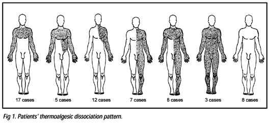
Figure 1 shows the patients' thermoalgesic dissociation pattern. Using the score scale, each patient was evaluated preoperatively and postoperatively toward obtaining a percentage of recovery or aggravation. The recovery percentage of operated patients is shown in Figure 2.
The comparative analysis of signs and symptoms of syringomyelia and Chiari malformation, using the statistical test of variance analysis at the reliability index of 95%, showed that, in relation to symptoms, there is no difference in the level of recovery by surgical treatment between both syndromes. With respect to signs, the response to treatment is not similar for both syndromes, with predomination of recovery from syringomyelia.
Most frequent complications from surgical treatment included: pseudomeningocele (23.3%), meningitis (6.6%), and cerebellar ptosis (1.6%). Among the patients with pseudomeningocele, 1 needed surgery, and 3 were treated by lumbar drainage.
The study of herniated cerebellar tonsils revealed cell loss and vacuolation in molecular layer with the occurrence of moderate quantity of amyloid bodies. Cell loss was also noted in Purkinje and granular layers. Other histological findings included gliosis, pia arachnoid proliferation, and vascular thickness.
DISCUSSION
The study of pathological conditions arising from the syringomyelic syndrome sometimes confuses with the pathogenic analysis of spinal cord cavitations. As such, when analyzing a group of patients with CM and SM, one is very often faced with several data common to the pathogenesis of both conditions.
The osseous malformation most typically found in this study was the basilar impression (BI) occurring in 43 subjects (71.6%). In this work, the term basilar impression is used to describe the craniovertebral anomaly consisting of atlanto-axoidean portion penetration in the posterior fossa, without taking into account whether the process etiology is primary or acquired8. The incidence of BI varied greatly in the several series found in literature. Milhorat et al.9 evaluated 238 patients with CM associated with SM and proved the occurrence of BI in 32 subjects, corresponding to 13.4% of total. Caetano de Barros et al.10 and Gonçalves da Silva11 called the attention to the high incidence of BI in cases studies in Northeastern Brazil, what is confirmed in our series.
The second most frequent osseous malformation found at craniocervical joint level was the atlanto-occipital fusion found in 18 patients, corresponding to 30% of total cases.
Following the initial study of Gustafson & Oldberg12, which correlate BI and SM, a number of works associated occipitocervical transition malformations, in particular BI and CM, with spinal cord cavitations. The analysis of literature indicated a simultaneous occurrence of SM and CM and/or BI in several series, which ranged between 6%, in Caetano de Barros et al.10 series and 83.7%, Gardner & Angel13 series. In the present study of 129 cases, 60 patients presented simultaneously SM and CM, corresponding to 46.5% of total cases.
Syringomyelia diagnosis was confirmed in all our 60 cases. Magnetic resonance image was applied to 41 patients in this study (68.3%). In 100% of subjects tested, the examination made CM diagnosis possible. Patients with associated BI submitted to MRI had their diagnosis confirmed, with the advantage of enabling the study of the relationships between structures of the region, in particular the direction of the odontoid tip when facing the anterior face of the brainstem. Control MRI indicated the occurrence of a cisterna magna, as highlighted by Sahuquillo et al.14, which was inexistent or reduced before the surgery, as well as the reduction or abatement of the syringomyelic cavity occurring in all of the cases, however remaining residual cavitations in spinal cord15. An important spinal cord abatement was found in a patient submitted to MRI several years after surgery, suggesting spinal cord atrophy.
Most common symptoms found at the disease onset were those related to difficulty in performing activities requiring soft and coordinate hand movements. Reduced strength or unilateral or bilateral muscular atrophy of the distal portion of upper limbs was present in 56.6% of cases. Sequentially, cervical pain (26.6%) and headache (20%) were the initial symptoms most frequently found after upper limp paresthesia and paresis. Earliest treated patients had suffered from the disease for 6 months at surgery time. The longest time of the disease affecting a patient was 21 years. The average disease time in all 60 cases under study was 6.2 years. In Matsumoto & Symon16 series, the average disease time was 7.6 years, while in Dyste et al.17 series it was 5.4 years.
To enable the postoperative evaluation of patients with CM associated with SM, a protocol was prepared, where symptoms and signs from each condition could be listed separately7.
In this study, symptoms listed as originated from compression at occipitocervical joint level included: headache, diplopia, dysphagia, dizziness, drop attacks, and hearing disorders.
Headache showed weight or sharp pain characteristics extending unilaterally or bilaterally from the cervical region to the forehead or retrorbital region. This headache is probably caused by the effect of compression and traction from herniated cerebellar tonsils on the first sensitive cervical roots, and by circulatory CSF disturbances9. Symptoms listed as originated from spinal cord cavitation included: limb and chest pain, muscular weakness, sudoresis disorders, and thermoalgesic sensibility dissociation. Pain from SM is a frequent symptom18. Pain may be deeply burning, central or significantly similar to causalgia19. In all cases under study, the deep burning pain was the most frequent.
In this study, muscular weakness was analyzed as a symptom of SM and a sign that could originate from both CM and SM. The reason for this double evaluation, also included in Bindall et al.20 study, is due to the fact that many patients understand atrophy or even dysesthesia as muscular weakness. Muscular weakness, as a symptom, was present in 50% of patients with CM and SM, and in 80% of patients with symptomatology from spinal cord cavitation alone in Bindall et al.20 series. This weakness occurred in 60% of Dyste et al.17 cases and in 80% of Gonçalves da Silva et al.21 cases. In the series under study, muscular weakness was the most frequent symptom found in 58 patients, corresponding to 96.6% of the cases.
Thermoalgesic dissociation occurred in 52 patients, or 86.6% of subjects of this study. The type of dissociation predominating in 17 patients was " suspended" or shawl pattern dissociation. Most diverse distributions were found in other 35 patients, suggesting that spinal cord cavitation can very often be eccentric or even multiple (Fig 1).
Symptoms attributed to both CM and SM, which were included in the protocol, are: nasal speech, pain in the face, sexual impotence, staggering gait, neck pain, and dysesthesia. Sexual impotence was found in 41.1% of Gonçalves da Silva et al.21 cases, in 39% of CM and BI cases studied by Caetano de Barros et al.10, and in 15 of 51 male patients with CM associated with SM, corresponding to 29.4% of Milhorat et al.9 series. In our series, sexual impotence of several intensity levels was found in 20 of 28 male patients, corresponding to 71.4% of cases. Dysesthesia, which was initially more frequent in upper limb extremities, may occur anywhere in the body. In this study, dysthesia was present in 59 patients, corresponding to 98.3% of cases. Evaluated signs attributed to CM included: nystagmus, hypoglossal disorder, hypoacusia, and impaired gag and palatal reflexes.
In this study, 8 patients corresponding to 13.3% of cases, presented hypoglossal lesion. Hypoacusia was found in 15 patients who presented the symptom of "reduced hearing" . Evaluated signs from spinal cord cavitation included: hyporeflexia of lower limbs, muscular atrophy, fasciculations, hypotonia of upper limbs, and hypotonia of lower limbs.
Muscular atrophy in one or both limbs is common at the disease onset18. In Dyste et al.17 series, upper limp atrophy was found in 32% of cases. Milhorat et al.9 found atrophy in episodes of SM associated with CM in 10.5% of patients, however this atrophy was found in only 0.7% of episodes of CM without SM. In Bindall et al.20 series, atrophy was detected in 40% of episodes of SM, but no occurrence was found in CM without SM. In this study, atrophy was present in 44 patients, corresponding to 73.3% of cases.
Signs attributed to CM and SM include: spinal injury, muscular weakness, hyperreflexia of upper limps, hyperreflexia of lower limbs, and proprioception impairment.
Muscular weakness, as a sign that can be evaluated and quantified by the practitioner, was present in 59 patients of this series, corresponding to 98% of cases. This sign was also present in 91.6% of Taricco & Pereira22 cases, in 60% of Dyste et al.17 cases, in 20% of brainstem compression cases, in 83.3% of cases of brainstem compression combined with SM, and in 100% of cases with only SM symptomatology of Bindall et al.20 series.
Proprioception impairment may be caused by direct brainstem compression in CM or syringomyelic cavity, when it extends to posterior cords. In case of SM associated with CM and/or BI, treatment aims to decompress the occipitocervical joint with the objective of eliminating the mechanisms that, upon changing the circulation of CSF, produce and perpetuate syringomyelia.
Compression at occipitocervical transition level caused by herniated tonsils in patients with CM led this work to adopt the surgical technique of craniocervical decompression with cerebellar tonsillectomy23. Cerebellar tonsillectomy, using the technique described by Jules Bertrand in 1973 and later modified by Batzdorf24, was adopted for all patients in this study, including sometimes tonsil coagulation and retraction or subpial aspiration of herniated cerebellar tissue, and sometimes the total amputation of the herniated portion of tonsils.
In patients presenting symptomatic BI associated with equally symptomatic CM and SM, occipitocervical fixation by metallic rods with bone grafting was performed as a preparation for eventual transoral odontoid removal. The occurrence of osseous malformation together with impaired long tracts, sensitive syndrome, impaired cranial nerves function, and cerebellar syndrome are indicative of changes in foramen magnum at both anterior and posterior level10.
Postoperatively, statistical analysis was performed considering the patients' improvement or aggravation. Results from analysis of CM symptoms showed an accentuated statistical improvement. All symptoms improved to p = 0, except for reduced hearing that also improved statistically, but not to p = 0 but rather to p > 0.013. All SM symptoms improved statistically to the reliability index of 100% (p = 0). All signs associated with CM improved statistically, being nystagmus the most significantly improved. At analyzing the SM signs, it was found that only hyporeflexia of upper limbs did not improve, and that, in spite of statistical improvement of muscular atrophy (p < 0.03), this improvement was not as significant as that of other signs (hyporeflexia of lower limbs, fasciculations, and hypotonia of upper and lower limbs). Regarding SM and CM symptoms, only sexual impotence did not improve statistically p > 0.1). With respect to signs attributed to both syndromes, only hyperreflexia of upper limbs did not improve (p > 0.06). Upon analyzing the score system established for this study, it was found that patients achieved a score ranging between 18 points (least symptomatic) and 50 points (more symptomatic) at the preoperative stage. At the postoperative stage, 5 points was the best result compared to 55 points for the worst result. Most patients of this study are in the improvement range of 45-60% in relation to their disease (Fig 2).
The score system was efficient to evaluate the percentage of each patient's improvement or aggravation, and enabled a fast clinical evaluation of such patients' progress, as they very often insist on an unimproved sign or symptom, and make the interpretation of results difficult. With the score system, this type of problem is minimized, and evaluation can be more objective.
The comparison of both syndromes and the application of the variance analysis at the reliability index of 95% in terms of symptoms, showed that there is no difference in improvement intensity between SM and CM. In relation to signs, there was a difference in improvement intensity in both syndromes, predominantly in syringomyelia.
Pseudomeningocele was the most frequent surgical complication in patients of this series. Fourteen patients presented postoperative occipital pseudomeningocele. One patient required surgery to correct the problem, while three patients were treated by CSF drainage. Six patients underwent percutaneous punctures, followed by cyst aspiration. In all other cases, there was a spontaneous regression of cysts over a period ranging between 4 days and 2 weeks. Four subjects of the study presented postoperative bacterial meningitis. All these patients recovered without sequels attributable to meningeal infection.
Hernia of the cerebellar biventral lobe is seldom reported in the literature, though described by Williams25 by the name of cerebellar slump. Batzdorf24 called the condition of " cerebellar ptosis" , and he called the attention to the fact that this condition could alone compress the occipitocervical transition and cause persisting pressoric craniocervical dissociation. Holly & Batzdorf26 introduced the suboccipital cranioplasty technique to correct the problem. In this study, we detected one case of cerebellar ptosis, which was corrected surgically.
In the series of patients of Banerji & Millar27, mortality rate reached 5%. In the group studied by Matsumoto & Symon16, total mortality rate was 3.1%. In Logue & Edwards28 series, mortality rate was 1.3%; and in Taricco & Pereira's22, it was 7.6%. Bindall et al.20 study indicated no deaths, like in Batzdorf24 series, which adopts the same surgery technique of craniovertebral decompression with tonsillectomy used in this study. In this series, no death was reported among 69 patients operated with combined CM and SM (9 patients did not meet the criteria for inclusion in this study). It should be pointed out, however, that all 60 patients of the study were selected from a universe of 129 patients with malformations at occipitocervical joint level, operated between 1981 and 2000. In this universe of patients, there were 9 deaths (6.9%), all occurred in the 1980's, when the current postoperative care was not available yet.
CONCLUSIONS
We conclude that craniocervical decompression with tonsillectomy and duramater graft proved an effective method for the treatment of SM and CM.
The comparative analysis of sings and symptoms of SM and those of CM showed that in relation to symptoms, there is no difference in the level of recovery by surgical treatment between both syndromes. With respect to signs, there is a predomination of recovery from SM. All CM signs and symptoms improved. Syringomyelia signs and symptoms also improved except for "upper limb hiporeflexia" which did not improve. Signs and symptoms attributed to both SM and CM, also, improved, except for "upper limb hyperreflexia" and "sexual impotence" that failed to improve.
The score system made clear that half of the subjects showed improved signs and symptoms in the 40-60% range.
Received 1 August 2003, received in final form 18 September 2003.
Accepted 5 November 2003.
Dr. José Arnaldo Motta de Arruda - Lab. de Neurologia Experimental e Neurofisiologia/DFF/FM/UFC - Rua Coronel Nunes de Melo 1127 - 60430-270 Fortaleza CE - Brasil. E-mail: arnaldo_arruda@uol.com.br.
- 1.Lotbinière ACJ. Historical considerations. In Anson JA, Benzel EC, Awad IA (eds). Neurosurgical topics: syringomyelia and Chiari malformations. Illinois: American Association of Neurologic Surgeons, 1997:1-26.
- 2. Ballantine HT, Ojemann RG, Drew JH. Syringomyelia. In Krayenbühl H, Maspes PE, Sweet WH (eds). Progress in neurological surgery. New York: Karger, 1971:227-245.
- 3. Barnett HJM, Foster JB, Hudgson P. Syringomyelia. Major problems in neurology. London: Saunders, 1973.
- 4. Carmel PW, Markesbery WR. Early descriptions of the Arnold-Chiari malformation: the contribution of John Cleland. J Neurosurg 1972;37:543-547.
- 5. Penfield W, Coburn DF. Arnold-Chiari malformation and its operative treatment: case report. Arch Neurol Psychiatry 1938;40:328-336.
- 6. Koehler PJ. Chiari's descriptions of cerebellar ectopy (1891): with a summary of Cleland's and Arnold's contributions and some early observations on neural-tube defects. J Neurosurg 1991;75:823-826.
- 7. Arruda JAM, Costa CMC, Tella OI Jr. Avaliação de pacientes com siringomielia associada à malformação de Chiari: proposta de um protocolo multicêntrico. J Bras Neurocirurg 1999;10:93-96.
- 8. Canelas MH, Tenuto RA, Zaclis J, Cruz OR. Malformações occípito-cervicais: a propósito de vinte novos casos. Arq Neuropsiquiatr 1956;1:1-27.
- 9. Milhorat TH, Chou MW, Trinidad EM, et al. Chiari I malformation redefined: clinical and radiographic findings for 364 symptomatic patients. Neurosurgery 1999;44:1005-1017.
- 10. Caetano de Barros M, Farias W, Ataíde L, Lins L. Basilar impression and Arnold-Chiari malformation: a study of 66 cases. J Neurol Neurosurg Psychiatry 1968;31:596-605.
- 11. Gonçalves da Silva JAG. Basilar impression and Arnold-Chiari malformation: surgical findings in 209 cases. Neurochirurgie 1992;35:189-195.
- 12. Gustafson WA, Oldberg E. Neurologic significance of platybasia. Arch Neurol Psychiatry 1940;44:1184-1198.
- 13. Gardner WJ, Angel J. The mechanism of syringomyelia and its surgical correction. Clin Neurosurg 1958;6:131-140.
- 14 . Sahuquillo J, Rubio E, Poca MA, Rovira A, Rodriguez-Baeza A, Cevere C. Posterior fossa reconstruction, a surgical technique for the treatment of Chiari I malformation and Chiari I / syringomyelia complex: preliminary results and magnetic resonance imaging quantitative assessment of hindbrain migration. Neurosurgery 1994;35:874-885.
- 15. Barkovich AJ, Wippold FJ, Sherman JL, Citrin CM. Significance of cerebellar tonsillar position on MR. AJNR 1986;7:795-799.
- 16. Matsumoto T, Symon L. Surgical management of syringomyelia: Current results. Surg Neurol 1989;32:258-265.
- 17. Dyste GN, Menezes AH, Vangilder JC. Symptomatic Chiari malformation: an analysis of presentation, management, and long-term outcome. J Neurosurg 1989; 71:159-168.
- 18. Foster JB. Neurology of syringomyelia. In Batzdorf U (ed). Syringomyelia: current concepts in diagnosis and treatment. Baltimore: Williams & Wilkins, 1991:91-115.
- 19. Todor RD, Harrison TMMU, Milhorat TH. Pain and syringomyelia: a review. Disponível em: <http://www. Neurosurg.Focus 8 (3): Article 11, 2000>. Acesso em: 5 abr. 2000.
- 20. Bindall AK, Dunsker SB, Tew JM Jr. Chiari I malformation: classification and management. Neurosurgery 1995;37:1069-1074.
- 21. Gonçalves da Silva JAG, Cantisani JUF, Brito JCF, et al. Impressão basilar, Arnold-Chiari e siringomielia: análise de 20 casos operados. Arq Bras Neurocirurg 1987;6:77-95.
- 22. Taricco MA, Pereira WC. Tratamento cirúrgico da siringomielia: associada à malformação de Chiari do tipo I. Arq Bras Neurocirurg 1997;16:47-60.
- 23. Gonçalves da Silva JAG, Holanda MMA. Basilar impression, Chiari malformation and syringomyelia. Arq Neuropsiquiatr 2003;61:368-375.
- 24. Batzdorf U. Syringomyelia related to abnormalities at the level of the craniovertebral junction. In Batzdorf U (ed). Syringomyelia: current concepts in diagnosis and treatment. Baltimore: Williams & Wilkins, 1991:163-182.
- 25. Williams B. A critical appraisal of posterior fossa surgery for communicating syringomyelia. Brain 1978;101:223-250.
- 26. Holly LT, Batzdorf U. Management of cerebellar ptosis following craniovertebral decompression for Chiari I malformation. J Neurosurg 2001;94:21-26.
- 27. Banerji NK, Millar JHD. Chiari malformation presenting in adult life. Its relationship to syringomyelia. Brain 1974;97:157-168.
- 28. Logue V, Edwards MR. Syringomyelia and its surgical treatment: an analysis of 75 patients. J Neurol Neurosurg Psychiatry 1981;44:273-284.
Publication Dates
-
Publication in this collection
23 June 2004 -
Date of issue
June 2004
History
-
Accepted
05 Nov 2003 -
Reviewed
18 Sept 2003 -
Received
01 Aug 2003