Abstracts
Headache as a chief complaint is rare in the paediatric emergency room. Actually, very seldom cases secondary to life threatening conditions as non-traumatic subarachnoid haemorrhage have been reported. A child with severe headache and nuchal rigidity and no other abnormalities on the physical examination is reported. Magnetic resonance angiography and cerebral angiography disclosed a ventricular arteriovenous malformation in the choroid plexus, supplied by the anterior choroidal artery, classified according to Spetzler grading system as grade 3 (deep venous drainage: 1; eloquence area: 0 and size: 2). The differences in the clinical presentations of the central nervous system arteriovenous malformation between children and adults are discussed.
secondary headache in children; arteriovenous malformation of the brain; pediatric stroke
Cefaléia como queixa principal raramente ocorre num serviço de emergência pediátrica. Quando isso acontece, casos de cefaléia secundária que trazem risco de vida, tais como a hemorragia subaracnóide são raramente relatados. Apresentamos o caso de uma criança que apresentou cefaléia de forte intensidade associada a rigidez de nuca, sem outras anormalidades no exame físico. A angioressonância e angiografia digital evidenciaram malformação arteriovenosa na topografia do plexo coróide do ventrículo lateral direito, nutrida pela artéria coroidéia anterior, grau III na classificação de Spetzler (drenagem venosa profunda: 1; área de eloqüência: 0 e tamanho: 2). Nós discutimos as diferenças na apresentação clínica das malformações arteriovenosas encefálicas nas crianças e adultos.
cefaléia secundária; malformação arteriovenosa do encéfalo; malformação arteriovenosa ventricular
Ventricular arteriovenous malformation bleeding: a rare cause of headache in children. Case report
Hemorragia cerebral secundária a malformação artério-venosa ventricular: uma causa rara de cefaléia na infância. Relato de caso
Hilton Mariano da SilvaI; Luciano Ricardo França da SilvaII; Eric Homero Albuquerque PaschoalII; Feres Eduardo Aparecido Chaddad NetoII; Carlos Alberto BordiniI; José Geraldo SpecialiI
IDepartment of Neurology, Psychiatry, and Medical Psychology, São Paulo University School of Medicine at Ribeirão Preto, Brazil (FMRP/USP)
IIDepartment of Neurology and Neurosurgery of Mário Gatti Hospital, Campinas, SP, Brazil
ABSTRACT
Headache as a chief complaint is rare in the paediatric emergency room. Actually, very seldom cases secondary to life threatening conditions as non-traumatic subarachnoid haemorrhage have been reported. A child with severe headache and nuchal rigidity and no other abnormalities on the physical examination is reported. Magnetic resonance angiography and cerebral angiography disclosed a ventricular arteriovenous malformation in the choroid plexus, supplied by the anterior choroidal artery, classified according to Spetzler grading system as grade 3 (deep venous drainage: 1; eloquence area: 0 and size: 2). The differences in the clinical presentations of the central nervous system arteriovenous malformation between children and adults are discussed.
Key words: secondary headache in children, arteriovenous malformation of the brain, pediatric stroke.
RESUMO
Cefaléia como queixa principal raramente ocorre num serviço de emergência pediátrica. Quando isso acontece, casos de cefaléia secundária que trazem risco de vida, tais como a hemorragia subaracnóide são raramente relatados. Apresentamos o caso de uma criança que apresentou cefaléia de forte intensidade associada a rigidez de nuca, sem outras anormalidades no exame físico. A angioressonância e angiografia digital evidenciaram malformação arteriovenosa na topografia do plexo coróide do ventrículo lateral direito, nutrida pela artéria coroidéia anterior, grau III na classificação de Spetzler (drenagem venosa profunda: 1; área de eloqüência: 0 e tamanho: 2). Nós discutimos as diferenças na apresentação clínica das malformações arteriovenosas encefálicas nas crianças e adultos.
Palavras-chave: cefaléia secundária, malformação arteriovenosa do encéfalo, malformação arteriovenosa ventricular.
A headache due to a hemorrhagic stroke is a life-threatening clinical condition rarely seen in a paediatric emergency department1,2. When it occurs, an arteriovenous malformation should be ruled out. The arteriovenous malformation of the brain (AVM) accounts for 30% to 50% of such hemorrhagic strokes in children3-6 and it has been associated with a 25% mortality rate7. On the other hand, the ventricular location is found in only 4% of the AVM in childhood8; and in 1,3% of the adult's AVM9. We report a child with a history of sudden headache secondary to a bleeding of a ventricular arteriovenous malformation.
CASE
A 9-year-old previously healthy girl was admitted to the Emergency Room with an eight-hour history of sudden onset of severe headache. The pain was pulsatile and bilateral and not accompanied by other symptoms. There is no history of migraine, epilepsy or stroke. Parents reported that soon after the onset of the headache the patient became drowsy for about one hour. No trigger factor was identified. On the neurological examination, the patient was alert and well oriented with no other abnormalities but mild nuchal rigidity.
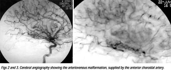
Computed tomography of the brain revealed hemorrhage in the right lateral ventricle (Fig 1) and gadolinium-enhanced magnetic resonance imaging study of the brain disclosed a heterogeneous lesion in the mesial portion of the right temporal lobe, above and inside the temporal horn of the lateral ventricle. The lesion extended until the subependimary area of the trigono of the right ventricle. The lesion was hypointense on T1 and T2-weighted images and enhanced with the contrast. Other hyperintense T1 and T2-weighted images lesions were seen in the right lateral ventricle suggesting bleeding. Magnetic resonance angiography and cerebral angiography disclosed an arteriovenous malformation in part of the choroid plexus, supplied by the anterior choroidal artery (Figs 2 and 3). The AVM was classified according to Spetzler grading system as grade 3 (deep venous drainage: 1; eloquence area: 0 and size: 2).
A surgical procedure was done resulting in an almost complete excision of the AVM and without sequelae. The patient remains asymptomatic after one year of follow-up.
This report was approved by the Hospital Ethics Committee and her parents signed the informed consent.
DISCUSSION
Headache is a very common symptom in children10,11, affecting approximately 82.9% of the Brazilian population ranging between 10 to 18 years of age12. Despite such high prevalence, headache as a chief complaint is rare in the paediatric emergency room. Actually, very seldom cases secondary to life threatening conditions as non-traumatic subarachnoid haemorrhage (NTSH) are seen1,2. Accordingly, the data regarding the occurrence of NTSH in childhood are scant. Previous reports suggest that an arteriovenous malformation of the brain is the most frequent cause of this condition in children13, a condition by far more prevalent in adults14.
The clinical presentations of AVM in children also differ considerably from those in adults. There is a high propensity (80%) for the AVM childhood to present bleeding8, what is higher than that reported for adults9,15-17. Likewise, epilepsy was reported in 12-18%of the AVM' s children series7,18 and in 16 to 53% of the adult patients19,20. In neonates, AVM has been recognized as a cause of life-threatening congestive heart failure21.
Several authors have reported that the prognosis was not so good in children with AVM in comparison to adults7,18,22. Conversely, a better prognosis was suggested for purely intraventricular haemorrhage arteriovenous malformation as observed in our patient by some reports23. One factor that had a dramatic impact on the diagnosis and treatment of AVM was the development of the modern neuroimaging techniques24. The treatment, however, remains a challenging matter. Endovascular embolization, radiosurgery, surgical excision or a multimodality approach have been used to treat this condition, however studies are not conclusive yet25.
This is an interesting case report of a rare condition that causes headache in children. The particular localization of the AVM produces a headache associated with nuchal rigidity without other abnormality on the neurological examination.
Received 30 September 2003, received in final form 9 January 2004. Accepted 6 February 2004.
Dr. José Geraldo Speciali - Departamento de Neurologia (FMRP/USP) - Avenida Bandeirantes 3900 - 14049-900 Ribeirão Preto SP - Brasil.
- 1. Kan L, Nagelberg J, Maytal J. Headaches in a pediatric emergency department: etiology, imaging, and treatment. Headache 2000;40:25-29.
- 2. Burton LJ, Quinn B, Pratt-Cheney JL, Pourani M. Headache etiology in a pediatric emergency department. Pediatr Emerg Care 1997;13:1-4.
- 3. Broderick J, Talbot GT, Prenger E, Leach A, Brott T. Stroke in children within a major metropolitan area: the surprising importance of intracerebral hemorrhage. J Child Neurol 1993;8:250-255.
- 4. Celli P, Ferrante L, Palma L, Cavedon G. Cerebral arteriovenous malformations in children: clinical features and outcome of treatment in children and in adults. Surg Neurol 1984;22:43-49.
- 5. Gold AP, Challenor UB, Gilles FH. Report of joint committee for stroke facilities: XI. Strokes in children. Stroke 1973;4:835-894.
- 6. Lanthier S, Carmant L, David M, Larbrisseau A, De Veber G.Stroke in children: the coexistence of multiple risk factors predicts poor outcome Neurology 2000;54:371-378.
- 7. Kondziolka D, Humphreys RP, Hoffman HJ, Hendrick EB, Drake JM. Arteriovenous malformations of the brain in children: a forty - year experience. Can J Neurol Sci 1992;19:40-45.
- 8. Humphreys RP. Hemorrhagic stroke in childhood. J Pediatric Neurosci 1986;2:1-10.
- 9. Jomin M, Lesoin F, Lozes G. Prognosis for arteriovenous malformations of the brain in adults based on 150 cases. Surg Neurol 1985;23:362-366.
- 10. Bille B. Migraine in school children. Acta Paediatr 1962;51:1-151.
- 11. Deubner DC. An epidemiologic study of migraine and headache in 10-20 year olds. Headache 1977;17:173-180.
- 12. Barea LN, Tannhauser M, Rotta NT. An epidemiological study of headache among children and adolescents of southern Brazil. Cephalalgia 1996;16:545-549.
- 13. Schoenberg VS, Mellinger JF, Schoenberg DG. Cerebrovascular disease in infants and children: a study of incidence, clinical features, and survival. Neurology 1978;28:763.
- 14. Mayberg MR, Batjer HH, Dacey R, et al. Guidelines for the management of aneurysmal subarachnoid hemorrhage: a statement for healthcare professionals from a special writing group of the Stroke Council, American Heart Association. Stroke 1989;25:2315-2328.
- 15. Guidetti B, Delitala A. Intracranial arteriovenous malformations: conservative and surgical treatment. J Neurosurg 1983;53:149-152.
- 16. Heros RC, Korosue K, Diebold PM. Surgical excision of cerebral arteriovenous malformations: late results. Neurosurgery 1990;26:578-579.
- 17. Itoyama Y, Uemura S, Ushio Y, et al. T. Natural course of unoperated intracranial arteriovenous malformations: study of 50 cases. J Neurosurg 1989;71:805-809.
- 18. Gerosa MA, Cappellotto P, Licata C, Iraci G, Pardatscher K, Fiore DL. Cerebral arteriovenous malformations in children (56 cases). Childs Brain 1981;8:356-371.
- 19. The Arteriovenous Malformation Study Group. Arteriovenous malformations of the brain in adults. N Engl J Med 1999;340:1812-1818.
- 20. Guiotoku C M, Arruda W O, Ramina R, Pedrozo A A, Meneses M S. Arteriovenous malformations of the central nervous system: a review of 53 cases. Arq Neuro psiquiatr 1999;57:452-456.
- 21. Hara H, Burrows PE, Flodmark O, Terbrugge K, Humphreys R. Neonatal superficial cerebral arteriovenous malformations. Pediatric Neurosurg 1994;20:126-136.
- 22. Mori K, Murata T, Hashimoto N, Handa H. Clinical analysis of arteriovenous malformations in children. Childs Brain 1980;6:13-25.
- 23. Hartmann A, Mast H, Mohr J P, et al. Morbidity of intracranial hemorrhage in patients with cerebral arteriovenous malformation. Stroke 1998;29:931-934.
- 24. Humphreys RP, Hoffman HJ, Drake JM, Rutka JT. Choices in the 1990s for the management of pediatric cerebral arteriovenous malformations. Pediatric Neurosurg 1996;25:277-285.
- 25. Ogilvy CS, Stieg PE, Awad I, et al. Recommendations for the management of intracranial arteriovenous malformations: a statement for healthcare professionals from a special writing group of the stroke council, American Stroke Association. Stroke 2001;32:1458-1471.
Publication Dates
-
Publication in this collection
20 July 2004 -
Date of issue
June 2004
History
-
Received
30 Sept 2003 -
Reviewed
09 Jan 2004 -
Accepted
06 Feb 2004