Abstracts
Objective
To investigate the correlation between the Alberta Program Early CT Score (ASPECTS) and the Scandinavian Stroke Scale (SSS) for the evaluation of neurological impairment in patients with acute stroke.
Method
59 patients with a first acute ischemic stroke were evaluated. The ASPECTS were evaluated by 2 neurologists at admission and by another neurologist after 48 hours. The NIHSS and SSS was applied to determinate stroke severity. Correlations and agreements were analysed statistically by Spearman and Kappa tests.
Results
ASPECTS was correlated with National Institute of Health Stroke Scale (NIHSS) at admission (r = -0.52; p < 0.001) and SSS (r = 0.50; p < 0.001). The ASPECTS and SSS items were most correlated with arm (r = 0.52; p < 0.001) and hand (r = 0.49; p < 0.001) motor power, and speech (r = 0.51; p < 0.001). The SSS of 25.5 shows sensitivity (68%) and specificity (72%) when associated with ASPECTS ≤ 7.
Conclusion
The SSS can predict worst neurological impairment when associated with lower values of ASPECTS.
stroke; neuroimaging; outcome measures; scales
Objetivo
Investigar a relação entre o Alberta Program Early CT Score (ASPECTS) e a Scandinavian Stroke Scale (SSS) para avaliação da incapacidade neurológica de pacientes na fase aguda do acidente vascular cerebral (AVC).
Método
59 pacientes com diagnóstico de primeiro AVC foram avaliados. O ASPECTS foi avaliado por 2 neurologistas na admissão e por outro neurologista após 48 horas. O National Institute of Health Stroke Scale (NIHSS) e SSS foram aplicadas para determinar a gravidade do AVC. As correlação e concordâncias foram analisadas estatisticamente pelos testes de Spearman e Kappa.
Resultados
ASPECTS foi correlacionado com o NIHSS na admissão (r = -0,52; p < 0,001) e SSS (r = 0,50; p < 0,001). O ASPECTS e os itens do SSS que mais se relacionaram foram força do braço (r = 0,52; p < 0,001), da mão (r = 0,49; p < 0,001) e fala (r = 0,51; p < 0,001). A pontuação da SSS de 25,5 mostrou sensibilidade (68%) e especificidade (72%) quando associado ao ASPECTS ≤ 7.
Conclusão
A SSS pode predizer pior incapacidade neurológica quando associado a baixos valores do ASPECTS.
acidente vascular cerebral; neuroimagem; medidas de desfecho funcional; escalas
In Brazil as well as worldwide, stroke is among the leading causes of the functional
disability and death in the adult population11 Go AS, Mozaffarian D, Roger VL, Benjamin EJ, Berry JD, Borden WB et
al. Heart Diseases and Stroke Statistics – 2013 Update: a report from the
American Heart Association. Circulation. 2013;127(1):e6-245.
http://dx.doi.org/10.1161/CIR.0b013e31828124ad
http://dx.doi.org/10.1161/CIR.0b013e3182...
,
22 Lotufo PA, Benseñor IM. Trends of stroke subtypes mortality in Sao
Paulo, Brazil (1996-2003). Arq Neuropsiquiatr. 2005;63(4):951-5.
http://dx.doi.org/10.1590/S0004-282X2005000600009
http://dx.doi.org/10.1590/S0004-282X2005...
,
33 Langhorne P, Coupar F, Pollock A. Motor recovery after stroke: a
systematic review. Lancet Neurol. 2009;8(8):741-54.
http://dx.doi.org/10.1016/S1474-4422(09)70150-4
http://dx.doi.org/10.1016/S1474-4422(09)...
,
44 Nichols-Larsen DS, Clark PC, Zeringue A, Greenspan A, Blanton S.
Factors influencing stroke survivors’ quality of life during subacute recovery.
Stroke. 2005;36(7):1480-4.
http://dx.doi.org/10.1161/01.STR.0000170706.13595.4f
http://dx.doi.org/10.1161/01.STR.0000170...
,
55 Saposnik G; Del Brutto OH. Stroke in South America: A systematic
review of incidence, prevalence, and stroke subtypes. Stroke. 2003;34(9):
2103-7. http://dx.doi.org/10.1161/01.STR.0000088063.74250.DB
http://dx.doi.org/10.1161/01.STR.0000088...
. The increasing importance of imaging studies in recent years has led to the
appearance of objective scales for evaluation of severity, such as the Alberta Program
Early CT Score (ASPECTS), which subdivides the territory of the middle cerebral artery
(MCA) into ten standardized regions evaluated by cranial CT66 Barber PA, Demchuk AM, Zhang J, Buchan AM. Validity and reliability
of a quantitative computed tomography score in predicting outcome of hyperacute
stroke before thrombolytic therapy. ASPECTS Study Group. Alberta Stroke
Programme Early CT Score. Lancet. 2000;355(9216):1670-4.
http://dx.doi.org/10.1016/S0140-6736(00)02237-6
http://dx.doi.org/10.1016/S0140-6736(00)...
,
77 Puetz V, Dzialowski I, Hill MD, Demchuk AM. The Alberta Stroke
Program Early CT Score in clinical practice: what have we learned?. Int J
Stroke. 2009;4(5):354-64.
http://dx.doi.org/10.1111/j.1747-4949.2009.00337.x
http://dx.doi.org/10.1111/j.1747-4949.20...
.
The ASPECTS is a precise and practical method for evaluating stroke in the acute phase
and is indicated especially in the first 6 hours after the initial event to detect early
changes in the hyperacute phase and predict the possibility of haemorrhagic
transformation, by outlining the severity of the ischemic area88 Pexman JHW, Barber PA, Hill MD, Sevick RJ, Demchuk AM, Hudon ME, et
al. Use of the Alberta Stroke Program Early CT Score (ASPECTS) for assessing CT
scanS in patients with acute stroke. AJNR Am J Neuroradiol.
2001;22(8):1534-42.
,
99 Lin K, Rapalino O, Law M, Babb JS, Siller KA, Pramanik BK. Accuracy
of the Alberta Stroke Program Early CT Score during the first 3 hours of middle
cerebral artery stroke: comparison of noncontrast CT, CT angiography source
images, and CT perfusion. AJNR Am J Neuroradiol. 2008;29(5):931-6.
http://dx.doi.org/10.3174/ajnr.A0975
http://dx.doi.org/10.3174/ajnr.A0975...
,
1010 Mak HK, Yau KK, Khong Pl, Ching AS, Cheng PW, Au-Yeung PK et al.
Hipodensity of >1/3 middle cerebral artery territory versus Alberta Stroke
Programme Early CT Score (ASPECTS) comparison of two methods of quantitative
evaluation of Early CT changes in hyperacute ischemic Stroke in the community
setting. Stroke. 2003;34(5):1194-6.
http://dx.doi.org/10.1161/01.STR.0000069162.64966.71
http://dx.doi.org/10.1161/01.STR.0000069...
. Recently we validated the SSS for the Brazilian population and found it useful
for evaluating severity in stroke patients in our country1111 Luvizutto GJ, Monteiro TA, Braga G, Pontes-Neto OM, de Lima Resende
LA, Bazan R. Validation of the scandinavian stroke scale in a multicultural
population in Brazil. Cerebrovasc Dis Extra. 2012;2(1):121-6.
http://dx.doi.org/10.1159/000345948
http://dx.doi.org/10.1159/000345948...
. This study aimed to investigate the correlation between the ASPECTS and the
Scandinavian Stroke Scale (SSS) to evaluate the severity of neurological deficit in
Brazilian patients with acute stroke involving the middle cerebral artery (MCA), to
relate each item on the SSS scale with ASPECTS and to identify whether there is some
cutoff value of SSS that maximizes the sensitivity and specificity of the severity in a
worst neurological impairment. The first hypothesis of the study is that lower values of
ASPECTS are positively correlated with lower SSS values, especially with specific items
from the scale that evaluates the MCA territory.
METHOD
The study is a prospective series of cases that includes 59 individuals, of both
sexes, admitted to the Stroke Unit of the Botucatu Medical School in the period from
March to December of 2012 with a diagnosis of ischemic stroke – confirmed by
clinical examination and noncontrast CT scan – in middle cerebral artery territory
confirmed by the classification of BAMFORD1212 Bamford J, Sandercock P, Dennis M, Burn J, Warlow C. Classification
and natural history of clinically identifiable subtypes of cerebraL infarction.
Lancet. 1991;337(8756):1521-6.
http://dx.doi.org/10.1016/0140-6736(91)93206-O
http://dx.doi.org/10.1016/0140-6736(91)9...
, and excluding patients with ischemic stroke in the anterior or posterior
cerebral artery territory, haemorrhagic stroke, prior stroke, presenting a modified
Rankin Scale (mRS) score prior to admission ≥ 1, previous diagnosis of dementia or
patients that were eligible for and received any type of treatment by cerebral
reperfusion therapy (intra-arterial or intravenous).
At the admission in the stroke unit, the first procedure was clinical attention by
means of neurological examination and National Institutes of Health Stroke Scale
(NIHSS) by neurovascular physician of the Stroke Unit; then a noncontrast CT scan
was obtained for clinical procedures and determination of the ASPECTS by 2
neurologists from the stroke team at admission independently. The new evaluation was
applied after 48 hours by another expert neurologist to determine the neurological
impairment severity through NIHSS1313 Cincura C, Pontes-Neto OM, Neville IS, Mendes HF, Menezes DF,
Mariano DC et al. Validation of the Natinal Institutes of Health Stroke Scale,
modified Rankin Scale and Barthel Index in Brazil: the role of cultural
adaptation and structured interviewing. Cerebrovasc Dis. 2009;27(2):119-22.
http://dx.doi.org/10.1159/000177918
http://dx.doi.org/10.1159/000177918...
, and the quantitative severity of paralysis and incapacity according to
SSS1111 Luvizutto GJ, Monteiro TA, Braga G, Pontes-Neto OM, de Lima Resende
LA, Bazan R. Validation of the scandinavian stroke scale in a multicultural
population in Brazil. Cerebrovasc Dis Extra. 2012;2(1):121-6.
http://dx.doi.org/10.1159/000345948
http://dx.doi.org/10.1159/000345948...
. These scales were applied without knowledge of the previous ASPECTS value
at admission and performed by a neurologist certified in these scales. The study was
approved by the Ethics Committee on Human Research of our Institution and all the
patients or their guardians signed terms of free and informed consent to be
included.
Statistical analysis
The concordance between the 2 neurologists for ASPECTS scoring at admission was evaluated by the Kappa coefficient. The relation among SSS, NIHSS and ASPECTS was assessed by the Spearman correlation and linear regression model considering the potential confounders (age, sex, topography of lesion and NIHSS at admission). The test of Shapiro-Wilk was performed to indicate whether the linear regression was adequate. The correlation between the items SSS and ASPECTS was measured by means of the Spearman correlation. The cutoff value of SSS that maximizes the sensitivity and specificity of the severity of the worst neurological impairment was estimated by constructing ROC curves using the ASPECTS. Significance was set at p less than 0.05. Statistical analyses were performed using the software SPSS v.15.0.
RESULTS
Based on the exclusion criteria, 151 individuals had been excluded from the study: 42 patients received a cerebral reperfusion therapy, 14 presented previous diagnosis of dementia, 28 had a prior stroke, 38 presented haemorrhagic stroke, 8 had an ischemic stroke in posterior cerebral artery territory, 12 had an ischemic stroke in anterior cerebral artery territory and 9 presented a mRS > 1 at admission. The 59 studied patients were comprised of 17 males and 42 females, with mean age of 66 years. The concordance between the 2 neurologists for ASPECTS was – Kappa coefficient = 0.706; p < 0.001. The ASPECTS values ranged from 5 to 10 (median = 9), NIHSS varied from 0 to 25 (median = 8); after 72 hours the NIHSS values ranged from to 0 to 31 (median = 6), while SSS varied from 4 to 58 (median = 34). At admission, the higher the ASPECTS, the lower the NIHSS values, -r = -0.56. After 48 hours the ASPECTS score was negatively correlated with NIHSS (-0.52) but positively related to SSS (r = 0.50).
Table 1 shows that ASPECTS accounts for 63.5% of the SSS variability. For each unit of increase in ASPECTS, there is a corresponding rise of 1.57 points in the SSS median considering the potential confounders. The p-value (p = 0.17) obtained by the Shapiro-Wilk test signifies that the adjustment was appropriate.
The correlations between ASPECTS and SSS items (Table 2) were assessed for consciousness (r = 0.23; p = 0.082), eye movements (r = 0.23; p = 0.074), arm motor power (r = 0.52; p < 0.001), hand motor power (r = 0.49; p < 0.001), leg motor power (r = 0.33; p = 0.010), orientation (r = 0.28; p = 0.031), speech (r = 0.51; p < 0.001), facial palsy (r = 0.21; p = 0.108), gait (r = 0.18; p = 0.167) and maximal score (r = 0.50; p = 0.001).
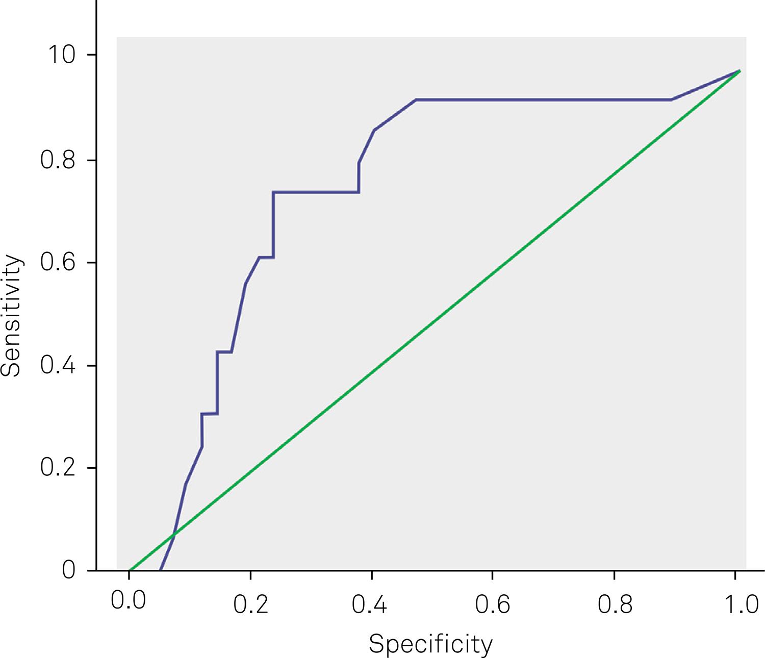
The Scandinavian Stroke Scale score of 25.5 shows sensitivity and specificity of 68% and 72%, respectively, when associated with ASPECTS ≤ 7 for the worst neurological impairment (Figure 1). Among the items on the Scandinavian Stroke Scale, speech presented sensitivity and specificity, respectively, of 72% and 78% (Figure 2).
Sensitivity and specificity of speech item on Scandinavian Stroke Scale with ASPECTS values.
DISCUSSION
In the present study, the ASPECTS values have demonstrated a closer association with
the severity scales to identify neurological deficit and acute-phase outcome. The
medical literature reports that low ASPECTS values were correlated with a high NIHSS
score and a reduced long-term functional capacity1414 Aviv RI, Mandelcorn J, Chakraborty S, Gladstone D, Malham S,
Tomlinson G et al. Alberta Stroke Program Early CT Scoring of CT perfusion in
early stroke visualization and assessment. AJNR Am J Neuroradiol.
2007;28(10):1975-80. http://dx.doi.org/10.3174/ajnr.A0689
http://dx.doi.org/10.3174/ajnr.A0689...
,
1515 Menon BK, Puetz V, Kochar P, Demchuk AM. ASPECTS and other
neuroimaging scores in the triage and prediction of outcome in acute stroke
patients. Neuroimaging Clin N Am. 2011;21(2):407-23.
http://dx.doi.org/10.1016/j.nic.2011.01.007
http://dx.doi.org/10.1016/j.nic.2011.01....
,
1616 Alexander LD, Pettersen JA, Hopyan JJ, Sahlas DJ, Black SE.
Long-term prediction of functional outcome after stroke using the Alberta Stroke
Program Early Computed Tomography Score in the subacute stage. J Stroke
Cerebrovasc Dis. 2012;21(8):737-44.
http://dx.doi.org/10.1016/j.jstrokecerebrovasdis.2011.03.010
http://dx.doi.org/10.1016/j.jstrokecereb...
.
The kappa coefficient presented between the two neurologists in our study shows
moderate results. The interagreement concordance values of ASPECTS change with a
clinical experience, but is highly recommended for the clinical practice in
neurovascular88 Pexman JHW, Barber PA, Hill MD, Sevick RJ, Demchuk AM, Hudon ME, et
al. Use of the Alberta Stroke Program Early CT Score (ASPECTS) for assessing CT
scanS in patients with acute stroke. AJNR Am J Neuroradiol.
2001;22(8):1534-42.. In study to analyses the interagreement confiability of ASPECTS to
intra-arterial stroke treatment selection, the coefficient of correlation was 0.77,
and when ASPECTS was dichotomized in ≤ 7 or > 7, the value was 0.531717 Gupta AC, Schaefer PW, Chaudhry ZA, Leslie-Maxwi TM, Chandra RV,
González RG et al. Interobserver reliability of baseline noncontrast CT Alberta
Stroke Program Early CT Score for intra-arterial stroke treatment selection.
AJNR Am J Neuroradiol. 2012;33(6):1046-9.
http://dx.doi.org/10.3174/ajnr.A2942
http://dx.doi.org/10.3174/ajnr.A2942...
. These findings suggest that the ASPECTS is dependent evaluator and the
concordance rate in our study is in agreement with the international studies.
The NIHSS is the principle scale utilized in the acute phase for its specific items
that define the neurological severity and predict the prognosis starting at the
admission of the patient77 Puetz V, Dzialowski I, Hill MD, Demchuk AM. The Alberta Stroke
Program Early CT Score in clinical practice: what have we learned?. Int J
Stroke. 2009;4(5):354-64.
http://dx.doi.org/10.1111/j.1747-4949.2009.00337.x
http://dx.doi.org/10.1111/j.1747-4949.20...
,
1313 Cincura C, Pontes-Neto OM, Neville IS, Mendes HF, Menezes DF,
Mariano DC et al. Validation of the Natinal Institutes of Health Stroke Scale,
modified Rankin Scale and Barthel Index in Brazil: the role of cultural
adaptation and structured interviewing. Cerebrovasc Dis. 2009;27(2):119-22.
http://dx.doi.org/10.1159/000177918
http://dx.doi.org/10.1159/000177918...
. In our study, we observed a strong association between the ASPECTS scores
and NIHSS values at admission into the stroke unit, which reflects the relation
between the ischemic area and severity of neurological deficit.
Diverse authors report that ASPECTS is closely associated with the functional outcome
of the dichotomous approach (> 7 vs. ≤ 7), having limited performance when only
its value is evaluated in isolation to define the prognosis77 Puetz V, Dzialowski I, Hill MD, Demchuk AM. The Alberta Stroke
Program Early CT Score in clinical practice: what have we learned?. Int J
Stroke. 2009;4(5):354-64.
http://dx.doi.org/10.1111/j.1747-4949.2009.00337.x
http://dx.doi.org/10.1111/j.1747-4949.20...
. In our study, we observed a mild association between ASPECTS values and the
scales of evaluation the neurological impairment in the acute phase of stroke1818 Demchuk AM, Coutts SB. Alberta Stroke Program Early CT Score in
acute stroke triage. Neuroimaging Clin N Am. 2005;15(2):409-19.
http://dx.doi.org/10.1016/j.nic.2005.06.008
http://dx.doi.org/10.1016/j.nic.2005.06....
,
1919 Demchuk AM, Hill MD, Barber PA, Silver B, Patel SC, Levine SR.
Importance of early ischemic computed tomography changes using ASPECTS in NINDS
rtPA Stroke Study. Stroke. 2005;36(10):2110-5.
http://dx.doi.org/10.1161/01.STR.0000181116.15426.58
http://dx.doi.org/10.1161/01.STR.0000181...
. For each ASPECTS score increase at hospital admission, there was a
1.5-point improvement in SSS, a determinative indicator in the neurological
impairment after the acute phase of stroke.
The highest correlation of ASPECTS with the SSS items was with the upper limbs
(strength of the arm and hand) and speech. Diverse studies emphasize that the
territory irrigated by the MCA presents greater cortical representation for the hand
and speech, given that brachial paresis, global aphasia and comprehension disorders
are found with greater frequency in infarcts in these territories2020 Oliveira FF, Damasceno BP. A topographic study on the evaluation of
speech and language in the acute phase of a first stroke. Arq Neuropsiquiatr.
2011;69(5):790-8.
http://dx.doi.org/10.1590/S0004-282X2011000600013
http://dx.doi.org/10.1590/S0004-282X2011...
,
2121 Pikula A, Stefanidou M, Romero JR, Kase CS. Pure motor upper limb
weakness and infarction in the precentral gyrus: mechanisms of stroke. J Vasc
Interv Neurol. 2011;4(1):10-3..
The SSS is currently utilized to predict the severity of the neurological
impairment2222 Oxbury JM, Greenhall RC, Grainger KM. Predicting the outcome of
stroke: acute stage after cerebral infarction. BMJ. 1975;3(5976):125-7.
http://dx.doi.org/10.1136/bmj.3.5976.125
http://dx.doi.org/10.1136/bmj.3.5976.125...
,
2323 Scandinavian Stroke Study Group: Multicenter trial of hemodilution
in ischemic stroke: background and study protocol. Stroke. 1985;16(5):885-90.
http://dx.doi.org/10.1161/01.STR.16.5.885
http://dx.doi.org/10.1161/01.STR.16.5.88...
,
2424 De Haan R, Horn J, Limburg M, Van Der Meulen J, Bossuyt P. A
comparison of five stroke scales with measures of disability, handicap, and
quality of life. Stroke. 1993;24(8):1178-81.
http://dx.doi.org/10.1161/01.STR.24.8.1178
http://dx.doi.org/10.1161/01.STR.24.8.11...
. When ASPECTS was associated with total SSS score, it was observed that the
values below 25.5 presented higher sensitivity and specificity for scores ≤ 7, a
finding demonstrating that values below this endpoint should not be considered
alarming from the neurological and functional point of view, with this association
being demonstrated in a manner unreported by the literature.
Limitations arising in our study included not delimiting confounding factors for acute-phase outcome such as levels of glycaemia and haemoglobin, as well as its small sample size of patients. Despite the limits of the study, ASPECTS represents a useful system for detecting the ischemic area, besides being a consistent predictor of prognosis. Given that cranial tomography is performed in Brazil and several other countries on the majority of acute-phase stroke patients, ASPECTS must be proposed as a tool for objective evaluation due to its low cost and high benefit. However, we emphasize the need for extensive training with the scales and the interpretation of images by the stroke team.
In conclusion, the Scandinavian Stroke Scale showed better sensitivity and specificity when associated with lower values of ASPECTS, especially in speech, for predicting worst early neurological impairment, while the SSS demonstrated correlation with speech and upper-limb functions during the acute phase of a stroke involving the middle cerebral artery.
References
-
1Go AS, Mozaffarian D, Roger VL, Benjamin EJ, Berry JD, Borden WB et al. Heart Diseases and Stroke Statistics – 2013 Update: a report from the American Heart Association. Circulation. 2013;127(1):e6-245. http://dx.doi.org/10.1161/CIR.0b013e31828124ad
» http://dx.doi.org/10.1161/CIR.0b013e31828124ad -
2Lotufo PA, Benseñor IM. Trends of stroke subtypes mortality in Sao Paulo, Brazil (1996-2003). Arq Neuropsiquiatr. 2005;63(4):951-5. http://dx.doi.org/10.1590/S0004-282X2005000600009
» http://dx.doi.org/10.1590/S0004-282X2005000600009 -
3Langhorne P, Coupar F, Pollock A. Motor recovery after stroke: a systematic review. Lancet Neurol. 2009;8(8):741-54. http://dx.doi.org/10.1016/S1474-4422(09)70150-4
» http://dx.doi.org/10.1016/S1474-4422(09)70150-4 -
4Nichols-Larsen DS, Clark PC, Zeringue A, Greenspan A, Blanton S. Factors influencing stroke survivors’ quality of life during subacute recovery. Stroke. 2005;36(7):1480-4. http://dx.doi.org/10.1161/01.STR.0000170706.13595.4f
» http://dx.doi.org/10.1161/01.STR.0000170706.13595.4f -
5Saposnik G; Del Brutto OH. Stroke in South America: A systematic review of incidence, prevalence, and stroke subtypes. Stroke. 2003;34(9): 2103-7. http://dx.doi.org/10.1161/01.STR.0000088063.74250.DB
» http://dx.doi.org/10.1161/01.STR.0000088063.74250.DB -
6Barber PA, Demchuk AM, Zhang J, Buchan AM. Validity and reliability of a quantitative computed tomography score in predicting outcome of hyperacute stroke before thrombolytic therapy. ASPECTS Study Group. Alberta Stroke Programme Early CT Score. Lancet. 2000;355(9216):1670-4. http://dx.doi.org/10.1016/S0140-6736(00)02237-6
» http://dx.doi.org/10.1016/S0140-6736(00)02237-6 -
7Puetz V, Dzialowski I, Hill MD, Demchuk AM. The Alberta Stroke Program Early CT Score in clinical practice: what have we learned?. Int J Stroke. 2009;4(5):354-64. http://dx.doi.org/10.1111/j.1747-4949.2009.00337.x
» http://dx.doi.org/10.1111/j.1747-4949.2009.00337.x -
8Pexman JHW, Barber PA, Hill MD, Sevick RJ, Demchuk AM, Hudon ME, et al. Use of the Alberta Stroke Program Early CT Score (ASPECTS) for assessing CT scanS in patients with acute stroke. AJNR Am J Neuroradiol. 2001;22(8):1534-42.
-
9Lin K, Rapalino O, Law M, Babb JS, Siller KA, Pramanik BK. Accuracy of the Alberta Stroke Program Early CT Score during the first 3 hours of middle cerebral artery stroke: comparison of noncontrast CT, CT angiography source images, and CT perfusion. AJNR Am J Neuroradiol. 2008;29(5):931-6. http://dx.doi.org/10.3174/ajnr.A0975
» http://dx.doi.org/10.3174/ajnr.A0975 -
10Mak HK, Yau KK, Khong Pl, Ching AS, Cheng PW, Au-Yeung PK et al. Hipodensity of >1/3 middle cerebral artery territory versus Alberta Stroke Programme Early CT Score (ASPECTS) comparison of two methods of quantitative evaluation of Early CT changes in hyperacute ischemic Stroke in the community setting. Stroke. 2003;34(5):1194-6. http://dx.doi.org/10.1161/01.STR.0000069162.64966.71
» http://dx.doi.org/10.1161/01.STR.0000069162.64966.71 -
11Luvizutto GJ, Monteiro TA, Braga G, Pontes-Neto OM, de Lima Resende LA, Bazan R. Validation of the scandinavian stroke scale in a multicultural population in Brazil. Cerebrovasc Dis Extra. 2012;2(1):121-6. http://dx.doi.org/10.1159/000345948
» http://dx.doi.org/10.1159/000345948 -
12Bamford J, Sandercock P, Dennis M, Burn J, Warlow C. Classification and natural history of clinically identifiable subtypes of cerebraL infarction. Lancet. 1991;337(8756):1521-6. http://dx.doi.org/10.1016/0140-6736(91)93206-O
» http://dx.doi.org/10.1016/0140-6736(91)93206-O -
13Cincura C, Pontes-Neto OM, Neville IS, Mendes HF, Menezes DF, Mariano DC et al. Validation of the Natinal Institutes of Health Stroke Scale, modified Rankin Scale and Barthel Index in Brazil: the role of cultural adaptation and structured interviewing. Cerebrovasc Dis. 2009;27(2):119-22. http://dx.doi.org/10.1159/000177918
» http://dx.doi.org/10.1159/000177918 -
14Aviv RI, Mandelcorn J, Chakraborty S, Gladstone D, Malham S, Tomlinson G et al. Alberta Stroke Program Early CT Scoring of CT perfusion in early stroke visualization and assessment. AJNR Am J Neuroradiol. 2007;28(10):1975-80. http://dx.doi.org/10.3174/ajnr.A0689
» http://dx.doi.org/10.3174/ajnr.A0689 -
15Menon BK, Puetz V, Kochar P, Demchuk AM. ASPECTS and other neuroimaging scores in the triage and prediction of outcome in acute stroke patients. Neuroimaging Clin N Am. 2011;21(2):407-23. http://dx.doi.org/10.1016/j.nic.2011.01.007
» http://dx.doi.org/10.1016/j.nic.2011.01.007 -
16Alexander LD, Pettersen JA, Hopyan JJ, Sahlas DJ, Black SE. Long-term prediction of functional outcome after stroke using the Alberta Stroke Program Early Computed Tomography Score in the subacute stage. J Stroke Cerebrovasc Dis. 2012;21(8):737-44. http://dx.doi.org/10.1016/j.jstrokecerebrovasdis.2011.03.010
» http://dx.doi.org/10.1016/j.jstrokecerebrovasdis.2011.03.010 -
17Gupta AC, Schaefer PW, Chaudhry ZA, Leslie-Maxwi TM, Chandra RV, González RG et al. Interobserver reliability of baseline noncontrast CT Alberta Stroke Program Early CT Score for intra-arterial stroke treatment selection. AJNR Am J Neuroradiol. 2012;33(6):1046-9. http://dx.doi.org/10.3174/ajnr.A2942
» http://dx.doi.org/10.3174/ajnr.A2942 -
18Demchuk AM, Coutts SB. Alberta Stroke Program Early CT Score in acute stroke triage. Neuroimaging Clin N Am. 2005;15(2):409-19. http://dx.doi.org/10.1016/j.nic.2005.06.008
» http://dx.doi.org/10.1016/j.nic.2005.06.008 -
19Demchuk AM, Hill MD, Barber PA, Silver B, Patel SC, Levine SR. Importance of early ischemic computed tomography changes using ASPECTS in NINDS rtPA Stroke Study. Stroke. 2005;36(10):2110-5. http://dx.doi.org/10.1161/01.STR.0000181116.15426.58
» http://dx.doi.org/10.1161/01.STR.0000181116.15426.58 -
20Oliveira FF, Damasceno BP. A topographic study on the evaluation of speech and language in the acute phase of a first stroke. Arq Neuropsiquiatr. 2011;69(5):790-8. http://dx.doi.org/10.1590/S0004-282X2011000600013
» http://dx.doi.org/10.1590/S0004-282X2011000600013 -
21Pikula A, Stefanidou M, Romero JR, Kase CS. Pure motor upper limb weakness and infarction in the precentral gyrus: mechanisms of stroke. J Vasc Interv Neurol. 2011;4(1):10-3.
-
22Oxbury JM, Greenhall RC, Grainger KM. Predicting the outcome of stroke: acute stage after cerebral infarction. BMJ. 1975;3(5976):125-7. http://dx.doi.org/10.1136/bmj.3.5976.125
» http://dx.doi.org/10.1136/bmj.3.5976.125 -
23Scandinavian Stroke Study Group: Multicenter trial of hemodilution in ischemic stroke: background and study protocol. Stroke. 1985;16(5):885-90. http://dx.doi.org/10.1161/01.STR.16.5.885
» http://dx.doi.org/10.1161/01.STR.16.5.885 -
24De Haan R, Horn J, Limburg M, Van Der Meulen J, Bossuyt P. A comparison of five stroke scales with measures of disability, handicap, and quality of life. Stroke. 1993;24(8):1178-81. http://dx.doi.org/10.1161/01.STR.24.8.1178
» http://dx.doi.org/10.1161/01.STR.24.8.1178
Publication Dates
-
Publication in this collection
17 Apr 2015 -
Date of issue
May 2015
History
-
Received
06 Oct 2014 -
Received
09 Jan 2015 -
Accepted
29 Jan 2015