Abstracts
OBJECTIVE: To perform a comparative assessment of the clinical and epidemiological aspects of testicular tumors in childhood and adolescence. METHODS: Retrospective analysis of medical records of patients with testicular or paratesticular neoplasms. Patients under 10 years were classified as children and patients between 10 and 20 were classified as adolescents. The obtained results were compared through the two-sample test for proportions: non-parametric Mann-Whitney test and log-rank test. RESULTS: 60 patients were admitted in the period from January 1992 to July 2009: 34 children and 26 adolescents with testicular or paratesticular neoplasms. The main manifestations were testicular tumor and scrotal pain. Pain complaints were more common in adolescents (p = 0.006), who presented a mean time from disease onset to diagnosis of 4.9 months, longer than children, who presented a period of 2.3 months from disease onset to diagnosis (p = 0.01). Histological types were as follows: germ cell tumors in 32/60 (53%), rhabdomyosarcomas (RMSs) in 23/60 (38.3%), and other in 5/60 (8.3%). Adolescents presented a higher incidence of RMSs, lymph node metastases (p = 0.003) and distant metastases (p = 0.035). Differences in survival rates among the studied patients were not statistically significant, the only indicative being that survival in RMS cases is longer for children (p = 0.072). CONCLUSIONS: Compared to children, adolescents with testicular tumor presented longer time from disease onset to diagnosis, aggressive histological type and advanced illness at diagnosis, despite the small sample analyzed.
Testicular neoplasms; child; adolescent
OBJETIVO: Avaliar comparativamente aspectos clínicos e epidemiológicos dos tumores de testículo na infância e adolescência. MÉTODOS: Análise retrospectiva dos prontuários de pacientes com neoplasias de testículo ou paratesticulares. Foram classificados como crianças os menores de 10 anos e como adolescentes os pacientes entre 10 e 20 anos. Os resultados obtidos foram comparados por meio do teste para duas proporções: teste não paramétrico de Mann-Whitney e teste de log-rank. RESULTADOS: No período de janeiro de 1992 a julho de 2009, foram admitidos 60 pacientes: 34 crianças e 26 adolescentes com neoplasias de testículo ou paratesticulares. As principais manifestações foram tumor e dor na bolsa escrotal. A queixa de dor foi mais comum em adolescentes (p = 0,006). Estes apresentaram tempo médio de história de 4,9 meses, mais prolongado do que crianças, com 2,3 meses (p = 0,01). Os tipos histológicos encontrados foram: tumores de células germinativas em 32/60 (53%), rabdomiossarcomas (RMSs) em 23/60 (38,3%) e outros em 5/60 (8,3%). Os adolescentes apresentaram maior frequência de RMSs, metástases em linfonodos (p = 0,003) e a distância (p = 0,035). As diferenças na sobrevida dos pacientes estudados não foram estatisticamente significantes, havendo apenas indicativo de que a sobrevida, nos casos de RMS, é maior nas crianças (p = 0,072). CONCLUSÕES: Os adolescentes com tumor testicular apresentaram maior tempo de história, tipo histológico agressivo e doença avançada ao diagnóstico quando comparados às crianças, a despeito da pequena amostra.
Neoplasias testiculares; criança; adolescente
ORIGINAL ARTICLE
IAcadêmico de Medicina, Universidade Federal de São Paulo (UNIFESP), São Paulo, SP, Brazil. Bolsista, Conselho Nacional de Desenvolvimento Científico e Tecnológico (CNPq) e Programa Institucional de Bolsas de Iniciação Científica (PIBIC-UNIFESP)
IILivre-docente, Pediatria, UNIFESP, São Paulo, SP, Brazil
IIIMestre, Ciências, UNIFESP, São Paulo, SP, Brazil
IVDoutora, Medicina, UNIFESP, São Paulo, SP, Brazil
Correspondence
ABSTRACT
OBJECTIVE: To perform a comparative assessment of the clinical and epidemiological aspects of testicular tumors in childhood and adolescence.
METHODS: Retrospective analysis of medical records of patients with testicular or paratesticular neoplasms. Patients under 10 years were classified as children and patients between 10 and 20 were classified as adolescents. The obtained results were compared through the two-sample test for proportions: non-parametric Mann-Whitney test and log-rank test.
RESULTS: 60 patients were admitted in the period from January 1992 to July 2009: 34 children and 26 adolescents with testicular or paratesticular neoplasms. The main manifestations were testicular tumor and scrotal pain. Pain complaints were more common in adolescents (p = 0.006), who presented a mean time from disease onset to diagnosis of 4.9 months, longer than children, who presented a period of 2.3 months from disease onset to diagnosis (p = 0.01). Histological types were as follows: germ cell tumors in 32/60 (53%), rhabdomyosarcomas (RMSs) in 23/60 (38.3%), and other in 5/60 (8.3%). Adolescents presented a higher incidence of RMSs, lymph node metastases (p = 0.003) and distant metastases (p = 0.035). Differences in survival rates among the studied patients were not statistically significant, the only indicative being that survival in RMS cases is longer for children (p = 0.072).
CONCLUSIONS: Compared to children, adolescents with testicular tumor presented longer time from disease onset to diagnosis, aggressive histological type and advanced illness at diagnosis, despite the small sample analyzed.
Keywords: Testicular neoplasms, child, adolescent.
Introduction
In pediatric oncology, testicular tumors are examples of neoplasms that present a bimodal pattern of incidence, with a peak for children and another for adolescents.1-3 Although rare, these tumors have diverse clinical, epidemiological and histological characteristics and distinct biological behavior for different age ranges.4 These tumors are difficult to diagnose in adolescents because diagnosis depends on the patients complaints and willingness to seek medical assistance.
Testicular tumors are rare and correspond to 1-2% of solid pediatric tumors in males.5 According to the Surveillance, Epidemiology, and End Results (SEER) program, from the U.S. National Cancer Institute, the incidence of testicular tumors was 1.27 per 100,000 in patients younger than 20 years in 2008.6 According to the Brazilian National Cancer Institute (Instituto Nacional do Câncer, INCA), 4.11% of all cancers diagnosed in male patients aged up to 18 years in the municipality of São Paulo were caused by malignant gonadal neoplasms, at an incidence rate of 0.9 per 100,000; 30.3% of these cases affected children aged 0-9, and 69.7% adolescents aged 10-18.7 A Brazilian study based on hospital records revealed that germ cell neoplasms and other gonadal tumors accounted for 2.9% of the total number of tumors diagnosed in males.8
Testicular tumors are a heterogeneous group of neoplasms with distinct biological behavior, different histological types and variable prognosis. They are divided into two main categories: germ cell tumors (GCTs) and non-germ cell tumors, derived from the stroma or spermatic cord.9 Rhabdomyosarcomas (RMSs), malignant tumors originated from primitive mesenchymal cells, are the ones most frequently found in the paratesticular region.
Considering that there are no publications in Brazil reporting on clinical and epidemiological differences between testicular tumors in children and adolescents, the objective of the present study was to perform a comparative assessment between these two age groups at a Brazilian pediatric oncology center. In the international literature, some small sample studies similar to this one were found; with sample sizes ranging from 11 to 68 patients.10-12 A similar study that compared the profiles of histological subtypes of testicular tumors between children and adolescents included 31 patients.3
Methods
This study performed a retrospective review of the medical records of patients with testicular or paratesticular neoplasms admitted between January 1992 and July 2009 to a Brazilian University Hospital specialized in pediatric oncology. Inclusion criteria were as follows: age equal to or lower than 20 and anatomopathological exam confirming the diagnosis of testicular neoplasm. The study excluded non-primary solid testicle tumors (metastases) and lymphoproliferative neoplasms (lymphoma and leukemia). A total of 65 patients were admitted during the studied period and five were excluded from the analysis: three had started the treatment at a different health service and had insufficient data on the records, and two were older than 20 at diagnosis. Patients were classified as children (under 10 years) and adolescents (10-20). The analysis of the records included clinical presentation, age at diagnosis, time since disease onset, histological type of neoplasm, metastases at diagnosis, alpha-fetoprotein tumor markers (AFP) and human chorionic gonadotropin (β-HCG), type of treatment and disease course.
Patients were staged according to the TNM system. However, because of the small sample analyzed, a simplified classification was chosen: localized tumors and metastatic tumors. Children and adolescents were compared using the two-sample test for proportions: non-parametric Mann-Whitney test and log-rank test, depending on the variable analyzed. The overall survival curves were estimated using the Kaplan-Meier method. Follow-up time was considered to be from the day of the surgery or anatomopathological exam until the date of the last evaluation or death.
The confidence level used was 95% and the programs used were Minitab 14 and XLSTAT 2010. This study was approved by the Research Ethics Committee of the Institution where it was conducted.
Results
Of the 60 patients with testicular or paratesticular tumor, 34 were children aged between three months and nine years, with a mean of 2.3 years and a median of 1.9 years. Among the 26 adolescents, ages ranged between 10 years and 19 years and six months, with a mean of 16.3 and a median of 17 years. Regarding laterality, 36 patients (61%) presented tumor in the left testicle, 23 (39%) in the right testicle and one patient did not present this information on the records.
The clinical manifestations of children with testicular tumor were the following: scrotal tumor in 30/32 (93.8%), fever in 3/32 (9.4%), scrotal pain in 2/32 (6.3%), local hyperemia in 2/32 (6.3%), extratesticular pain in 1/32 (3.1%) and weight loss in 1/32 (3.1%). Two children did not present this information on the records.
The clinical manifestations of adolescents with testicular tumor were as follows: scrotal tumor in 25/26 (96.2%), scrotal pain in 9/26 (34.6%), extratesticular pain in 4/26 (15.4%), local hyperemia in 3/26 (11.5%), weight loss in 3/26 (11.5%), fever in 1/26 (3.8%) and other in 8/26 (30.8%). Concerning the frequency of clinical presentation among children and adolescents, the result of the analysis was statistically significant only for the presence of scrotal pain (p = 0.006). It should be highlighted that patients reported one or more of the symptoms and one of the adolescents presented cryptorchidism. In five cases (8.6%), four children and one adolescent, the tumors were asymptomatic and were diagnosed in routine medical examinations.
Within the child group, the mean time from onset of symptoms to diagnosis was from 2 to 3 months, and the median was 2 months (2-month standard deviation). In the adolescent group, the mean time from onset of symptoms to diagnosis was 4.9 months, with a median of 4 months (3.7- month standard deviation). Adolescents presented a longer period of disease duration than children (p = 0.01).
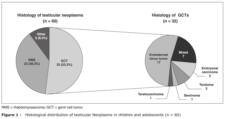
The distribution of the tumors by histological type was as follows: 32 (53%) GCTs, 23 (38.3%) RMSs and five tumors of other histological types: three stromal cell tumors, one desmoplastic and one undifferentiated (Figure 1).
GCTs occurred in 22 children and 10 adolescents with the following subtypes: 17 cases of endodermal sinus, seven mixed, three embryonal carcinomas, three teratomas, one seminoma and one teratocarcinoma. The most frequent tumor among children was the endodermal sinus tumor, in 17/22 (77.3%) and, among adolescents, the mixed tumor, in 7/10 (70%). RMSs affected eight children and 15 adolescents, and other tumors affected four children and one adolescent. The age distribution of GCTs and RMSs was bimodal, with a peak for children younger than 4 and another for adolescents (Figure 2).
At diagnosis, the involvement of regional lymph nodes occurred in 10/34 (24.4%) children and in 17/26 (65.4%) adolescents. In GCT patients, 8/22 (36.4%) children and 8/10 (80%) adolescents presented regional metastases. Adolescents with GCT presented higher incidence of lymph nodes metastases in comparison to the children (p = 0.007, proportions test). Among RMS patients, lymph node involvement in adolescents occurred in 8/15 (53.5%) and 1/8 (12.5%) in children. Adolescents with RMS presented a higher incidence of lymph node metastases than the children (p = 0.054, proportions test). Considering all the cases analyzed, children with testicular tumors presented a lower incidence of lymph node metastases (p = 0.003, proportions test).
In GCT patients (children and adolescents), the main sites of distant metastases were lung and liver. In RMS patients, distant metastases occurred only in adolescents, in the following sites: lung, in 5/15 (33.3%); bones, in 2/15 (13.3%); and bone marrow, in 1/15 (6.7%). Considering all cases analyzed, children with testicular tumors presented a lower incidence of non-lymph node metastases (p = 0.035, proportions test) while compared to adolescents with neoplasms in the same site.
AFP was high in 21/28 (75%) patients with GCT at diagnosis, and the β-HCG levels were high in 8/20 (40%). All endodermal sinus tumors presented AFP elevation at diagnosis. In patients with mixed tumors, three presented high AFP and β-HCG, one presented high AFP, and two presented high β-HCG. The AFP levels were not measured in four GCT patients and the levels of β-HCG were not measured in 12 patients. The treatment of all patients was surgical (unilateral orchiectomy) and 47/60 (78.3%) received adjuvant therapy. Among adolescents, only in one case of a mixed GCT patient, there was no need for adjuvant therapy. Among children, 12/34 (35.3%) were treated only with surgery. Chemotherapy was administered to all RMS patients after the surgical removal of the tumor.
A 5-year overall survival rate was estimated for patients with testicular tumor: GCT (85.5%) and RMS (79.5%). The confidence interval used was 95%. In GCT patients, the overall survival rates were 89.5 and 75% for children and adolescents, respectively (p = 0.429) (Figure 3). For patients with testicular RMS, the 5-year survival rate was estimated in 100%, and for adolescents, in 65% (p = 0.072) (Figure 4). Of 60 patients assessed in the study, nine died: three children (one desmoplastic tumor and two GCTs) and six adolescents (four RMSs and two GCTs). The causes of death were tumor progression in all cases. Due to the small sample presented, patients with other histological types were not analyzed.
Discussion
The presence of tumor in the scrotum was the most frequent manifestation among testicle neoplasms and occurred in 55/58 (94.8%) patients, which is consistent with the literature on the subject.5,12 An interesting finding was the report of scrotal pain in 11/58 (19%), more common in adolescents than in children (p = 0.006), since tumors are usually asymptomatic and without inflammation signs.5,12 Of 161 children with scrotal tumor evaluated by Yuan et al., 72% presented neoplasms, 13.7% presented malformations and 14.3% presented inflammatory lesions.13
In the present analysis, one adolescent presented cryptorchidism, which is one of the main risk factors for the development of testicle neoplasms.14 No cases with associated hydrocele were found in the present study, though literature reports that hydrocele may be associated with testicular tumor in 15 to 50% of cases.4 It should be highlighted that this study was a retrospective analysis with a limited sample size.
Adolescents presented a mean time from disease onset to diagnosis of 4.9 months, which was significantly higher (p = 0.01) than the mean time for the children. In childhood, the mean time from disease onset to diagnosis was 2.3 months. This difference in diagnosis time may be due to the lower frequency of medical routine examinations among adolescents and also due to the fact that parents, who are responsible for the hygiene care of their children, would be more attentive to physical changes, being able to promptly identify changes such as an enlarged scrotum.
AFP levels were high in all GCT patients with the endodermal sinus subtype, what is in line with findings in literature.12,15-17 Beta-subunit human chorionic gonadotropin was high in five out of seven patients with mixed tumors. This indicator is normally high in patients with mixed tumors who present clones of syncytiotrophoblastic cells and occasionally high in embryonal carcinoma subtype tumors. β-HCG was also high in three out of five patients with embryonal carcinoma in the study of Taskinen et al.16,17
In the patients with testicle tumor analyzed, the most frequent histological type was the GCT (53%), what is in accordance with findings in literature.12,18 Among the GCTs, the most common histological subtypes in childhood are endodermal sinus tumors and teratomas, while seminomas, embryonal carcinomas and mixed tumors are more common in post-puberty.19 The endodermal sinus tumor was the most common GCT, accounting for 28.3% of total. These tumors occurred in children with a mean age of 1.7 years, accordingly to findings in other publications.12,18 In those publications, the percentages of this histological type were 25 and 45.1% respectively.12,18 Teratomas affected young children, with a mean age a little higher than the one for the endodermic sinus tumor, which was 3.4 years, in accordance with the literature.5,12,17 Some studies suggest that the teratoma is the most common type of tumor in childhood, and it may be underreported due to its benign character.5,12,20,21 The mixed GCTs were diagnosed only in adolescents and accounted for the majority of GCTs in this age group (70%). Terenziani et al. found similar results: of 31 patients with GCT analyzed, the 10 which presented endodermal sinus tumor were children, and the 11 affected by mixed tumors were adolescents.3
RMS was the second most frequent histological subtype (38.3%) and affected patients with higher mean age at diagnosis, what is in line with two other similar studies.12,18 The fact that this neoplasm affects older patients could have influenced the larger incidence in our study, once it corresponded to approximately 19%12,18 in the two former studies mentioned. In fact, in the Metcalfe et al. study, which analyzed only patients aged < 12 years, RMS accounted for 27% of total, while among the studied patients, from the same age group, the RMS totaled 27.8%.5
Only three patients (5%) were affected by stromal tumor, while in literature its frequency ranges from 4 to 11%.5,12,15,18 In one of the affected adolescents, this tumor presented aggressive behavior with distant metastases, although it is usually described as having a good prognosis and rarely manifesting through endocrine-related alterations.22 In the present series, desmoplastic tumor in the paratesticular region was diagnosed in one patient. Desmoplastic tumors affect mainly the abdominal region, and reports of tumors in the paratesticular region are very rare in literature.23
The percentage of metastases of any kind was 50% for GCTs and 39.1% for RMSs. In the studies of Sugita et al. and Cifti et al., more metastases occurred among the RMSs.12,18 However, this comparison in not accurate, once these studies presented a higher percentage of benign tumor and did not present mixed tumors, which were quite metastatic in the present study (5/7).12,18 Age was an important factor to predict the presence of neoplastic dissemination, once adolescents presented a higher incidence of lymph node involvement (p = 0.003) and distant metastases (p = 0.035). In relation to the RMS, the adolescents presented a higher incidence of local and distant metastasis than the children, proving that for the same histological type, biological behavior may be different.
Concerning survival, it was longer for GCT patients than for RMS patients and it was longer for children, for both histological types. However, in none of these comparisons the differences were statistically significant, the only indicative being that survival for RMS patients is longer for children (p = 0.072). The overall survival, estimated in 5 years for all the GCTs in children, was 89.5%, similar to the result found in the literature on endodermal sinus tumor, which was approximately 85%.15
Adolescents with testicular tumor presented longer time since disease onset, aggressive histological type and advanced illness at diagnosis in comparison to children, despite the small sample. Given the scenario, it is considered relevant that the adolescents be encouraged to perform self-exams at regular medical examinations.24
Acknowledgements
The present study was funded by the Brazilian Scientific Initiation Scholarships Program (Programa Institucional de Bolsas de Iniciação Científica - PIBIC), Brazilian National Council for Scientific and Technological Development (Conselho Nacional de Desenvolvimento Científico e Tecnológico - CNPq).
References
- 1. Schneider DT, Calaminus G, Koch S, Teske C, Schmidt P, Haas RJ, et al. Epidemiologic analysis of 1,442 children and adolescents registered in the German germ cell tumor protocols. Pediatr Blood Cancer. 2004;42:169-75.
- 2. Poynter JN, Amatruda JF, Ross JA. Trends in incidence and survival of pediatric and adolescent patients with germ cell tumors in the United States, 1975 to 2006. Cancer. 2010;116:4882-91.
- 3. Terenziani M, Piva L, Spreafico F, Salvioni R, Massimino N, Luksch R, et al. Clinical stage I nonseminomatous germ cell tumors of the testis in childhood and adolescence: an analysis of 31 cases. J Pediatr Hematol Oncol. 2002;24:454-8.
- 4. Ahmed HU, Arya M, Munner A, Mushtaq I, Sebire NJ. Testicular and paratesticular tumours in the prepubertal population. Lancet Oncol. 2010;11:476-83.
- 5. Metcalfe PD, Farivar-Mohseni H, Farhat W, McLorie G, Khoury A, Bägli DJ. Pediatric testicular tumors: contemporary incidence and efficacy of testicular preserving surgery. J Urol. 2003;170:2412-6.
- 6. Fast Stats: An interactive tool for access to SEER cancer statistics [Internet]. Bethesda: Surveillance Research Program, National Cancer Institute. http://seer.cancer.gov/faststats Acesso: 21/08/2011.
- 7. Instituto Nacional de Câncer (Brasil), Coordenação de Prevenção e Vigilância de Câncer. Câncer na criança e no adolescente no Brasil: dados dos registros de base populacional e de mortalidade. Rio de Janeiro: INCA; 2008. 220 p.
- 8. da Silva DB, Pires MM, Nassar SM. Câncer pediátrico: análise de um registro hospitalar. J Pediatr (Rio J). 2002;78:409-14.
- 9. Eble JN, Sauter G, Epstein JI, Sesterhenn IA, editors. World Health Organization classification of tumours: pathology and genetics of tumours of the urinary system and male genital organs. Lyon: IARC Press; 2004.
- 10. Oottamasathien S, Thomas JC, Adams MC, DeMarco RT, Brock JW 3rd, Pope JC 4th. Testicular tumours in children: a single-institutional experience. BJU Int. 2007;99:1123-6.
- 11. Bujons A, Caffaratti J, Pascual M, Angerri O, Garat JM, Villavicencio H. Testicular tumours in infancy and children. Actas Urol Esp. 2011;35:93-8.
- 12. Sugita Y, Clarnette TD, Cooke-Yarborough C, Chow CW, Waters K, Hutson JM. Testicular and paratesticular tumors in children: 30 years experience. Aust N Z J Surg. 1999;69:505-8.
- 13. Yuan X, Wei G, Lin T, He D, Li X. Uncommon pediatric painless scrotal masses: a puzzle of pediatricians and urologists. Int Urol Nephrol. 2010;42:979-84.
- 14. Manecksha RP, Fitzpatrick JM. Epidemiology of testicular cancer. BJU Int. 2009;104:1329-33.
- 15. Ross JH, Rybicki L, Kay R. Clinical behavior and a contemporary management algorithm for prepubertal testis tumors: a summary of the Prepubertal Testis Tumor Registry. J Urol. 2002;168:1675-8.
- 16. Ross JH, Kay R. Prepubertal testis tumors. Rev Urol. 2004;6:11-8.
- 17. Taskinen S, Fagerholm R, Aronniemi J, Rintala R, Taskinen M. Testicular tumors in children and adolescents. J Pediatr Urol. 2008;4:134-7.
- 18. Ciftci AO, Bingöl-Koloğlu M, Senocak ME, Tanyel FC, Büyükpamukçu M, Büyükpamukçu N. Testicular tumors in children. J Pediatr Surg. 2001;36:1796-801.
- 19. Bahrami A, Ro JY, Ayala AG. An overview of testicular germ cell tumors. Arch Pathol Lab Med. 2007;131:1267-80.
- 20. Shukla AR, Woodard C, Carr MC, Huff DS, Canning DA, Zderic SA, et al. Experience with testis sparing surgery for testicular teratoma. J Urol. 2004;171:161-3.
- 21. Pohl HG, Shukla AR, Metcalf PD, Cilento BG, Retik AB, Bagli DJ, et al. Prepubertal testis tumors: actual prevalence rate of histological types. J Urol. 2004;172:2370-2.
- 22. Cecchetto G, Alaggio R, Bisogno G, Virgone C, Dall'Igna P, Terenziani M, et al. Sex cord-stromal tumors of the testis in children. A clinicopathologic report from the Italian TREP project. J Pediatr Surg. 2010;45:1868-73.
- 23. Rais H, Elmansouri F, Belaabidia B, Essadki O, Oussehal A, Sarf I. Paratesticular desmoplasic small round cell tumour. Case report with literature review. Cancer Radiother. 2010;14:111-4.
- 24. Coates V, Françoso LA, Beznos GW, coordenadores. Medicina do adolescente. 2nd ed. São Paulo: Sarvier; 2003. 731 p.
Testicular tumors in children and adolescents
Publication Dates
-
Publication in this collection
12 Mar 2012 -
Date of issue
Feb 2012
History
-
Received
06 July 2011 -
Accepted
17 Oct 2011