Abstracts
Two cases of Chronic Pulmonary Histoplasmosis are reported and other eleven cases, collected from Brazilian literature, are commented. After being clinically cured, one of our patients presented an Aspergillus fungus ball inside a cavitation in the wall of which H. capsulatum was disclosed. Comments are also done on the diagnosis of the mycosis
Histoplasmosis; Histoplasma capsulatum; Lung; Cavitation; Aspergillus fumigatus; Fungus ball; Transthoracic biopsy
Apresentam-se dois casos de Histoplasmose Pulmonar Crônica e outros onze casos da literatura brasileira são comentados. Após cura clínica, um de nossos pacientes apresentou bola fúngica aspergilar intracavitária, na parede da cavidade foi idenificado H. capsulatum. Comentam-se aspectos diagnósticos da micose
CHRONIC PULMONARY HISTOPLASMOSIS IN BRAZIL: REPORT OF TWO CASES WITH CAVITATION DIAGNOSED BY TRANSTHORACIC NEEDLE BIOPSY
Luiz Carlos SEVERO(1 (1 ) Pesquisador do CNPq. (2 ) Hospital Sanatório Partenon (HSP), Secretaria da Saúde, RS, Brasil (3 ) Bolsista da FAPERGS. (4 ) Instituto Especializado em Pesquisa e Diagnóstico (IPD), Santa Casa. (5 ) Pavilhão Pereira Filho (PPF), Santa Casa de Misericórdia de Porto Alegre, RS, Brasil Correspondence to: Dr. Luiz Carlos Severo. Laboratório de Microbiologia Clínica, IPD-Santa Casa, Annes Dias 285 90020-090 Porto Alegre, RS, Brazil. Fax: 55512281566 R 3604. E-mal: severo@santacasa.tche.br ), Carlos Fernando Carvalho RIZZON(2 (1 ) Pesquisador do CNPq. (2 ) Hospital Sanatório Partenon (HSP), Secretaria da Saúde, RS, Brasil (3 ) Bolsista da FAPERGS. (4 ) Instituto Especializado em Pesquisa e Diagnóstico (IPD), Santa Casa. (5 ) Pavilhão Pereira Filho (PPF), Santa Casa de Misericórdia de Porto Alegre, RS, Brasil Correspondence to: Dr. Luiz Carlos Severo. Laboratório de Microbiologia Clínica, IPD-Santa Casa, Annes Dias 285 90020-090 Porto Alegre, RS, Brazil. Fax: 55512281566 R 3604. E-mal: severo@santacasa.tche.br ), Eliane Wurdig ROESCH(3 (1 ) Pesquisador do CNPq. (2 ) Hospital Sanatório Partenon (HSP), Secretaria da Saúde, RS, Brasil (3 ) Bolsista da FAPERGS. (4 ) Instituto Especializado em Pesquisa e Diagnóstico (IPD), Santa Casa. (5 ) Pavilhão Pereira Filho (PPF), Santa Casa de Misericórdia de Porto Alegre, RS, Brasil Correspondence to: Dr. Luiz Carlos Severo. Laboratório de Microbiologia Clínica, IPD-Santa Casa, Annes Dias 285 90020-090 Porto Alegre, RS, Brazil. Fax: 55512281566 R 3604. E-mal: severo@santacasa.tche.br ), Flávio de Mattos OLIVEIRA(4 (1 ) Pesquisador do CNPq. (2 ) Hospital Sanatório Partenon (HSP), Secretaria da Saúde, RS, Brasil (3 ) Bolsista da FAPERGS. (4 ) Instituto Especializado em Pesquisa e Diagnóstico (IPD), Santa Casa. (5 ) Pavilhão Pereira Filho (PPF), Santa Casa de Misericórdia de Porto Alegre, RS, Brasil Correspondence to: Dr. Luiz Carlos Severo. Laboratório de Microbiologia Clínica, IPD-Santa Casa, Annes Dias 285 90020-090 Porto Alegre, RS, Brazil. Fax: 55512281566 R 3604. E-mal: severo@santacasa.tche.br ) & Nelson da Silva PORTO(5 (1 ) Pesquisador do CNPq. (2 ) Hospital Sanatório Partenon (HSP), Secretaria da Saúde, RS, Brasil (3 ) Bolsista da FAPERGS. (4 ) Instituto Especializado em Pesquisa e Diagnóstico (IPD), Santa Casa. (5 ) Pavilhão Pereira Filho (PPF), Santa Casa de Misericórdia de Porto Alegre, RS, Brasil Correspondence to: Dr. Luiz Carlos Severo. Laboratório de Microbiologia Clínica, IPD-Santa Casa, Annes Dias 285 90020-090 Porto Alegre, RS, Brazil. Fax: 55512281566 R 3604. E-mal: severo@santacasa.tche.br )
SUMMARY
Two cases of Chronic Pulmonary Histoplasmosis are reported and other eleven cases, collected from Brazilian literature, are commented. After being clinically cured, one of our patients presented an Aspergillus fungus ball inside a cavitation in the wall of which H. capsulatum was disclosed. Comments are also done on the diagnosis of the mycosis.
KEYWORDS: Histoplasmosis; Histoplasma capsulatum; Lung; Cavitation; Aspergillus fumigatus; Fungus ball; Transthoracic biopsy.
INTRODUCTION
Chronic Pulmonary Histoplasmosis results from exogenous primary infection or reinfection with the propagules of Histoplasma capsulatum var. capsulatum of an adult patient presenting a lung structural defect. The most frequent predisposing factor has been bullous or centrilobular emphysema, commonly in heavy smokers. Frequently signs and symptoms of chronic obstructive bronchopulmonary disease are associated. The lesions are limited to the lungs. Early and later on clinical and radiological features are similar to those of chronic pulmonary tuberculosis5, 6, 10.
Only eleven cases of chronic pulmonary histoplasmosis have been reported in Brazil. Two new cases will be added in this report. In one of the patients an Aspergillus fungus ball complicated the disease.
CASE REPORTS
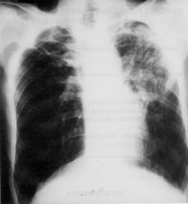
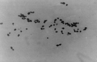
Case 1 A 62 years-old white man living in Santiago (RS) was admitted on Oct. 17, 1988 with a 7-months history of cough, purulent expectoration, hemoptysis, fever, anorexia, dyspnea and weight loss. The patient was a heavy smoker (30 cigarettes daily for 50 years) and a moderate drinker. His medical history was significant for a partial gastrectomy 22 years before. On physical examination diffuse crackles were heard in both lungs; abdominal examination revealed a median supraumbilical scar of abdominal surgery; and liver edge was palpable 3 cm bellow the costal margin. A chest X-ray revealed emphysema, scattered nodules in both lungs, small cavities int he aplical posterior segment of the left upper lobe, and a cavitation in the right upper lobe, with adjacent pleural thickenning (Fig. 1). Tuberculin skin test was negative. No acid-fast bacilli were observed in smeared sputum and in the sediment of the bronchoalveolar lavage; but, small, oval, budding yeast cells and pseudohyphae were disclosed in Grocott stained smears of both these specimens (Fig. 2). A transthoracic cutting needle biopsy was, then, performed; in H&E stained sections a fibrotic margin enclosing a palisade of epithelioid cells surrounding a necrotic focus, containing leukocytes, were seen; budding oval yeast cells are disclosed within the necrotic focus by Grocott stain. H. capsulatum var. capsulatum and Candida albicans were isolated from the sputum and the sediment of bronchoalveolar lavage planted on BHI and Sabouraud, incubated, respectively 35° C and 25° C; but, only H. capsulatum grew up in the cultures from biopsied tissue. The patient received ketocanozole, 200 mg twice daily, for three weeks, and, was discharged on 200 mg daily.
Fig. 1 Case one, X-ray shows emphysema, multiple nodules in both lungs and cavitary lesion in both apex, surrounded by pleural thikening at right.
Fig. 2 Case one, silver-stained smear of sputum shows yeast cells and pseudohyphae of (Grocott × 100).
Two months later he continued treatment with ketoconazole, but his respiratory condition did not improve completely. The chest X-ray (Jan. 09, 89) of the patient revealed partial regression of the cavitary lesion in the rigth upper lobe.
Seven months later (May, 29, 89) the patient was readmitted complaining of hemoptysis, weight loss and dyspnea. Physical examination was normal except for decreased breath sounds in the right upper lung field. The chest roentgenogram revealed markedly regression of pleural thickening and in the thickness of the walls of cavities in both upper lobes. In addition to ketoconazole, he received amphotericin (0,5 mg/kg/day) for 40 days. The patient improved and was discharged on ketoconazole (200 mg twice daily).
Two and half years later (25.11.91) the patient was again hospitalized complaining of frequent and severe hemoptysis, productive cough, fever (38° C), dyspnea, and weight loss. Chest X-ray disclosed apical opacities in both lungs and a round mass within a cavity in the upper left lobe (Fig. 3). An immunodiffusion test revealed precipitin bands against A. fumigatus aspergillin. A lobectomy was performed. At cut section of the ressected lobe a thick walled cavity, measuring 6 cm in diameter, limited by smooth irregular wall was observed; brownish friable masses, weighing 5 g lied inside the cavity; fibrosis, anthracosis, nodules and micronodules were also seen in the adjacent lung parenchyma. Microscopic examination of histological sections of the nodules and micronodules, stained by H&E, revealed necrotizing tuberculoid granulomas surrounded by a thin layer of hyalinized tissue; oval budding yeast cells, compatible with H. capsulatum were disclosed in the center of the necrotic foci in Grocott stained sections. Histological sections of the masses removed from the cavity revealed that they consisted of a tangled network of hyaline septate hyphae, 3 a 5 µm in width. Small fragments of these masses were inoculated on to Sabouraud dextrose agar slants and incubated at both room temperature and at 37° C; luxurious growth of A. fumigatus were obtained.
Fig. 3 Case one, X-ray shows bilateral apical opacity and rounded mass of soft tissue density (fungus ball), at least, at left.
Post operative course: after surgery the patients condition deteriorated (pleural bacterial empyema) and he died.
Case 2 This patient was a 64 year-old white man living in Itaqui (RS), admitted to our hospital on February, 1990. Since four months ago, because he presented cough, mucopurulent sputum, fever, weight loss, night sweats, and dyspnea he was treated for a presumed tuberculosis, in spite of negative tuberculin skin test and negative results of microscopic examination of sputum smears for acid-fast bacilli. The patient was a heavy smoker (40 cigarettes daily) and moderate drinker for 48 years. Physical examination revealed a tall, slim man with pectus excavatum. Diffuse crackles were heard in both lungs. A chest roentgenogram showed emphysema, cavitary lesions in both upper lobes and bronchogenic spread to lower lobes (Fig. 4). Multiple sputum smears revealed no acid-fast bacilli. A transthroracic needle biopsy was performed; small oval yeast-like cells were disclosed in Grocott stained smear (Fig. 5). The isolates obtained on Sabouraud chloramphenicol medium incubated at room temprature were identified as H. capsulatum var. capsulatum by its dimorphic presentation in subcultures on BHI incubated at 37° C. Immunodiffusion test revealed M band. The patient improved on ketoconazole (200 mg twice daily) treatment during 18 months.
Fig. 4 Case two, X-ray shows emphysema and cavitary lesions in both upper lobes and bronchogenic spread in lower lobes.
Fig. 5 Case two, microscopic examination of the transthoracic needle biopsy, shows in smear yeast cells of H. capsulatum (Grocott × 250).
On year after later, the patient presented again cough, mucopurulent sputum and weight loss. A chest X-ray showed greater thickening of the walls of the apical cavities. Small oval yeast-like cells were again disclosed, but in sputum smears stained by Grocott; and again H. capsulatum was isolated from sputum cultures. The patients was then treated with itraconazole (100 mg/day) for 6 months; in spite of his recovery the drug was mantained for more 7 months.
Follow-up: on October 95 the chest X-ray showed only fibrotic scars (Fig. 6) and the patient remained in good conditions 3 years after stopping antifungal therapy (November 96).
Fig. 6 X-ray shows fibrotic scar in a symptoms-free treated patient (Case 2).
COMMENTS
Eleven cases of Chronic Pulmonary Histoplasmosis (CPH) were reported in Brazil (Table 1). The rarity of the diagnosis of CPH in Brazil can be explained partly by its clinical and radiological similarities with tuberculosis. On the other hand, mycological diagnostic facilities are not available in many places of the vast territory of Brazil. Therefore, pointing out that CPH is overlooked.
Eleven cases of chronic pulmonary histoplasmosis reported in Brazil.
Diagnosis: M = microscopy, C = culture, I = animal inoculation.
IDh: immunodiffusion test to histoplasmosis.
The first step to recognize CPH is to consider it in the differential diagnosis. Serologic test can be used as screening test for histoplasmosis; immunodiffusion is the best because complement fixation crossreaction does occur. However, H band sometimes may not be present. Mycological diagnosis may be based on the observation of the fungus in clinical specimens and its isolation and identification in culture5, 6, 10.
Expectoration is an usual symptom in CPH, so sputum is the easiest specimen to be examined. However, H. capsulatum may be recognized if sputum is smeared and stained by Gomori-Grocott technique. Nevertheless, fungus cells may be scarce or another yeast-like elements (Candida spp.) may be present, leading to misdiagnosing it (Fig. 2). For that reason, culure, using proper media and proper incubation temperature for the isolation and recognition of the fungus, is necessary.
Clinical specimen of CPH can be obtained with transthoracic needle biopsy4. Tissue sections (case 1) or necrotic material (case 2) of part of the specimen may be used for microscopic examination, another part for culturing the fungus.
Two and half years after been treated with antifungals, patient (case 1) presented a fungus ball by A. fumigatus. The diagnosis was suggested by chest X-ray8, presumed by immunodiffusion and proved mycologically. Active lesions of histoplasmosis were also detected in the patients lesions. This is the first case reported in Brazil, presenting such association.
RESUMO
Histoplasmose pulmonar crônica no Brasil: relato de dois casos com cavitação diagnosticados por biópsia transtorácica
Apresentam-se dois casos de Histoplasmose Pulmonar Crônica e outros onze casos da literatura brasileira são comentados. Após cura clínica, um de nossos pacientes apresentou bola fúngica aspergilar intracavitária, na parede da cavidade foi idenificado H. capsulatum. Comentam-se aspectos diagnósticos da micose.
ACKNOWLEDGEMENTS
Our sincere appreciation is expressed to Dr. A. T. Londero (UFSM, Santa Maria, RS) for revision of the manuscript.
Recebido para publicação em 27/12/1996
Aceito para publicação em 23/09/1997
- 1. ALECRIM, I.; DODGE, C. W. & TEIXEIRA, H. First cases of histoplasmosis diagnosed in Recife, Brasil. Mycopathologia (Den Haag), 21: 204-207, 1963.
- 2. CAPONE, D. Histoplasmose pulmonar crônica Rio de Janeiro, 1992. (Tese de Mestrado Universidade Federal do Rio de Janeiro.)
- 3. CHAGAS, P. R. S. Pesquisa de agentes e anticorpos fúngicos em pacientes com pneumopatias a esclarecer Săo Paulo, 1982. (Tese de Mestrado Escola Paulista de Medicina.)
- 4. FRASER, R. S. Transthoracic needle aspiration. The benign diagnosis. Arch. Path. Lab. Med., 115: 751-761, 1991.
- 5. GOODWIN Jr., R. A.; OWENS, F. T.; SNELL, J. D. et al. Chronic pulmonary histoplasmosis. Medicine (Baltimore), 55: 413-452, 1976.
- 6. LOEWEN, D. F.; PROCKNOW, J. J. & LOOSLI, C. G. Chronic active pulmonary histoplasmosis with cavitation. A clinical and laboratory study of thirteen cases. Amer. J. Med., 28: 252-280, 1960.
- 7. PAULA, A.; BLUNDI, E.; McCLURE, V. & PERNAMBUCO, P. Histoplasmose pulmonar. Tratamento pela anfotericina B. Brasil.-méd., 73: 13-18, 1959.
- 8. SCHWARZ, J.; BAUM, G. L. & STRAUB, M. Cavitary histoplasmosis complicated by fungus ball. Amer. J. Med., 31: 692-700, 1961.
- 9. WANKE, B. Histoplasmsoe. Estudo epidemiológico, clínico e experimental Rio de Janeiro, 1985. (Tese de Doutorado Universidade Federal do Rio de Janeiro.)
- 10. WHEAT, J. L.; WASS, J.; NORTON, J.; KOHLER, R. B. & FRENCH, M. L. V. Cavitary histoplasmosis occurring during two large urban outbreaks. Analysis of clinical, epidemiologic, roentgenographic, and laboratory features. Medicine, 63: 201-209, 1984.
Publication Dates
-
Publication in this collection
04 Feb 1999 -
Date of issue
Sept 1997
History
-
Accepted
23 Sept 1997 -
Received
27 Dec 1996