Abstracts
Cysticercosis is a condition that occurs when man is infested by the larvae of Taenia solium, acting as an intermediate host instead of definitive. Oral cysticercosis is a rare event, and it represents a difficulty in clinical diagnosis. A case of oral cysticercosis in a 23-year-old white female who presented a painless swelling in the dorsal portion of the tongue is reported. An excisional biopsy was performed and histopathological examination revealed a cystic cavity containing the tapeworm.
Oral cysticercosis; Taenia solium; Cysticercus cellulosae; Taeniasis
A cisticercose é uma doença que ocorre quando o indivíduo é infectado pela larva da Taenia solium, atuando como hospedeiro intermediário ao invés de definitivo. A cisticercose em cavidade oral é rara e seu diagnóstico clínico é difícil. Neste trabalho, é relatado um caso de cisticercose oral em paciente de 23 anos, sexo feminino que apresentou um crescimento indolor na região de dorso de língua. Foi realizada uma biópsia excisional e o exame histopatológico revelou uma cavidade cística apresentando em seu interior a larva.
CASE REPORT
Oral cysticercosis: case report and review of the literature
Cisticercose oral: relato de caso e revisão da literatura
Fernando Melhem EliasI; Marília Trierveiler MartinsII; Rodrigo ForondaI; Waldyr Antônio JorgeI; Ney Soares de AraújoII
IOral and Maxillofacial Surgery, University Hospital, University of São Paulo, São Paulo, SP, Brasil
IIOral Pathology Department, School of Dentistry, University of São Paulo, São Paulo, SP, Brasil
Correspondence Correspondence to Ney Soares de Araújo Disciplina de Patologia Bucal Faculdade de Odontologia da USP 05508-900 São Paulo, SP, Brasil Phone/fax: +55 11 3091 7894 e-mail: nsaraujo@usp.br.
SUMMARY
Cysticercosis is a condition that occurs when man is infested by the larvae of Taenia solium, acting as an intermediate host instead of definitive. Oral cysticercosis is a rare event, and it represents a difficulty in clinical diagnosis. A case of oral cysticercosis in a 23-year-old white female who presented a painless swelling in the dorsal portion of the tongue is reported. An excisional biopsy was performed and histopathological examination revealed a cystic cavity containing the tapeworm.
Keywords: Oral cysticercosis; Taenia solium; Cysticercus cellulosae; Taeniasis.
RESUMO
A cisticercose é uma doença que ocorre quando o indivíduo é infectado pela larva da Taenia solium, atuando como hospedeiro intermediário ao invés de definitivo. A cisticercose em cavidade oral é rara e seu diagnóstico clínico é difícil. Neste trabalho, é relatado um caso de cisticercose oral em paciente de 23 anos, sexo feminino que apresentou um crescimento indolor na região de dorso de língua. Foi realizada uma biópsia excisional e o exame histopatológico revelou uma cavidade cística apresentando em seu interior a larva.
INTRODUCTION
Platyhelminthes have a life cycle characterized by two stages; first as a larva, and then as an adult worm, besides an egg phase. Each of these phases requires a different host.
The adult Taenia solium, or pork tapeworm, lives in the small intestine of man, its definitive host. Infestation by Taenia solium is common in areas where pig breeding is not controlled and sanitation is inadequate. Latin America, Southern Africa, India, Southeast Asia and Eastern Europe are the most frequent locations of occurrence17.
The parasitic infestation by the pork tapeworm larval stage, the Cysticercus cellulosae, is called cysticercosis, and results from the ingestion of tapeworm eggs through contaminated food and water or dirty hands. Highest incidence countries are Brazil, Chile, Ecuador, Mexico, South Africa, East Africa and India17.
The most frequent sites of cysticercosis occurrence are subcutaneous layers, brain, muscles, heart, liver, lungs, and peritoneum4,33.
Although the exact incidence is still unknown, oral cysticercosis is considered a rare event and a precise clinical diagnosis is not usually established16. We present a case of cysticercosis on the tongue of a Brazilian female, as well as a review of cases reported in the literature.
CASE REPORT
A 23-year-old white female presented with a swelling on the tongue. During anamnesis, the patient reported that the lesion was painless and had been present for about three years, showing a slow rate of growth. Past medical history was noncontributory.
On physical examination, the patient had a submucous nodule on the left border of the tongue. The nodule was soft and painful under palpation, measuring about 15 mm, and was lined by normal mucosa (Fig. 1). No palpable lymph nodes were present in the head and neck region.
Under a clinical diagnosis of a benign neoplasm of neural origin, an excisional biopsy was performed under local anesthesia. During the surgical procedure, the lesion could be seen as encapsulated (Fig. 2).
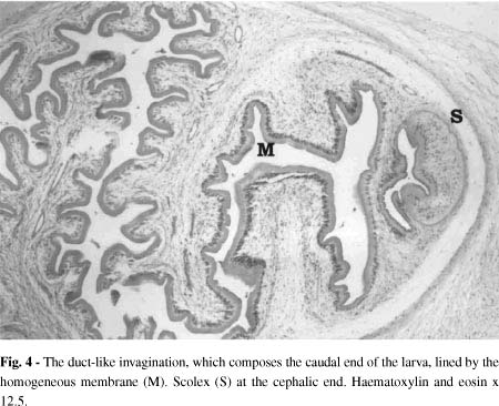
Microscopic examination revealed a capsule of fibrous connective tissue surrounding a cystic cavity, which contained the Cysticercus cellulosae, the Taenia solium larval form (Fig. 3). The capsule showed intense inflammatory infiltrate, consisting mainly of lymphocytes and plasma cells. Groups of eosinophils were seen throughout the connective tissue. The larva was composed of a scolex, where a sucker could be identified, and a duct-like invaginated segment - the caudal end. Both larva and cystic structure were lined by a homogeneous eosinophilic membrane (Fig. 4 and 5). No areas of dystrophic calcification were present in the specimen. Based on these findings, a diagnosis of cysticercosis was made.
After diagnosis, complete blood count and stool examination were performed, and resulted normal. To eliminate the possibility of neurocysticercosis, a computarized tomography of the head and an electroencephalogram were performed and no other parasitosis focus was found. The patient was scheduled for regular check-ups but was subsequently lost to follow-up.
REVIEW
The tapeworm is composed of the scolex (head) and proglottids (caudal end). Each proglottid contains from 40,000 to 60,000 eggs, which are released through faeces. The pig becomes infected by ingesting the eggs from the ground contamined by human faeces and develops the larval form. By consuming inadequately cooked infected pork, man acquires the larva, which becomes, inside the small intestine of the host, an adult worm, the Taenia sp4,26. Humans can have taeniasis for long periods and contaminate environment continuously4.
When man ingests the eggs, he becomes the intermediate host, a role usually played by the pig, and a larval form will develop. The ingestion of Taenia solium eggs happens through ingestion of fecally contaminated vegetables, food or water, as well as self-contamination, by reflux of the proglottid from the intestine into the stomach or contamined hands22,26,30. The eggs develop into oncospheres that penetrate intestine wall and - via lymphatic or vascular circulation - reach a destination, where larvae develop and become the cysticerci or "bladder worm", a fluid-filled cyst22,32.
Once a person becomes the host of Cysticercus cellulosae, cysticercosis can develop in various organs and tissues22. The most serious involvement is that of the central nervous system, followed by ocular involvement, usually the only ones which are symptomatic4,8. Generalized symptoms include headache, fever and myalgia. Patients with neurocysticercosis can present with several signs and symptoms, being the most frequent seizures, increased intracranial pressure, obstructive hydrocephalus, meningitis, and mental disorders11,32.
In spite of the abundance of muscular tissue in the oral and maxillofacial region, this is not a frequent site of occurrence for cysticercosis. We have found sixty-five cases reported in the literature1-3,6-16,18-20,22-24,26-32. The age was reported in sixty-four cases and varied from three to seventy years, with a mean age of 23.7 years. The most frequently affected decade was the third (32%), followed by the fourth decade (20.6%). There was an equal distribution between genders. The most frequently involved site were the tongue (42.15%), followed by the lips (26.15%) - with the lower lip lounging 64.7% of those - and the buccal mucosa (18.9%). Several reports1,24,27,28,30,32 revealed multiple foci in the same patient.
Usually, the patient complaint is a swelling. Although pain is not a frequent feature, it had been reported in secondarily infected cases13. In the case here reported the patient presented pain under palpation but not spontaneous pain. It was suggested that the lesion on the tongue could interfere with movement, causing discomfort during speaking and eating27.
Differential diagnosis of oral lesion depends on the site involved. In this case, in which the aspect was of a solitary nodule on the tongue, lined by normal mucosa, the differential diagnosis would be benign neoplasm of neural tissue origin, as neurofibroma and benign schwannoma, granular cell myoblastoma, vascular neoplasm, vascular leiomyoma, fibroma, and lipoma.
Histopathological examination makes up a diagnosis of cysticercosis by the detection of a cystic space containing the Cysticercus cellulosae. The scolex has four suckers and a double crown of rostellar hooklets13. A duct-like invaginated segment, lined by a homogeneous anhistic membrane, composes the caudal end. The eosinophilic membrane that lines the capsule is double-layered, consisting of an outer acellular and an inner sparsely cellular layer15. After a period within three and five years the larva dies and the cyst undergoes calcification10.
Currently other diagnostic tools as radiologic imaging and serology can be used. Besides normal radiographic examination other modalities of imaging exams are very effective in the detection of cysticerci, as computerized tomography and magnetic resonance21. SARAN et al.26 proposed the use of fine-needle aspiration cytology, which identifies the tegument layer of the larva, to help the clinician in planning the treatment.
Immunodetection of cysticercosis can be achieved in sera, cerebrospinal fluid and saliva, by ELISA (enzyme-linked immunosorbent) assay or EITB (enzyme-linked immunoelectrotransfer blot)4, but it is important to consider that individuals living in an endemic area may have antibodies because of an exposure instead of an established infection5. SANCHEZ et al.25, performing EITB in patients from endemic areas with brain lesion compatible with neurocysticercosis, found a similar proportion of seropositive and seronegative groups.
Every case of oral cysticercosis should be thoroughly investigated to determine the involvement of multiple foci, since there is a high incidence of such feature11.
Drugs as praziquantel and albendazole are potent antihelminthics used in the treatment of cysticercosis22, replacing niclosamide, which was the drug of choice for the treatment of the disease for a long period7,13. Drugs should be used especially in cases where surgical treatment is risky or not possible, as in neurocysticercosis. As the patient in the present case had no other occurrence of cysticercosis or any other symptoms, no additional treatment was prescribed except the periodic follow-ups.
It is important to consider the diagnosis of cysticercosis in oral solitaire nodular lesions presenting in patients living in an endemic area. This case emphasizes the role of the dentist in the detection of a disease that can have more serious involvement, as well as the importance of routine histological examination.
Received: 27 September 2004
Accepted: 4 January 2005
- 1. BEDI, T.R.; PANDHI, R.K. & BHUTANI, L.K. - A case of cysticercosis cellulosae involving the oral cavity. Int. J. Derm., 13: 188-189, 1974.
- 2. CZERNIC, S. - Cysticercosis de la levre et des extremites superieures. Canad. J. Otolaryng., 1: 68-72, 1972.
- 3. FAZAKERLEY, M.W. & WOOLGAR, J.A. - Cysticercosis cellulosae. An unusual cause of a labial swelling. Brit. dent. J., 170: 105-106, 1991.
- 4. FLISSER, A.; PLANCARTE, A.; CORREA, D. et al. - New approaches in the diagnosis of Taenia solium cysticercosis and taeniasis. Ann. Parasit. hum. comp., 65: 95-98, 1990.
- 5. GARCIA, H.H.; HARRISON, L.J.; PARKHOUSE, R.M. et al - A specific antigen-detection ELISA for the diagnosis of human neurocysticercosis. The Cysticercosis Working Group in Peru. Trans. roy. Soc. trop. Med. Hyg., 92: 411-414, 1998.
- 6. GUPTA, S.C. & GUPTA, S.C. - Cysticercosis of the tongue. Ear Nose Throat J., 74: 174, 177-178, 1995.
- 7. HANSEN, L.S. & ALLARD, R.H. - Encysted parasitic larvae in the mouth. J. Amer. dent. Ass., 108: 632-636, 1984.
- 8. INDIRA, C.; RAMESH, V. & MISRA, R.S. - Association of oral cysticercosis and post kala azar dermal leishmaniasis. Int. J. oral maxillofac. Surg., 19: 266-267, 1990.
- 9. JAIN, R.K.; GUPTA, O.P. & ARYYA, N.C. - Cysticercosis of the tongue. J. Laryng., 103: 1227-1228, 1989.
- 10. KINNMAN, J.; CHI, C.H. & PARK, J.H. - Cysticercosis in otolaryngology. Arch. Otolaryng., 102: 144-147, 1976.
- 11. LUSTMANN, J. & COPELYN, M. - Oral cysticercosis. Review of the literature and report of 2 cases. Int. J. oral Surg., 10: 371-375, 1981.
- 12. MAHINDRA, S.; DALJIT, R.; SOHAIL, M.A. & MAHESHWARI, H.B. - Cysticercosis in the practice of otolaryngology. Acta oto-laryng. (Stockh.),92: 189-191, 1981.
- 13. MISHRA, Y.C. & NADKARNI, N. - Oro-facial cysticercosis. Ann. Dent., 47: 23-25, 1988.
- 14. NIGAM, S.; SINGH, T.; MISHRA, A. & CHATURVEDI, K.U. - Oral cysticercosis: report of six cases. Head Neck, 23: 497-499, 2001.
- 15. OSTROFSKY, M.K. & BAKER, M.A. - Oral cysticercosis: three case reports. J. dent. Ass. S. Afr., 30: 535-537, 1975.
- 16. PINSWASDI, P. & CHAROENSIRI, D.J. - Cysticercosis in labial tissue. Case report. Aust. dent. J., 42: 319-321, 1997.
- 17. PRABHU, S.R. - Oral diseases in the tropics Oxford, Oxford University Press, 1992. p. 126-129.
- 18. PUPPIN Jr., D.; CAVEGN, B.M. & DELMAESTRO, D. - Subcutaneous cysticercosis of the tongue mimicking a tumor. Int. J. Derm., 32: 818-819, 1993.
- 19. RAO, P.L.; RADHAKRISHNA, K. & KAPADIA, R.D. - Cysticercosis of the tongue. Int. J. Pediat. Otorhinolaryng., 20: 159-161, 1990.
- 20. REDDI, S.P.; MOLARES, M.J. & ADDANTE, R.R. - Solitary lesion in the masseter muscle. J. oral maxillofac. Surg., 59: 71-75, 2001.
- 21. RICHARDS Jr., F. & SCHANTZ, P.M. - Laboratory diagnosis of cysticercosis. Clin. Lab. Med., 11: 1011-1028, 1991.
- 22. ROMERO DE LEON, E. & AGUIRRE, A. - Oral cysticercosis. Oral Surg. oral Med. oral Path. oral Radiol. Endod., 79: 572-577, 1995.
- 23. ROSENCRANS, M. & BARAK, J. - Parasitic infection of the mouth. A case report of Cysticercus cellulosae N. Y. St. dent. J., 35: 271-273, 1969.
- 24. ROTH, B.; GOCHT, A. & METTERNICH, F.U. - Cysticercosis as a rare cause of a tumor of the tongue. Laryngorhinootologie, 82: 564-567, 2003.
- 25. SANCHEZ, A.L.; LINDBACK, J.; SCHANTZ, P.M. et al. - A population-based, case-control study of Taenia solium taeniasis and cysticercosis. Ann. trop. Med. Parasit., 93: 247-258, 1999.
- 26. SARAN, R.K.; RATTAN, V.; RAJWANSHI, A.; NIJKAWAN, R. & GUPTA, S.K. - Cysticercosis of the oral cavity: report of five cases and a review of literature. Int. J. paediat. Dent., 8: 273-278, 1998.
- 27. SHARMA, A.K.; MISRA, R.S.; MUKHERJEE, A.; RAMESH, V. & JAIN, R.K. - Oral cysticercosis. Int. J. oral maxillofac. Surg., 15: 349-351, 1986.
- 28. SOOKNUNDUN, M.; KACKER, S.K. & KAPILLA, K. - Cysticercosis of the oral cavity: a clinicopathological study of ten and a half years. J. Ind. dent. Ass., 58: 257-259, 1986.
- 29. SOUZA, P.E.; BARRETO, D.C.; FONSECA, L.M. et al. - Cysticercosis of the oral cavity: report of seven cases. Oral Dis., 6: 253-255, 2000.
- 30. TIMOSCA, G. & GAVRILITA, L. - Cysticercosis of the maxillofacial region. A clinicopathologic study of five cases. Oral Surg. oral Med. oral Path., 37: 390-400, 1974.
- 31. TOLEDO, O.A. & MARTINELLI, C. - Cysticercosis in the human alveolar mucosa: case report. Rev. Fac. Farm. Odont. Araraquara, 3: 129-132, 1969.
- 32. WEBB, D.J.; SEIDEL, J. & CORRELL, R.W. - Multiple nodules on the tongue of a patient with seizures. J. Amer. dent. Ass., 112: 701-702, 1986.
- 33. WORTMAN, P.D. - Subcutaneous cysticercosis. J. Amer. Acad. Derm., 25: 409-414, 1991.
Publication Dates
-
Publication in this collection
04 May 2005 -
Date of issue
Apr 2005
History
-
Accepted
04 Jan 2005 -
Received
27 Sept 2004