Abstracts
Two hundred and twenty three subjects from a Schistosoma mansoni low morbidity endemic area and nine hospitalized hepatosplenic patients were submitted to stool test and clinical examination and abdomen ultrasound assessments. According to stool examination and ultrasound results, they were grouped as follows: G1 - 63 Schistosoma mansoni egg-negative individuals; G2 - 141 egg-positive patients and without evidence of periportal fibrosis; G3 - 19 egg-positive patients with periportal echogenicity (3-6mm); and G4 - 9 hepatosplenic patients with periportal echogenicity (> 6mm). Hepatomegaly detected by physical examination of the abdomen evaluated in the midclavicular line was verified in G1, G2 and G3, respectively, in 11.1, 12.1 and 26.3%. In G1, G2 and G3, periportal thickening occurred only in schistosomal patients (8.5%). Mild pathological alterations in patients that cannot yet be detected by clinical examination were detectable in the liver by ultrasound and can be due to fibrosis. The degree of mild periportal fibrosis was diminished in 57.9% of patients 12 months after treatment of schistosomiasis with oxamniquine. At ultrasonography, the mean liver left lobe measurement of G3 was larger than that of G1, and that of G4 larger than that of G1 and G2. The mean size of the spleen of G4 was significantly larger than that of the other three groups, and that of G3 larger than that of G1 and G2.
Schistosomiasis mansoni; Ultrasonography; Periportal fibrosis
Duzentos e vinte e três indivíduos de área endêmica de baixa morbidade para esquistossomose e nove pacientes hospitalizados com a forma hepatoesplênica foram submetidos ao exame de fezes e clínico e à ultra-sonografia do abdômen. De acordo com os resultado dos exames de fezes e do ultra-som eles foram agrupados do seguinte modo: G1 - 63 indivíduos sem ovos de Schistosoma mansoni nas fezes; G2 - 141 indivíduos apresentando ovos de Schistosoma mansoni nas fezes, sem ecogenicidade periportal. G3 - 19 indivíduos com ovos de Schistosoma mansoni nas fezes e ecogenicidade periportal entre 3-6mm.; G4 - 9 pacientes hepatesplênicos com ecogenicidade periportal > 6mm. Pelo exame físico do abdômen, a hepatomegalia na linha hemiclavicular direita foi constatada em G1, G2 E G3, respectivamente, em 11,1, 12,1 e 26,3%. Nos grupos G1, G2 e G3, houve espessamento periportal somente em esquistossomáticos (8,5%). Alterações patológicas leves em pacientes, as quais não puderam ser detectadas pelo exame clínico, foram evidenciadas no fígado pelo ultra-som e podem ser devidas à fibrose. O grau de fibrose periportal leve foi diminuído em 57,9% dos pacientes 12 meses após tratamento da esquistossomose com oxamniquine. Na ultra-sonografia, a média da medida do lobo esquerdo do fígado dos indivíduos de G3 foi maior que a de G1 e, a de G4 maior que a de G1 e G2. O tamanho médio do baço de G4 foi significativamente maior que o dos outros grupos e o de G3 foi maior que o de G1 e G2.
Esquistossomose mansoni; Ultra-sonografia; Fibrose periportal
ARTICLE ARTIGO
Comparative clinical and ultrasound study of egg-negative and egg-positive individuals from Schistosoma mansoni low morbidity endemic areas, and hospitalized patients with hepatosplenic disease
Estudo clínico comparativo e ultra-sonografia entre indivíduos negativos e positivos para Schistosoma mansoni em áreas endêmicas de baixa morbidade e pacientes hospitalizados com doença hepatosplênica
Telcia V.B. MagalhãesI; Giovanni GazzinelliI, VII; Maria Carolina B. AlvarezI; F.C. Lima e SilvaI; Lucia Alves Oliveira FragaII; Alda Maria S. SilveiraII; Andrea GazzinelliIII; Jeffrey BethonyV; Philip LoVerdeVI; Iramaya R. CaldasVII; Rodrigo Correa-OliveiraVII; Aluízio PrataIV
ISanta Casa Hospital de Belo Horizonte, MG, Belo Horizonte, MG
IIUniversidade Vale do Rio Doce, Governador Valadares, MG
IIIEscola de Enfermagem da Universidade Federal de Minas Gerais, Belo Horizonte, MG
IVFaculdade de Medicina do Triângulo Mineiro, Uberaba, MG
VThe George Washington University Medical Center, Washington DC, USA
VIDepartment of Microbiology School of Medicine and Biomedical Sciences, Buffalo New York, USA
VIICentro de Pesquisas René Rachou da Fundação Oswaldo Cruz, Belo Horizonte, MG
Correspondence Correspondece to Prof. Giovanni Gazzinelli Av. Augusto de Lima 1715, Barro Preto 30190-002 Belo Horizonte, MG, Brazil Tel: 55 31 3295-3566, Fax: 51 31 3295-3115 e-mail: ggaz@cpqrr.fiocruz.br
ABSTRACT
Two hundred and twenty three subjects from a Schistosoma mansoni low morbidity endemic area and nine hospitalized hepatosplenic patients were submitted to stool test and clinical examination and abdomen ultrasound assessments. According to stool examination and ultrasound results, they were grouped as follows: G1 - 63 Schistosoma mansoni egg-negative individuals; G2 - 141 egg-positive patients and without evidence of periportal fibrosis; G3 - 19 egg-positive patients with periportal echogenicity (3-6mm); and G4 - 9 hepatosplenic patients with periportal echogenicity (> 6mm). Hepatomegaly detected by physical examination of the abdomen evaluated in the midclavicular line was verified in G1, G2 and G3, respectively, in 11.1, 12.1 and 26.3%. In G1, G2 and G3, periportal thickening occurred only in schistosomal patients (8.5%). Mild pathological alterations in patients that cannot yet be detected by clinical examination were detectable in the liver by ultrasound and can be due to fibrosis. The degree of mild periportal fibrosis was diminished in 57.9% of patients 12 months after treatment of schistosomiasis with oxamniquine. At ultrasonography, the mean liver left lobe measurement of G3 was larger than that of G1, and that of G4 larger than that of G1 and G2. The mean size of the spleen of G4 was significantly larger than that of the other three groups, and that of G3 larger than that of G1 and G2.
Key-words: Schistosomiasis mansoni. Ultrasonography. Periportal fibrosis.
RESUMO
Duzentos e vinte e três indivíduos de área endêmica de baixa morbidade para esquistossomose e nove pacientes hospitalizados com a forma hepatoesplênica foram submetidos ao exame de fezes e clínico e à ultra-sonografia do abdômen. De acordo com os resultado dos exames de fezes e do ultra-som eles foram agrupados do seguinte modo: G1 - 63 indivíduos sem ovos de Schistosoma mansoni nas fezes; G2 - 141 indivíduos apresentando ovos de Schistosoma mansoni nas fezes, sem ecogenicidade periportal. G3 19 indivíduos com ovos de Schistosoma mansoni nas fezes e ecogenicidade periportal entre 3-6mm.; G4 9 pacientes hepatesplênicos com ecogenicidade periportal > 6mm. Pelo exame físico do abdômen, a hepatomegalia na linha hemiclavicular direita foi constatada em G1, G2 E G3, respectivamente, em 11,1, 12,1 e 26,3%. Nos grupos G1, G2 e G3, houve espessamento periportal somente em esquistossomáticos (8,5%). Alterações patológicas leves em pacientes, as quais não puderam ser detectadas pelo exame clínico, foram evidenciadas no fígado pelo ultra-som e podem ser devidas à fibrose. O grau de fibrose periportal leve foi diminuído em 57,9% dos pacientes 12 meses após tratamento da esquistossomose com oxamniquine. Na ultra-sonografia, a média da medida do lobo esquerdo do fígado dos indivíduos de G3 foi maior que a de G1 e, a de G4 maior que a de G1 e G2. O tamanho médio do baço de G4 foi significativamente maior que o dos outros grupos e o de G3 foi maior que o de G1 e G2.
Palavras-chaves: Esquistossomose mansoni. Ultra-sonografia. Fibrose periportal.
Schistosoma mansoni has a wide geographical distribution in Africa, South America and the Caribbean. The disease is mainly due to eggs deposited in the host tissue by the adult female whose antigens induce granuloma formation and fibrosis, essentially in intestine and liver portal system. Most of the infected individuals living in endemic areas are asymptomatic but a few develop Symmers' periportal fibrosis of the liver, that results in the severe form of the disease with subsequent portal hypertension, splenomegaly, esophageal varices and recurrent hematemesis. The hepatointestinal and compensated hepatosplenic clinical forms represent early forms of the severe clinical disease found in the hospital (decompensated hepatosplenic)5. The sequential development of hepatosplenic disease was well demonstrated and anatomicopathologically proven15. These conclusions were obtained from clinical diagnosis based on liver left lobe enlargement, consistency, as well as spleen size detected by physical examination15. However, when ultrasound was used for abdominal evaluation in field surveys in endemic areas for schistosomiasis, it became clear that the physical examination of the patients did not always reflect the stage of the disease1. This conclusion is based on the thickness of periportal fibrosis as determined by ultrasound echogenicity which correlates with liver biopsies in pathological studies9. However, there is a variation on the assessment of images provided by ultrasound8, especially in mild pathology, frequently seen in field surveys. In these cases, the interpretation of ultrasound images has been discussed.
In order to identify the meaning of some findings, we compared several clinical and ultrasound parameters of distinct groups of schistosomiasis patients with an egg-negative group from the same endemic area. These groups were also compared with hospitalized patients with the severe hepatosplenic form of the disease.
MATERIAL AND METHODS
Studied population. This study was performed in two localities endemic for schistosomiasis (Dionísio and Melquiades) in the state of Minas Gerais, Brazil. A schistosomiasis program aimed at studying several aspects of the disease has been active in these two localities since 1995. The volunteers (n = 223) were submitted to parasitological, clinical and ultrasound examinations. Nine patients with a clinical diagnosis of hepatosplenic schistosomiasis from the Santa Casa Hospital of Belo Horizonte city (Brazil) were also included in this study.
It is important to mention that malaria is not endemic in these areas; therefore it is not a confounding disease. Protocols involving human subjects were approved by the institutional review and ethical committee of the Fundação Oswaldo Cruz (Fiocruz). Treatment was offered to all individuals independent of whether they consented to participate in the study.
Parasitological examination.Schistosoma mansoni infection was evaluated by individual fecal egg examinations determined by the Kato-Katz method on stool samples obtained on three consecutive days11 .
Clinical examination. Clinical examination of 223 subjects was performed by two physicians. One examined patients from Dionisio and, the other those from Melquiades. Both physicians were blind to the results of the feces and ultrasonographic examinations. Hepatosplenic patients were examined by other physicians.
At abdominal examination, palpation of the right lobe of the liver was evaluated in the midclavicular line and the left lobe in the epigastrium at inspiration; the enlargement, consistency and surface were recorded. The spleen was examined below left costal margin at inspiration.
Based on clinical criteria, the patients were considered as intestinal (liver <2 cm under the costal edge), hepatointestinal (liver palpable >2 cm under the costal edge) and hepatosplenic.
Patients with schistosomiasis were treated with oral oxamniquine, in a single dose of 15mg/kg.
Ultrasound. A conventional portable Hitachi EUB-200 ultrasound equipment (Tokyo, Japan) was used in the field study and a Sonoline Versa Plus, Siemens ultrasound with a transducer of 3.5 MHz for hospitalized patients. Liver size, portal-vein diameter, thickness of the walls of central and peripheral portal branches, spleen size and splenic vein diameters were assessed as described elsewhere1 9. Liver span was measured both in the midclavicular line and the midline. The liver was also examined for surface smoothness. Portal vein diameter was measured at its entrance into the liver and its bifurcation inside the liver. The spleen was evaluated by using oblique and longitudinal scanning of the left upper quadrant. The gallbladder was examined for wall thickness and stones. The periportal thickness was evaluated according to established criteria1 4 9, with few modifications. The two physicians performing ultrasound examinations were neither aware of the infection status nor clinical examinations of the patients undergoing ultrasonography. The echo enhancer contrast (Levovistâ ) was used as described by Albrecht et al2.
One year after treatment, some patients were again submitted to ultrasound evaluation.
Classification of studied population. According to the result of stool test for Schistosoma mansoni eggs and the presence of periportal fibrosis, the individuals in the endemic areas were classified into three groups.
Group 1 - Control subjects - S. mansoni egg-negative individuals at stool test. Group 2 - Patients eliminating S. mansoni eggs in the feces and without periportal thickening. Group 3 - Schistosoma mansoni egg-positive patients at stool test and periportal thickening at ultrasound examination. A Group 4 was added to these three groups, composed of patients from Santa Casa Hospital of Belo Horizonte diagnosed with schistosomiasis hepatosplenic form.
Statistical methods . The data were analyzed by least square analysis of variance using general linear models procedures. Tukey's test was used to determine if significant statistical differences (p <0.05) existed between groups in relation to hepatic left lobe, spleen size, portal and splenic vein diameters and central and peripheral echogenicity, after data transformation. Since it was not possible to use analysis of variance in the analysis of age and spleen vein diameter, Kruskall-Wallis and Mann-Whitney non-parametric tests, were used for group comparisons. To evaluate the relationships among several parameters the Spearman correlation coefficient was used. Variables before and after treatment were examined by Student's t-test. All the analyses were performed by using the SPSS software, release 8.0.
RESULTS
Characterization of the study sample based on clinical examination and echogenicity. Group 1-Consisted of 63 individuals without S. mansoni eggs in the feces and without thickening of the portal vein wall (periportal fibrosis). At clinical examination, the liver was palpable in 7 (11.1%) 2-7 cm under right costal edge at midclavicular line level. Group 2 - With 141 egg-positive patients without periportal fibrosis. In this Group there were 17 (12.1 %) patients with right liver lobe palpable 2-9cm under right costal edge. Group 3 - With 19 egg-positive patients with periportal fibrosis. The periportal thickening varied from 3 to 6mm, this being central in 15 and peripheral in 14. In only 5 (26.3 %) patients of this Group, the liver was palpable at right midclavicular line. Group 4 - Composed of 9 patients. From these, 4 had digestive hemorrhages and were hospitalized and 5 were cared for at the out-patient clinic. The liver was palpated in 7 (77.8%) of them and the spleen in 8 (88.8%). The central periportal thickening varied from 7 to 13mm and the peripheral one from 3.5 to 5mm.
With regard to the spleen, it was not palpated in any patient of Groups 1, 2 or 3 and in only one of Group 4. As to the clinical forms, in Group 2 there were 124 intestinal and 17 hepatointestinal, and in Group 3, respectively 14 and 5; there were no patients with the hepatosplenic form nor with hard or nodular liver in Groups 2 and 3. In Group 4, all patients were hepatosplenic.
In short, comparing the palpation of liver at right midclavicular line, 2cm or more under costal edge, with the presence of periportal fibrosis (central and/or peripheral), it was verified that hepatomegaly without periportal fibrosis occurred in 24 individuals (G1 and G2) and hepatomegaly in 5/19 with periportal fibrosis.
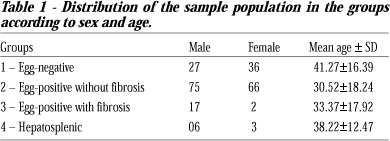
The age of the individuals varied from 8 to 77 years and the distribution according to age and sex in each group is presented in Table 1. Group 2 has the lowest mean age, but differed statistically only from Group 1. In relation to sex, the total sample is balanced with 54% male and 46% female. However, a statistical difference was observed in Group 3 of which 89.5% were male.
Ultrasound measurements of organ size, and vein diameter. The mean values for longitudinal and antero-posterior measurements of liver left lobe and spleen as well as the diameters of portal and splenic veins are presented in Table 2. The statistical differences among Groups 1, 2, 3 and 4 are shown in Figure 1. In the statistical analysis, the effect of sex and age were controlled by including these variables in the statistical model. The mean left lobe longitudinal measurement of G3 differed statistically only from Group 1 (control) and the mean antero-posterior measurement of G4 differed from G1 and G2. The mean enlargement of the spleen seems to be a better parameter than that of the liver to assess morbidity by physical examination. This is demonstrated by both measurements of the spleen, longitudinal and antero-posterior, of patients in G4 who differed from the other three groups. In addition, the mean longitudinal measurement of the spleen from G3 patients was significantly higher than those of G1 and G2 and lower than those of G4. The mean diameter of the portal vein of G3 was significantly larger than those of G1 and G2, but the mean diameter of G4 did not differ from G1 and G2. The mean diameter of the splenic vein of G4 differed statistically from G2, but not from the other two groups. Parametric test showed that both groups differed statistically (p < 0.05) from each other.
Doppler ultrasonography using an echo enhancer agent (Levovist) showed an improvement in the visualization of the portal hepatic veins, but did not present any additional information.
Ultrasound after treatment. One year after treatment, as shown in Table 3, there was a significant decrease in the measurements of portal and splenic vein diameters as well as in central echogenicity. After treatment, portal vein echogenicity was reduced in 11 (57.9%) patients, remained unaltered in 5 (26.3%) and increased in 3 (15.8%). In 14 patients, the periportal thickening after treatment became < 3mm, and in five it remained between 3-6 mm.
DISCUSSION
Among the volunteers of the populations in the endemic areas studied, in spite of the high prevalence of positive stool tests for Schistosoma mansoni, there was no patient with the hepatosplenic form of the disease. Thus, the areas are of low morbidity. No patient had hard nor nodular liver nor with prominent left lobe, which are the main clinical features for suspicion of liver with periportal fibrosis. However, even without clinical suspicion, periportal thickening was evidenced in 19 (8.5%) of the schistosomal individuals examined. It has long been known14 that Symmers' fibrosis could occur without splenomegaly and without portal hypertension and that this was frequent in endemic areas13 The ultrasound examination not only allows confirmation of the diagnosis but shows that the finding is still more frequent than it was expected to be12. And also, as can be seen in this work, it occurs even in some patients without clinical signs suggestive of Symmers' fibrosis.
Unlike that which occurred in hepatosplenic patients (G4), the echogenicity degree found in the endemic areas (G3) was slight. The mild periportal fibrosis and cellular infiltration can be seen in portal spaces and although more frequent in schistosomiasis3, it had been reported also in non schistosomiasis patients3 16. It is necessary to keep this in mind when the interpretation of the meaning of mild periportal thickness found in many people in endemic areas is considered. S. mansoni egg-negative individuals at stool test (G1), and the disappearance of this thickening in 57.9% of our patients after treatment (G3) suggest its connection with schistosomiasis.
In addition, the mean longitudinal measurement of the spleen of G3 patients was significantly higher than those of G1 and G2 and smaller than those of G4. Jesus et al10 observed in hepatosplenic patients a correlation between the echogenicity of the portal tract and spleen size. Our data also showed that the portal and splenic vein diameters were slightly larger in G3 and G4 groups but these results were not statistically significant.
Our studies are in agreement with previous studies by Doehring-Schwerdtfeger et al8, demonstrating that treatment induces significant decrease in morbidity, in the group of patients where severe morbidity has not yet developed. Whether mild periportal fibrosis is reinstalled when an individual is reinfected is still to be evaluated. It is also important to evaluate whether in patients with hepatosplenomegaly, the ultrasound confirms the same decrease in morbidity evidenced by clinical examination6.
Doppler ultrasonography using the echo enhancer agent (Levovist) showed an improvement in the visualization of the portal hepatic veins but did not add any additional relevant information.
In our study, the most precise data that allowed us to determine the stage of pathological development were from analyses of central and peripheral echogenicity of the periportal region and spleen size.
ACKNOWLEDGEMENTS
We thank Maria de Fátima da Silva, Marlucy Rodrigues Lima, Lilian Cardoso Moreira and Ivanete dos Santos Nascimento for field and laboratory expert technical assistance.
Recebido para publicação em 2/7/2003
Aceito em 6/8/2004
Supported by CNPq, FAPEMIG, NIH grant AI45451, UNDP/World Bank/WHO Special Program for Research and Training in Tropical Diseases.
- 1. Abdel-Wahab F, Esmat G, Farrag A, Elboraey WA, Strickland GT. Grading of hepatic schistosomiasis by the use of ultrasonography. American Journal of Tropical Medicine and Hygiene 46: 403-408, 1992.
- 2. Albrecht T, Blomley MJK, Cosgrove DO, Taylor-Robinson SD, Jayaram V, Eckersley R, Urbank A, Bretler-Barnes J, Patel N. Non invasive diagnosis of hepatic cirrhosis by transit-time analysis of an ultrasound contrast agent. The Lancet 353: 1579-1583, 1999.
- 3. Andrade Z, Prata A. Asymptomatic schistosomiasis studied by needle biopsy of the liver. American Journal of Tropical Medicine and Hygiene 12: 854-858, 1963.
- 4. Cerri GG, Alves VA, Magalhães A. Hepatosplenic schistosomiasis mansoni: ultrasound manifestations. Radiology 153: 777-780, 1984.
- 5. Colley DG, Garcia AA, Lambertucci JR., Parra JC, Katz N, Rocha RS, Gazzinelli G. Immune responses during human schistosomiasis. XII Differential responsiveness in patients with hepatosplenic disease. American Journal of Tropical Medicine and Hygiene 35: 793-802, 1986.
- 6. Dietze R, Prata A. Rate of reversion of hepatosplenic schistosomiasis after specific therapy. Revista da Sociedade Brasileira de Medicina Tropical 19: 69-73, 1986.
- 7. Doehring-Schwerdtfeger E, Abdel-Rahim IM, Kardorff R, Kaiser C, Franke D, Schlake J, Richter J, Elsheikh MQ, Ehrich JH. Ultrasonographical investigation of periportal fibrosis in children with Schistosoma mansoni infection: reversibility of morbidity twenty-three months after treatment with praziquantel. American Journal of Tropical Medicine and Hygiene 46: 409-415, 1992.
- 8. Doehring-Schwerdtfeger E, Kaiser C, Franke D, Kardorff R, Ali QM, Abdel-Rahim IM. Inter-observer variance in ultrasonographical assessment of Schistosoma mansoni-related morbidity in young school children. Acta Tropica 51: 85-88, 1992b.
- 9. Homeida M, Abdel-Gadir AF, Cheever AW, Bennett JL, Arbah BMO, Ibrahim SZ, Abdel-Salam IM, Dafalla AA, Nash T. Diagnosis of pathologically confirmed Symmers' periportal fibrosis by ultrasonography: A prospective blinded study. American Journal of Tropical Medicine and Hygiene 38: 86-91, 1988.
- 10. Jesus AR, Miranda DG, Miranda RG., Araújo I, Magalhães A, Bacellar M, Carvalho EM. Morbidity associated with Schistosoma mansoni infection determined by ultrasound in an endemic area of Brazil, Caatinga do Moura. American Journal of Tropical Medicine and Hygiene 63: 1-4, 2000.
- 11. Katz N, Chaves A, Pellegrino J. A simple device for quantitative stool thick smear technique in Schistosoma mansoni Revista do Instituto de Medicina Tropical de Sao Paulo 14: 397-400, 1972.
- 12. Lambertucci JR, Cota GF, Pinto-Silva RA, Serufo JC, Gerspacher-Lara R, Drummond SC, Antunes CM, Nobre V, Rayes A. Hepatosplenic schistosomiasis in field-based studies: a combined clinical and sonographic definition. Memórias do Instituto Oswaldo Cruz 96: 147-150, 2001.
- 13. Prata A. Infection with S. mansoni. In: Jordan P, Webbe G (eds) Schistosomiasis Epidemiology, Treatment and Control, William Heinemann Books Ltd, London, p 105-127, 1982.
- 14. Prata A, Andrade Z. Fibrose hepática de Symmers sem esplenomegalia. O Hospital 63: 617-623, 1963.
- 15. Prata A, Bina JC. Development of the hepatosplenic form of schistosomiasis. Gazeta de Medicina da Bahia 68: 49-60, 1968.
- 16. Schaffer F, Popper H. Non specific reactive hepatitis in aged and infirm people. American Journal Digestive Diseases 4: 389-399, 1959.
Correspondece to
Publication Dates
-
Publication in this collection
18 Jan 2005 -
Date of issue
Feb 2005
History
-
Received
02 July 2003 -
Accepted
06 Aug 2004