
Abstract
Background
Atherosclerosis, in some cases, is an asymptomatic condition, and it is important to know the degree of arterial impairment caused by plaques and its association with risk factors. Autopsy examination provides understanding of basic disease processes and assessment to data about macroscopic characteristic of atherosclerotic involvement.
Objective
To macroscopically assess and standardize atherosclerotic involvement of aorta, carotid and iliac arteries and compare with age, gender and causes of death.
Methods
We collected 53 aortic arteries, 53 right carotid arteries, 53 left carotid arteries, 53 right iliac arteries and 53 left iliac arteries. For this assessment, the extension of fatty streaks, atheromatous plaques, fibrosis and calcification were considered, being the reference to score the degree of atherosclerotic involvement. Many degrees of atherosclerosis and accurate values were observed for mild, moderate and severe classification. For statistical analysis, data were analyzed using the software GraphPad Prism® 7.0. Differences were considered statistically significant if p-value was less than 5% (p <0.05).
Results
Carotid arteries had greater atherosclerotic involvement compared to the other arteries (K = 15.73, p = 0.0004). Atherosclerosis was progressive and significant with increasing age (carotid arteries: t = 6.321; p <0.0001; aorta: U = 83.5; p <0.0001; iliac: U = 306; p <0.0001) and as cause of cardiovascular death (carotids: t = 5.047; p <0.0001; aorta: U = 98.5; p = 0.0068; iliac: U = 467.5; p = 0.0012).
Conclusion
Macroscopic assessment of atherosclerosis is an innovative and low-cost way of direct visualization of atherosclerotic plaques, enabling an association with risk factors such as increasing age and cardiovascular diseases, providing important data for clinical practice.
Cardiovascular Diseases; Atherosclerosis; Risk Factors; Asymptomatic Disease; Arteries; Autopsy; Heredity; Early Diagnosis
Resumo
Fundamento
A aterosclerose, em alguns casos, é uma condição assintomática, sendo necessário conhecer o grau de comprometimento arterial provocado pelas placas e sua associação com os fatores de risco. O exame de autópsia permite a compreensão dos processos básicos de doenças, assim como a avaliação e fornecimento de dados sobre a característica macroscópica do acometimento aterosclerótico.
Objetivo
Avaliar macroscopicamente e padronizar o acometimento aterosclerótico das artérias aorta, carótidas e ilíacas e comparar com a idade, o sexo e a causa de morte.
Métodos
Foram coletados 53 artérias aorta, 53 artérias carótida direita, 53 artérias carótida esquerda, 53 artérias ilíaca direita e 53 artérias ilíaca esquerda. Para essa avaliação, foi considerada a extensão de estrias lipídicas, de placas ateromatosas, de fibrose e de calcificação, as quais serviram de referência para pontuar a intensidade do acometimento aterosclerótico. Foram observados vários graus da aterosclerose e valores acurados para a classificação discreta, moderada e acentuada. Para a análise estatística, os dados foram analisados utilizando-se o software GraphPad Prism ® 7.0. As diferenças foram consideradas estatisticamente significativas quando “p” foi menor que 5% (p<0,05).
Resultados
As artérias carótidas apresentaram maior acometimento aterosclerótico em comparação às outras artérias avaliadas (K=15,73, p=0,0004). A ocorrência da aterosclerose se mostrou progressiva e significativa com o decorrer da idade (carótidas: t=6,321; p<0,0001; aortas: U=83,5; p<0,0001; ilíacas: U=306; p<0,0001) e na causa de morte cardiovascular (carótidas: t=5,047; p<0,0001; aortas: U=98,5; p=0,0068; ilíacas: U=467,5; p=0,0012).
Conclusão
A avaliação macroscópica da aterosclerose trata-se de uma forma inovadora e de baixo custo de avaliação através da visualização direta das placas ateroscleróticas, possibilitando uma associação com fatores de risco como idade avançada e doenças cardiovasculares, fornecendo dados importantes para a prática clínica.
Doenças Cardiovasculares; Aterosclerose; Fatores de Risco; Doenças Assintomáticas; Artérias; Autópsia; Hereditariedade; Diagnóstico Precoce
Introduction
Atherosclerosis is a multifactorial disease associated with hereditary factors, sex and lifestyle habits such as smoking, inadequate diet and sedentary lifestyle.11. Levula M, Oksala N, Airla N, Zeitlin R, Salenius JP, Järvinen O et al. Genes involved in systemic and arterial bed dependent atherosclerosis–Tampere Vascular study. PLoS One. 2012;7(4):e33787. Inflammation and deposition of lipids in the artery wall are involved in triggering and progression of atherosclerotic plaques11. Levula M, Oksala N, Airla N, Zeitlin R, Salenius JP, Järvinen O et al. Genes involved in systemic and arterial bed dependent atherosclerosis–Tampere Vascular study. PLoS One. 2012;7(4):e33787. leading to cardiovascular diseases with high incidence worldwide.22. Gargiulo P, Marsico F, Parente A, Paolillo S, Cecere M, Casaretti L et al. Ischemic heart disease in systemic inflammatory diseases. An appraisal. Int J Cardiol. 2014;170(3):286–90.
The early diagnosis of atherosclerosis as a predictor of coronary artery disease and acute myocardial infarction reduces morbidity and mortality associated with the disease. Studies explore prevalence and association of factors that contribute to risk stratification.33. Simão AF, Precoma DB, Andrade JP, Correa Filho H, Saraiva JF, Oliveira GM, et al; Sociedade Brasileira de Cardiologia. [I Brazilian Guidelines for cardiovascular prevention]. Arq Bras Cardiol. 2013;101(6 Suppl 2):1-63. Erratum in: Arq Bras Cardiol. 2014;102(4):415. Evaluation and early diagnosis in patients of these groups are important.
It is known that the autopsy exam is extremely important, as it allows the understanding of the basic processes of diseases.44. Abedinzadeh N, Pedram B, Sadeghian Y, Nodushan SM, Gilasgar M, Darvish M et al. A histopathological analysis of the epidemiology of coronary atherosclerosis: an autopsy study. Diagn Pathol. 2015;3(10):87. Studies have shown that in addition to eliminating risk factors associated with atherosclerosis, there are currently effective drugs for the treatment of this disease.55. Bergheanu SC, Bodde MC, Jukema JW. Pathophysiology and treatment of atherosclerosis. Neth Heart J. 2017;25(4):231-42. However, for the treatment to be effective, it is worthy to know the degree of arterial impairment caused by atherosclerotic plaques.66. Spence JD, Hackam DG. Treating arteries instead of risk factors: a paradigm change in management of atherosclerosis. Stroke. 2010;41(6):1193-9. This fact makes our assessment and data on the macroscopic characteristics even more valuable, as autopsy studies allow a broad and direct view of atherosclerosis.77. Suemoto CK, Ferretti-Rebustini REL, Rodriguez RD, Leite REP, Soterio L, Brucki SMD et al. Neuropathological diagnoses and clinical correlates in older adults in Brazil: A crosssectional study. PLoS Med. 2017;14(3):e1002267. , 88. Van der Kooy K, Seidell JC. Techniques for the measurement of visceral fat: a practical guide. Int J Obes Relat Metab Disord. 1993;17(4):187–96.
As atherosclerosis is a multifactorial cardiovascular disease, responsible for the development of serious illnesses, its macroscopic assessment in autopsy material is important to provide a reliable and standardized description of atherosclerotic plaque progression. The association of macroscopic aspects with risk factors provides epidemiological data for clinical practice. The objective of this study was to identify macroscopically the intensity of atherosclerotic involvement of aorta, carotid and iliac arteries and to compare the degree of involvement with risk factors such as age, gender and cause of death.
Methods
We evaluated 2931 autopsy protocols performed between 1963 and 2018. Based on these protocols, biological materials (carotid, aorta and iliac arteries) were selected from those patients who presented complete autopsy report, with information about age (included those over 18 years old), gender and the cause of death (cardiovascular or not). Cases with biological materials in inadequate conditions or with incomplete autopsy reports were excluded from the study. A sample of 53 autopsied patients was obtained from the anatomical specimen file of the Discipline of General Pathology, totalizing 53 aortic arteries, 53 right carotid arteries, 53 left carotid arteries, 53 right iliac arteries and 53 left iliac arteries. The study was developed in the Discipline of General Pathology of Federal University of Triângulo Mineiro and approved by the Research Ethics Committee of Federal University of Triângulo Mineiro with the Certificate of Presentation of Ethical Appreciation (CAAE) number: 56931816.4.0000.5154 in accordance with the resolution 466/2012.
Macroscopic evaluation of atherosclerosis
Three examiners evaluated macroscopic degree of atherosclerosis in aorta, right carotid, left carotid, right iliac and left iliac arteries, using criteria already described in the literature.99. Ferraz MLF, Nascimento DMS, Rorato JPH, Espindula AP, Oliveira LF, Ramalho LS et al. Correlation of lifetime progress of atherosclerosis and morphologic markers of severity in humans: new tools for a more sensitive evaluation. Clinics. 2012;67(9):1071-5. The progression of fatty streaks, atheromatous plaques, fibrosis and calcification on artery walls were a reference to score the degree of involvement ( Figure 1 ). A 12.0 cm non-millimeter line was made on a sheet ( Figure 2A ) and this line was used as a scale for atherosclerosis involvement.

– A) Aortic artery with fatty streaks. B Aorta with atheromatous plaques. C) Aorta with atheromatous plaques, fibrosis and calcifications.
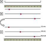
– A) A 12.0 cm non-millimeter line model used as a scale for measurement of atherosclerosis degree. B) Registration of the point on the non-millimeter scale, referring to the degree of involvement after evaluation of lesions. C) Measure of the distance from the 0.0 cm point to the marked point after finishing evaluations in all arteries.
After opening the artery, examiners observed the degree of lesions, then a point was recorded on the scale regarding the degree of involvement, the closer to 0.0 cm the lesser the involvement, and the closer to 12.0 cm the greater the involvement ( Figure 2B ).
After the end of all evaluations, the distance from the 0.0 cm point to the point marked on the scale by examiners was measured, in order to avoid interferences in classifications ( Figure 2C ). The intensity of atherosclerosis was classified as mild if the evaluation was from 0.1 cm to 4.0 cm; moderate, from 4.1cm to 7.0cm and accentuated, from 7.1cm to 12.0cm.99. Ferraz MLF, Nascimento DMS, Rorato JPH, Espindula AP, Oliveira LF, Ramalho LS et al. Correlation of lifetime progress of atherosclerosis and morphologic markers of severity in humans: new tools for a more sensitive evaluation. Clinics. 2012;67(9):1071-5. Many degrees of atherosclerosis and accurate values were observed in mild, moderate and accentuated classification ( Figure 3 ).
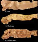
– A) Atherosclerosis in aortic artery assessed as mild. B) Atherosclerosis in aortic artery assessed as moderate. C) Atherosclerosis in aortic artery assessed as accentuated.
Statistical analysis
For statistical analysis, a database was created in the program Microsoft Excel ®. Data were analyzed using the software GraphPad Prism ® 7.0. To verify the type of distribution of variables, the statistical test of Kolmogorov-Smirnov (with Dallal-Wilkinson-Lillie for p-value). For continuous variables with normal distribution, the mean and standard deviation were presented, and for those with non-normal distribution, the median and interquartile range. We used the unpaired student’s t-test (t) for normal distribution and the Mann-Whitney (U) for non-normal distribution comparison between two groups. For the comparison of three groups, Kruskal-Wallis (H) test was used, followed by Dunn post-test. Spearman’s correlation coefficient (rS) for non-normal distribution was used for correlation. A p-value less than 0.05 (typically ≤ 0.05) was considered statistically significant.
Results
Regarding general distribution of the sample, data are described in Table 1 .
Carotid arteries showed greater atherosclerotic involvement than other arteries assessed (H = 15.73, p = 0.0004), with a significant difference found between carotid and iliac arteries (p = 0.0002). Variation in macroscopic degrees of atherosclerosis is described in Table 2 .
Macroscopic assessment and different degrees of macroscopic involvement of atherosclerosis in carotid, aortic and iliac arteries are shown in Figure 4 .

– A) Comparison of atherosclerosis macroscopic assessment in carotid, aortic and iliac arteries. B) Comparison between macroscopic evaluation of atherosclerosis in carotid, aortic and iliac arteries classified as mild (0 to 4 cm). C) Comparison between macroscopic assessment of atherosclerosis in carotid, aortic and iliac arteries classified as moderate (4.1 to 7 cm). D) Comparison between macroscopic assessment of atherosclerosis in the carotid, aortic and iliac arteries classified as accentuated (7.1 to 12 cm).
The occurrence of atherosclerosis was progressive and significant with age in carotid arteries (rS = 0.5133; p <0.0001), in aortic (rS = 0.716; p <0.0001) and in iliac arteries (rS = 0.7378; p <0.0001) ( Figure 5 ).
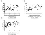
– A) Correlation between macroscopic evaluation of atherosclerosis in carotid arteries with age. B) Correlation between the macroscopic assessment of atherosclerosis in aortic arteries with age. C) Correlation between macroscopic assessment of atherosclerosis in iliac arteries with age.
Data regarding atherosclerosis macroscopic assessment and the variables analyzed are described in Table 2 .
Discussion
Using autopsy material we demonstrated atherosclerosis in different arterial beds, which are of great importance for the body’s blood supply. Macroscopic analysis is a way to understand disease development process, being a valid and accurate instrument for research, providing data to clinical practice as already demonstrated in other studies.99. Ferraz MLF, Nascimento DMS, Rorato JPH, Espindula AP, Oliveira LF, Ramalho LS et al. Correlation of lifetime progress of atherosclerosis and morphologic markers of severity in humans: new tools for a more sensitive evaluation. Clinics. 2012;67(9):1071-5.
10. Oliveira MS, Torquato BGS, da Silveira LAM, Juliano GR, Aguiar LS, Juliano GR et al. Evaluation of aortic changes in elderly people autopsied with acquired immunodeficiency syndrome. Surg Exp Pathol. 2018;1:7. - 1111. Nishizawa A, Suemoto CK, Farias-Itao DS, Campos FM, Silva KCS, Bittencourt MS et al. Morphometric measurements of systemic atherosclerosis and visceral fat: Evidence from an autopsy study. PLoS One. 2017;12(10):e0186630.
The present study demonstrated a greater involvement of carotid arteries, compared to aorta and iliac arteries, but with significant difference only between carotid and iliac arteries. Although atherosclerosis is a process that can affect the entire vascular tree, being found in any large or medium-sized arteries, the disease tends to be located in particular areas, such as aortoiliac, iliac-femoral segments or carotid arteries. Factors such as changes in blood flow, in extra vascular pressure and anatomical and biochemical features seem to explain the prevalence of lesions for these vessels.1212. Brasileiro Filho G. Bogliolo patologia geral. 5. ed. Rio de Janeiro: Guanabara Koogan;2013. Furthermore, carotid arteries are evaluated in several other studies for atherosclerosis degree and determination of cardiovascular diseases development.1313. Çetin M, Erdoğan T, Kiriş T, Özyildiz AG, Ergül E, Durakoğlugil E et al. Endothelial dysfunction, subclinical atherosclerosis and LDL cholesterol are the independent predictors of left atrial functions in hypertension. Int J Cardiovasc Imaging.2020;36(1):69-77.
14. Paraskevas KI, Sillesen HH. Carotid atherosclerosis markers and adverse cardiovascular events. Int J Cardiol. 2020;307:178. - 1515. Yoshida K, Yang T, Yamamoto YU, Kurosaki Y, Funaki T, Kikuchi T et al. Expansive carotid artery remodeling: possible marker of vulnerable plaque. J Neurosurg. 2019;1-6.
In general, atherosclerosis degree varied between mild and moderate. In lesions evaluated as mild, fatty streaks were present, which indicates the beginning of injury process. Although these lesions do not alter blood circulation as they do not obstruct vascular lumen, their location facilitates continuous lipid deposition and progression to atherosclerosis.1616. Lloyd-Jones DM, Nam BH, D’Agostino RB Sr, Levy D, Murabito JM, Wang TJ et al. Parental Cardiovascular Disease as a Risk Factor for Cardiovascular Disease in Middle-aged Adults: A Prospective Study of Parents and Offspring. JAMA. 2004;291(18):2204–11.
Elderly patients had significantly greater degree of atherosclerosis than non-elderly. Age was a significant predictor for atherosclerosis development.1717. Zavatta M, Mell MW. A national Vascular Quality Initiative database comparison of hybrid and open repair for aortoiliac-femoral occlusive disease. J Vasc Surg. 2018;67:199-205. , 1818. Hendriks EJ, Beulens JW, De Jong PA, Van Der Schouw YT, Sun WN, Wright CM et al. Calcification of the splenic, iliac, and breast arteries and risk of all-cause and cardiovascular mortality. Atherosclerosis. 2017 Apr;259:120-7. An increase in marked and asymptomatic atherosclerotic plaques was found in arteries from elderly patients,1919. Fernandez-Friera L, Penalvo Jl, Fernandez-Ortiz A, Ibanez B, Lopez-Melgar B, Laclaustra M. Prevalence, vascular distribution, and multiterritorial extent of subclinical atherosclerosis in a middle-aged cohort: The PESA (Progression of Early Subclinical Atherosclerosis) Study. Circulation. 2015;131(24):2104-13. as well as calcifications,2020. Chen HY, Engert JC, Thanassoulis G. Risk factors for valvular calcification. Curr Opin Endocrinol Diabetes Obes. 2019;26(2):1-7. which corroborates our findings.
Regarding gender, a higher degree of atherosclerosis was found in women, but without significant difference, which agrees with a similar study that used ultrasound to analyze atherosclerosis, in which authors found no differences between genders.2121. Yang YJ, Park HJ, Won KB, Chang HJ, Park GM, Kim YG, et al. Relationship between the optimal cut-off values of anthropometric indices for predicting metabolic syndrome and carotid intima-medial thickness in a Korean population. Medicine. 2019;98(42):e17620. A recent study proved that genders have diferente physiological responses to risk factors (smoking, obesity, diabetes and systemic arterial hypertension) and females are the most affected and sensitized with such aggressions. Although many studies showed higher rates of cardiovascular events in men, there are divergent researches showing female physiological response is more sensitive to risk factors, which contributes to the development or worsening of cardiovascular disease.2222. Cifkova R, Pitha J, Krajcoviechova A, Kralikova E. Is the impact of conventional risk factors the same in men and women? Plea for a more gender-specific approach. Int J Cardiol. 2019;286:214-9.
In the present study, patients who died due to cardiovascular causes had a significantly higher degree of atherosclerosis. The anatomopathological study of patients who died due to cardiovascular causes provides the best sample of population to study atherosclerosis.44. Abedinzadeh N, Pedram B, Sadeghian Y, Nodushan SM, Gilasgar M, Darvish M et al. A histopathological analysis of the epidemiology of coronary atherosclerosis: an autopsy study. Diagn Pathol. 2015;3(10):87. Cardiovascular diseases are directly associated with the occurrence of systemic atherosclerosis, asymptomatic in most cases,2323. Nakamura E, Sato Y, Iwakiri T, Yamashita A, Moriguchi-Goto S, Maekawa K et al. Asymptomatic Plaques of Lower Peripheral Arteries and Their Association with Cardiovascular Disease: An Autopsy Study. J Atheroscler Thromb. 2017;24(9):921-7. which makes prevention difficult, although extremely important.
As a post-mortem study, it has some limitations such as the absence of some data about patients’ lifestyle as medications, food, smoking, among other risk factors that are related to atherosclerosis development. In addition, some deaths occurred without atherosclerosis being previously investigated during patient’s hospitalization, which would be a good predictor for macroscopic evaluation accuracy. However, there are several positive points that strengthen the work, such as the direct and precise macroscopic evaluation through visualization of the entire plaque and the confirmation of association of intrinsic risk factors such as age and gender with cause of death that may have occurred due to extrinsic factors. In addition, several important arterial beds were collected (right and left carotid, right and left aorta and iliac) with similar results compared to risk factors and confirmed to be adequate sites for systemic atherosclerosis assessment.
Conclusions
Atherosclerosis is a progressive lesion throughout life, which affects different arterial beds, with carotid arteries being the most affected, constituting an adequate sites for studying and assessing atherosclerotic plaque progression. The study highlights the importance of assessing atherosclerosis and shows an innovative form of assessment, as it is possible to measure the macroscopic degree of impairment through direct visualization of atherosclerotic plaques and compare with risk factors that, in association, can contribute to plaque progression and development of other cardiovascular diseases. Advanced age, female gender and death due to cardiovascular causes contribute as risk factors for greater lipid accumulation in these arteries. Macroscopic evaluation is a low-cost, effective and standardized method for measuring atherosclerosis degree and allows a better understanding of cardiovascular events development at the time of autopsy, in addition to providing data for clinical practice.
Referências
-
1Levula M, Oksala N, Airla N, Zeitlin R, Salenius JP, Järvinen O et al. Genes involved in systemic and arterial bed dependent atherosclerosis–Tampere Vascular study. PLoS One. 2012;7(4):e33787.
-
2Gargiulo P, Marsico F, Parente A, Paolillo S, Cecere M, Casaretti L et al. Ischemic heart disease in systemic inflammatory diseases. An appraisal. Int J Cardiol. 2014;170(3):286–90.
-
3Simão AF, Precoma DB, Andrade JP, Correa Filho H, Saraiva JF, Oliveira GM, et al; Sociedade Brasileira de Cardiologia. [I Brazilian Guidelines for cardiovascular prevention]. Arq Bras Cardiol. 2013;101(6 Suppl 2):1-63. Erratum in: Arq Bras Cardiol. 2014;102(4):415.
-
4Abedinzadeh N, Pedram B, Sadeghian Y, Nodushan SM, Gilasgar M, Darvish M et al. A histopathological analysis of the epidemiology of coronary atherosclerosis: an autopsy study. Diagn Pathol. 2015;3(10):87.
-
5Bergheanu SC, Bodde MC, Jukema JW. Pathophysiology and treatment of atherosclerosis. Neth Heart J. 2017;25(4):231-42.
-
6Spence JD, Hackam DG. Treating arteries instead of risk factors: a paradigm change in management of atherosclerosis. Stroke. 2010;41(6):1193-9.
-
7Suemoto CK, Ferretti-Rebustini REL, Rodriguez RD, Leite REP, Soterio L, Brucki SMD et al. Neuropathological diagnoses and clinical correlates in older adults in Brazil: A crosssectional study. PLoS Med. 2017;14(3):e1002267.
-
8Van der Kooy K, Seidell JC. Techniques for the measurement of visceral fat: a practical guide. Int J Obes Relat Metab Disord. 1993;17(4):187–96.
-
9Ferraz MLF, Nascimento DMS, Rorato JPH, Espindula AP, Oliveira LF, Ramalho LS et al. Correlation of lifetime progress of atherosclerosis and morphologic markers of severity in humans: new tools for a more sensitive evaluation. Clinics. 2012;67(9):1071-5.
-
10Oliveira MS, Torquato BGS, da Silveira LAM, Juliano GR, Aguiar LS, Juliano GR et al. Evaluation of aortic changes in elderly people autopsied with acquired immunodeficiency syndrome. Surg Exp Pathol. 2018;1:7.
-
11Nishizawa A, Suemoto CK, Farias-Itao DS, Campos FM, Silva KCS, Bittencourt MS et al. Morphometric measurements of systemic atherosclerosis and visceral fat: Evidence from an autopsy study. PLoS One. 2017;12(10):e0186630.
-
12Brasileiro Filho G. Bogliolo patologia geral. 5. ed. Rio de Janeiro: Guanabara Koogan;2013.
-
13Çetin M, Erdoğan T, Kiriş T, Özyildiz AG, Ergül E, Durakoğlugil E et al. Endothelial dysfunction, subclinical atherosclerosis and LDL cholesterol are the independent predictors of left atrial functions in hypertension. Int J Cardiovasc Imaging.2020;36(1):69-77.
-
14Paraskevas KI, Sillesen HH. Carotid atherosclerosis markers and adverse cardiovascular events. Int J Cardiol. 2020;307:178.
-
15Yoshida K, Yang T, Yamamoto YU, Kurosaki Y, Funaki T, Kikuchi T et al. Expansive carotid artery remodeling: possible marker of vulnerable plaque. J Neurosurg. 2019;1-6.
-
16Lloyd-Jones DM, Nam BH, D’Agostino RB Sr, Levy D, Murabito JM, Wang TJ et al. Parental Cardiovascular Disease as a Risk Factor for Cardiovascular Disease in Middle-aged Adults: A Prospective Study of Parents and Offspring. JAMA. 2004;291(18):2204–11.
-
17Zavatta M, Mell MW. A national Vascular Quality Initiative database comparison of hybrid and open repair for aortoiliac-femoral occlusive disease. J Vasc Surg. 2018;67:199-205.
-
18Hendriks EJ, Beulens JW, De Jong PA, Van Der Schouw YT, Sun WN, Wright CM et al. Calcification of the splenic, iliac, and breast arteries and risk of all-cause and cardiovascular mortality. Atherosclerosis. 2017 Apr;259:120-7.
-
19Fernandez-Friera L, Penalvo Jl, Fernandez-Ortiz A, Ibanez B, Lopez-Melgar B, Laclaustra M. Prevalence, vascular distribution, and multiterritorial extent of subclinical atherosclerosis in a middle-aged cohort: The PESA (Progression of Early Subclinical Atherosclerosis) Study. Circulation. 2015;131(24):2104-13.
-
20Chen HY, Engert JC, Thanassoulis G. Risk factors for valvular calcification. Curr Opin Endocrinol Diabetes Obes. 2019;26(2):1-7.
-
21Yang YJ, Park HJ, Won KB, Chang HJ, Park GM, Kim YG, et al. Relationship between the optimal cut-off values of anthropometric indices for predicting metabolic syndrome and carotid intima-medial thickness in a Korean population. Medicine. 2019;98(42):e17620.
-
22Cifkova R, Pitha J, Krajcoviechova A, Kralikova E. Is the impact of conventional risk factors the same in men and women? Plea for a more gender-specific approach. Int J Cardiol. 2019;286:214-9.
-
23Nakamura E, Sato Y, Iwakiri T, Yamashita A, Moriguchi-Goto S, Maekawa K et al. Asymptomatic Plaques of Lower Peripheral Arteries and Their Association with Cardiovascular Disease: An Autopsy Study. J Atheroscler Thromb. 2017;24(9):921-7.
-
Study AssociationThis article is part of the thesis of master submitted by Mariana Oliveira, from Universidade Federal do Triângulo Mineiro.
-
Sources of Funding: This study was partially funded by CAPES, CNPq, FAPEMIG e FUNEPU
Publication Dates
-
Publication in this collection
14 June 2021 -
Date of issue
June 2021
History
-
Received
28 Nov 2019 -
Reviewed
31 Mar 2020 -
Accepted
20 May 2020


