ABSTRACT
The identification of female flesh flies was always considered a difficult task since morphological descriptions and keys for females are rare. Even in a forensic entomology framework, where females play a major role, female flesh flies are usually not identified. In order to fill this gap in Southern Brazil fauna we provide detailed descriptions and key for the female of nine species included in four genera: Microcerella halli (Engel), Oxysarcodexia paulistanensis (Mattos), Oxysarcodexia riograndensis (Lopes), Peckia (Euboettcheria) australis (Townsend), Peckia(Euboettcheria) florencioi (Prado and Fonseca), Peckia (Pattonella) intermutans (Walker), Peckia(Pattonella) resona (Lopes), Peckia (Sarcodexia) lambens (Wiedemann), and Sarcophaga(Bercaea) africa (Wiedemann). These species are distinguished mainly by genital characters as tergite 6 divided or undivided, presence of tergite 8, spermatheca morphology and vaginal plate shape.
Keywords
Forensic entomology;
Microcerella
;
Oxysarcodexia
;
Peckia
;
Sarcophaga
Introduction
Sarcophagidae Hagen, 1881 is widely distributed with about 3100 described species in 400 genera. Although it has worldwide geographic distribution, Sarcophagidae richness is remarkably concentrated in regions of tropical and warm temperate climate (Shewell, 1987Shewell, G.E., 1987. Sarcophagidae. In: McAlpine, J.F., Peterson, B.V., Shewell, G.E., Teskey, H.J., Vockeroth, J.R., Wood, D.M. (Eds.), Manual of Neartic Diptera, vol. 2. Monograph 108, Research Branch, Agriculture Canada, Otawa, pp. 1159–1186.; Pape, 1996Pape, T., 1996. A catalogue of Sarcophagidae of the world (Insecta: Diptera). Mem. Entomol. 8, Associated Publishers.) and in Neotropical region more than 800 species are found. There are three subfamilies, Miltogramminae, Paramacronychiinae and Sarcophaginae, but only Sarcophaginae has species of forensic and medical importance in the Neotropics (Pape, 1996Pape, T., 1996. A catalogue of Sarcophagidae of the world (Insecta: Diptera). Mem. Entomol. 8, Associated Publishers.).
The external morphology of most Sarcophaginae adults is extremely similar. The species share three black stripes pattern in the mesonotum, meron with bristles, undeveloped subscutellum, and abdomen checkered or spotted and medium to large size, ranging from 8 to 14 mm (de Carvalho and Mello-Patiu, 2008de Carvalho, C.J.B., Mello-Patiu, C.A., 2008. Key to the adults of the most common forensic species of Diptera in South America. Rev. Bras. Entomol. 52, 390-406.). Probably because of this morphological similarity and the lack of keys, this group is considered to be difficult for identification (Barros et al., 2008Barros, R.M., de Mello-Patiu, C.A., Pujol-Luz, J.R., 2008. Sarcophagidae (Insecta: Diptera) associados à decomposição de carcaças de Sus scrofa em área de cerrado do Distrito Federal, Brasil. Rev. Bras. Entomol. 52, 606-609.; Mulieri et al., 2010Mulieri, P.R., Mariluis, J.C., Patitucci, L.D., 2010. Review of the Sarcophaginae (Diptera: Sarcophagidae) of Buenos Aires Province (Argentina), with a key and description of a new species. Zootaxa 2575, 1-37.; Vairo et al., 2011Vairo, K.P., de Mello-Patiu, C.A., de Carvalho, C.J.B., 2011. Pictorial identification key for species of Sarcophagidae (Diptera) of potential forensic importance in southern Brazil. Rev. Bras. Entomol. 55, 333-347.).
Female flesh flies are much more abundant than males on carcasses. They use the corpse not only as source of food and mating site but also as larviposition site. In forensic entomology, the species that rear on corpses are considered the most important data source. The biological data from these species are essential to estimate the minimum post mortem interval (PMI), which corresponds to the period of insect activity on corpse (Tomberlin et al., 2011Tomberlin, J.K., Mohr, R., Benhow, M.E., Tarone, A.M., VanLaerhoven, S., 2011. A roadmap for bridging basic and applied research in forensic entomology. Annu. Rev. Entomol. 56, 401-421.). In addition to their use in applied sciences, such as forensic entomology, females have their own place in Sarcophagidae systematics and can provide important characters for mapping the group evolution (Lopes, 1941Lopes, H.S., 1941. Sôbre o aparelho Genital Feminino dos Sarcophagidae e sua importância na classificação (Diptera). Rev. Bras. Biol. 1, 215-221., 1958Lopes, H.S., 1958. Considerações sobre as espécies de Peckia Desvoidy, 1830 e de gêneros afins (Diptera, Sarcophagidae). An. Acad. Bras. Ciênc. 30, 211-243.; Tibana and de Mello-Patiu, 1985Tibana, R., de Mello-Patiu, C.A., 1985. O sintergito 6 + 7 nas fêmeas de Oxysarcodexia Townsend, 1917 (Diptera, Sarcophagidae). Rev. Bras. Biol. 45, 439-445.; de Mello-Patiu and Santos, 2001de Mello-Patiu, C.A., Santos, J.M., 2001. Nephochaetopteryx Townsend, 1934: descriptions and comparative morphological notes on the female terminalia (Diptera: Sarcophagidae). Stud. Dipterol. 8, 303-315.) although females are still unknown in many species. However, despite their importance, Sarcophagidae females are usually neglected in taxonomic and applied research.
In southern Brazil, forensic entomology is well disseminated (Vairo et al., 2015Vairo, K.P., Corrêa, R.C., Lecheta, M.C., Caneparo, M.F., Mise, K.M., de Carvalho, C.J.B., Almeida, L.M., Moura, M.O., 2015. Forensic use of a subtropical blowfly: the first case indicating minimum post-mortem interval (mPMI) in Southern Brazil and first record of Sarconesia chlorogaster from a human corpse. J. Forensic Sci. 1, 257-260.; Corrêa et al., 2014Corrêa, R.C., Moura, M.O., Almeida, L.M., 2014. Coleoptera associated with buried carrion: potential forensic importance and seasonal composition. J. Med. Entomol. 51, 1057-1066.) but there are no available keys for all necrophagous female flesh flies, making this group under utilized in forensic cases. Mulieri et al. (2010)Mulieri, P.R., Mariluis, J.C., Patitucci, L.D., 2010. Review of the Sarcophaginae (Diptera: Sarcophagidae) of Buenos Aires Province (Argentina), with a key and description of a new species. Zootaxa 2575, 1-37. provided a key to male and female adults of Sarcophaginae from Buenos Aires Province, including 39 species, that can be used partially to identify fauna from southern Brazil, but only four species herein analyzed were included among them. Nevertheless, a more detailed comparison of females of most species of forensic importance is essential to provide a greater number of characters and minimize the difficulties in the problematic task of female identification, especially by non-taxonomists, in medical, veterinary and forensic applications (de Carvalho and Mello-Patiu, 2008de Carvalho, C.J.B., Mello-Patiu, C.A., 2008. Key to the adults of the most common forensic species of Diptera in South America. Rev. Bras. Entomol. 52, 390-406.; Mulieri et al., 2010Mulieri, P.R., Mariluis, J.C., Patitucci, L.D., 2010. Review of the Sarcophaginae (Diptera: Sarcophagidae) of Buenos Aires Province (Argentina), with a key and description of a new species. Zootaxa 2575, 1-37.). Therefore, as a first step to fill this gap, we present a pictorial key for females of nine necrophagous species of Sarcophaginae from southern Brazil.
Material and methods
All species chosen met two criteria: can be reared in organic matter, thus being necrophagous, and have their geographic range reaching Southern Brazil. Those species are: Oxysarcodexia paulistanensis (Mattos, 1919), Microcerella halli (Engel, 1931), Peckia(Sarcodexia) lambens (Wiedemann, 1830), Peckia (Pattonella) resona(Lopes, 1935), Peckia (Pattonella) intermutans (Walker, 1861), Oxysarcodexia riograndensis (Lopes, 1946), Peckia(Euboettcheria) australis (Townsend, 1927), Peckia (Euboettcheria) florencioi (Prado and Fonseca, 1932) and Sarcophaga (Bercaea) africa(Wiedemann, 1824). The first five species have larvae already sampled on carcasses and/or human corpses (Salviano, 1996Salviano, R.J.B., 1996. Sucessão de Diptera Caliptrata em carcaça de Sus scrofa L. Dissertação de Mestrado. Universidade Federal Rural do Rio de Janeiro, Rio de Janeiro.; Moura et al., 1997Moura, M.O., de Carvalho, C.J.B., Monteiro-Filho, E.L.A., 1997. A preliminary analysis of insects of medico-legal importance in Curitiba, State of Paraná. Mem. Inst. Oswaldo Cruz 92, 269-274., 1998Moura, M.O., de Carvalho, C.J.B., Monteiro-Filho, E.L.A., 1998. Carrion attendant arthropods in southern Brazil. Cienc. Cult. 50, 377-381., 2005Moura, M.O., de Carvalho, C.J.B., Monteiro-Filho, E.L.A., 2005. Estrutura de Comunidades necrófagas: efeito da partilha de recursos na diversidade. Rev. Bras. Zool. 22, 1134-1140.; Carvalho and Linhares, 2001Carvalho, L.M.L., Linhares, A.X., 2001. Seasonality of insect succession and pig carcass decomposition in a natural Forest area in Southeastern Brazil. J. Forensic Sci. 46, 604-608.; Oliveira and Vasconcelos, 2010Oliveira, T.C., Vasconcelos, S.D., 2010. Insects (Diptera) associated with cadavers at the Institute of Legal Medicine in Pernambuco, Brazil: implications for forensic entomology. Forensic Sci. Int. 198, 97-102.; Vairo et al., 2011Vairo, K.P., de Mello-Patiu, C.A., de Carvalho, C.J.B., 2011. Pictorial identification key for species of Sarcophagidae (Diptera) of potential forensic importance in southern Brazil. Rev. Bras. Entomol. 55, 333-347.) and the last four have adults sampled in Paraná, Santa Catarina and Rio Grande do Sul and reared in laboratory with putrefied bovine meat.
Although 22 species of fleshflies with potential forensic importance were already registered in Southern Brazil (Vairo et al., 2011Vairo, K.P., de Mello-Patiu, C.A., de Carvalho, C.J.B., 2011. Pictorial identification key for species of Sarcophagidae (Diptera) of potential forensic importance in southern Brazil. Rev. Bras. Entomol. 55, 333-347.), in this present contribution we were interested in species that could be used to estimate the minimum post mortem interval, i.e., not only species attracted by carrion, but those species in which the larvae are reared on carcasses or corpses.
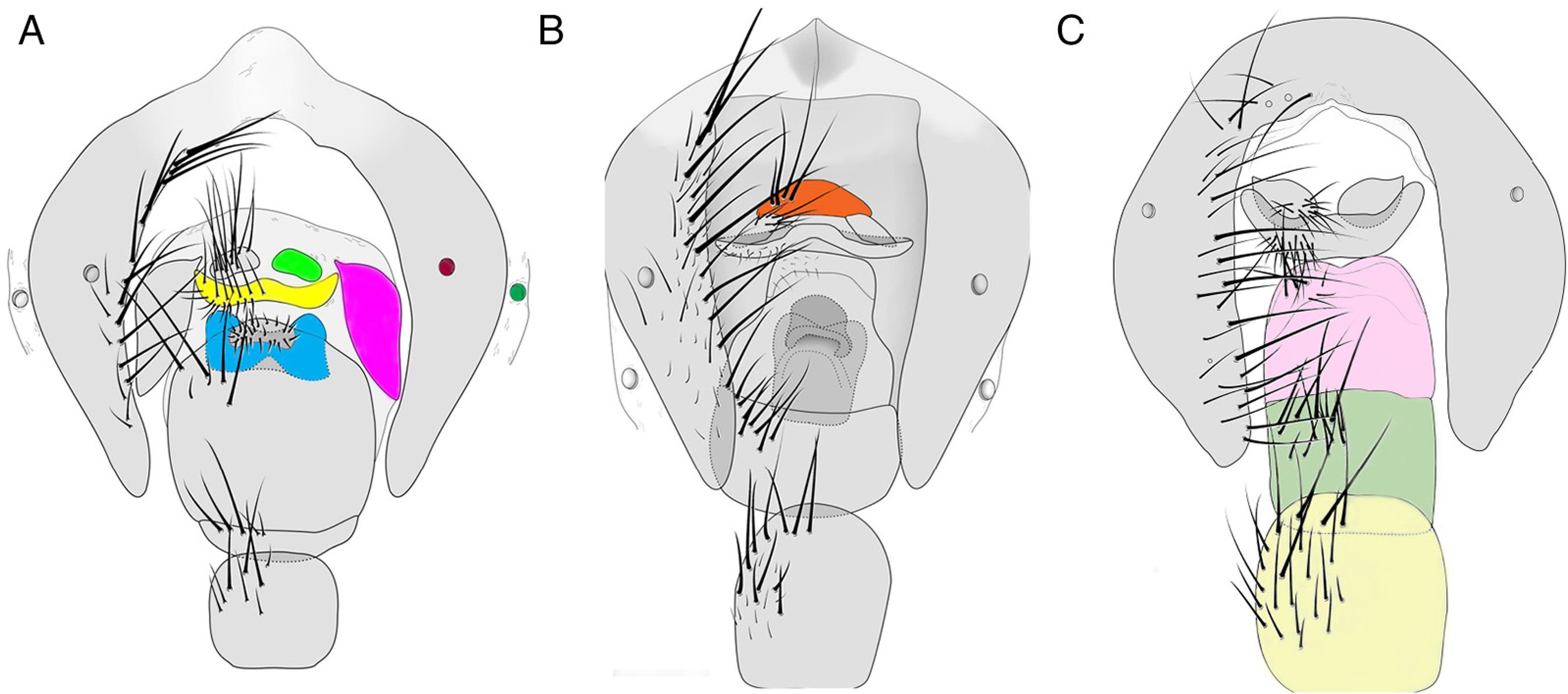
To start the colonies we collected specimens from Curitiba (Paraná), Campinas (São Paulo) and Bombas (Santa Catarina). Females were captured using a butterfly bait trap, which allows the researcher to choose flies in the field. All females were reared individually in small cages until larviposition, thus producing an isolineage. The larvae were reared in putrefied bovine meat until the emergence of adults. After the emergence, males were identified based on Vairo et al. (2011)Vairo, K.P., de Mello-Patiu, C.A., de Carvalho, C.J.B., 2011. Pictorial identification key for species of Sarcophagidae (Diptera) of potential forensic importance in southern Brazil. Rev. Bras. Entomol. 55, 333-347., thus ensuring the correct identification of females. Colonies were established and maintained at the Universidade Federal do Paraná, Centro Politécnico, Curitiba, Paraná, Brazil, except the colony of P. intermutans established at the Universidade Estadual de Campinas, Campinas, São Paulo, Brazil. The females were mounted and the abdomens removed and cleared in 10% potassium hydroxide, washed a few times in distilled water and immersed in 10% acetic acid. Photographs were taken with a Leica DFC 500 digital camera and Auto-Montage Pro Digital Imaging System (Syncropy), using a Leica MZ16 stereomicroscope. The illustrations were produced using drawing tube and edited with GIMP 2.8. Fig. 1 presents a general sketch of the female terminalia showing the main structures used in species identification. We adopted the terminology of Shewell (1987)Shewell, G.E., 1987. Sarcophagidae. In: McAlpine, J.F., Peterson, B.V., Shewell, G.E., Teskey, H.J., Vockeroth, J.R., Wood, D.M. (Eds.), Manual of Neartic Diptera, vol. 2. Monograph 108, Research Branch, Agriculture Canada, Otawa, pp. 1159–1186. for general morphology and Lopes (1939)Lopes, H.S., 1939. Contribuição ao conhecimento do gênero Helicobia Coquillett (Dipt. Sarcophagidae). Rev. Entomol. 10, 497-517. for “vaginal plate”. Synonymic information for each species is available in Pape (1996)Pape, T., 1996. A catalogue of Sarcophagidae of the world (Insecta: Diptera). Mem. Entomol. 8, Associated Publishers.. Updated distribution data after Pape (1996)Pape, T., 1996. A catalogue of Sarcophagidae of the world (Insecta: Diptera). Mem. Entomol. 8, Associated Publishers.are also provided (Barros et al., 2008Barros, R.M., de Mello-Patiu, C.A., Pujol-Luz, J.R., 2008. Sarcophagidae (Insecta: Diptera) associados à decomposição de carcaças de Sus scrofa em área de cerrado do Distrito Federal, Brasil. Rev. Bras. Entomol. 52, 606-609.; Barbosa et al., 2009Barbosa, R.R., de Mello-Patiu, C.A., Mello, R.P., Queiroz, M.M.C., 2009. New records of calyptrate dipterans (Fanniidae, Muscidae and Sarcophagidae) associated with the decomposition of domestic pigs in Brazil. Mem. Inst. Oswaldo Cruz 104, 923-926.; Rosa et al., 2009Rosa, T.A., Babata, M.L.Y., de Souza, C.M., de Souza, D., de Mello-Patiu, C.A., Mendes, J., 2009. Dípteros de Interesse Forense em Dois Perfis de Vegetação de Cerrado em Uberlândia, MG. Neotrop. Entomol. 38, 859-866.; Souza et al., 2011Souza, J.R.P., Esposito, M.C., Carvalho Filho, F.S., 2011. Composition, abundance and richness of Sarcophagidae (Diptera:Oestroidea) in forests and forest gaps with different vegetation cover. Neotrop. Entomol. 40, 20-27.; Vairo et al., 2011Vairo, K.P., de Mello-Patiu, C.A., de Carvalho, C.J.B., 2011. Pictorial identification key for species of Sarcophagidae (Diptera) of potential forensic importance in southern Brazil. Rev. Bras. Entomol. 55, 333-347., 2014Vairo, K.P., Ururahy-Rodrigues, A., Moura, M.O., de Mello-Patiu, C.A., 2014. Sarcophagidae (Diptera) with forensic potential in Amazonas: a pictorial key. Trop. Zool. 27, 140-152.; Buenaventura and Pape, 2013Buenaventura, E., Pape, T., 2013. Revision of the New World genus Peckia Robineau-Desvoidy (Diptera: Sarcophagidae). Zootaxa 3622, 1-87.). Vouchers are deposited in Coleção Entomológica Padre Jesus Santiago Moure, Universidade Federal do Paraná (DZUP) and Coleção Entomológica do Museu Nacional, Universidade Federal do Rio de Janeiro (MNRJ).
General morphology of female terminalia. (A) Oxysarcodexia paulistanensis (pink = tergite 8; green = cercus; yellow = hypoproct; blue = vaginal plate; dark red = spiracle 6; dark green = spiracle 7. (B) Peckia (Euboettcheria) florencioi (orange = epiproct). (C) Microcerella halli (light yellow = sternite 5; light green = sternite 6; light pink = sternites 7 + 8).
Results
The results are divided in an identification key that provides descriptions of each species with illustrations (Figs. 2–12).
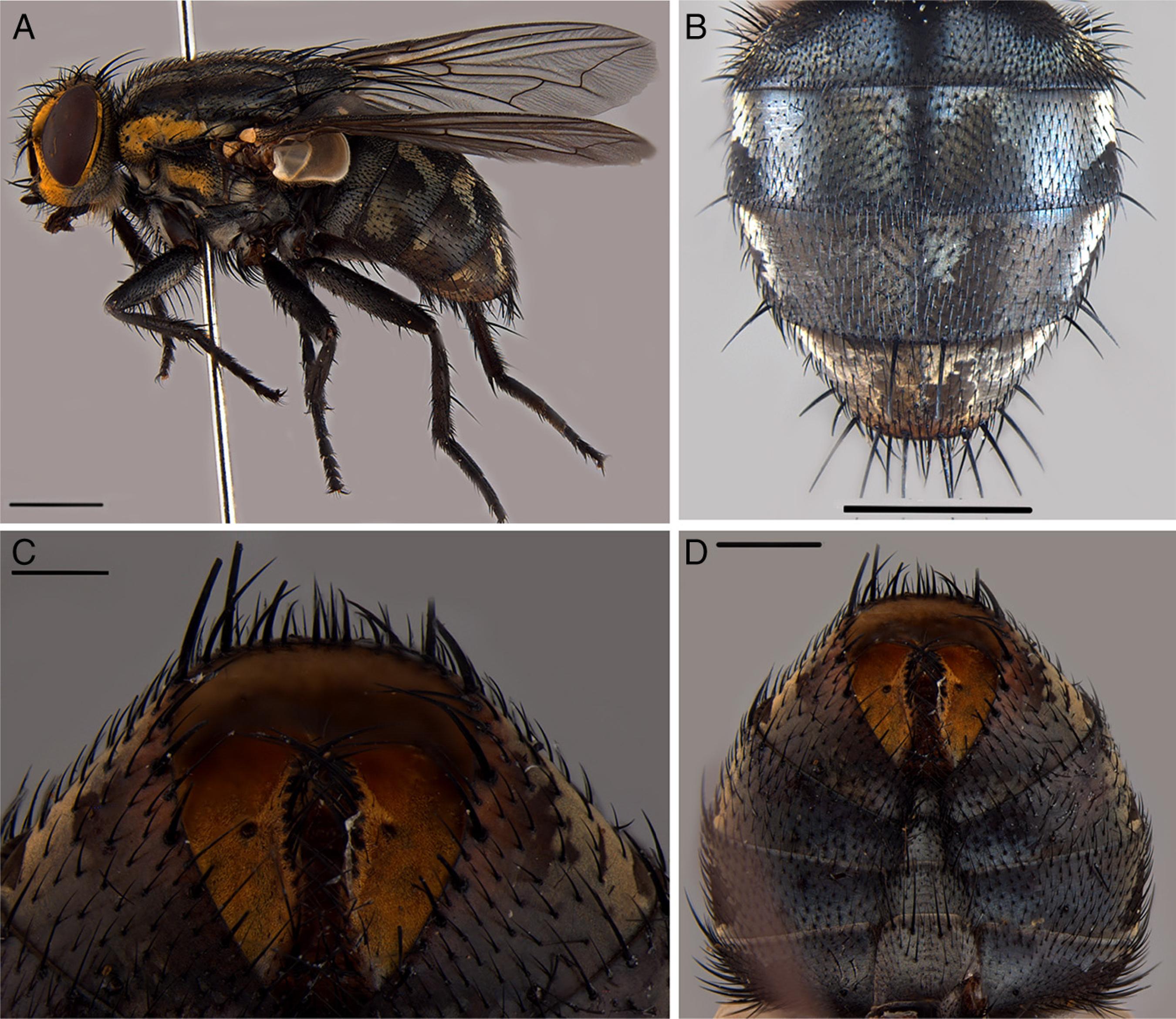
External female morphology of Oxysarcodexia paulistanensis. (A) Habitus, lateral view; scale: 2 mm; (B) abdomen, dorsal view; scale: 1 mm; (C) abdominal terminal segments, ventral view; scale: 0.5 mm; (D) abdomen, ventral view; scale: 1 mm.
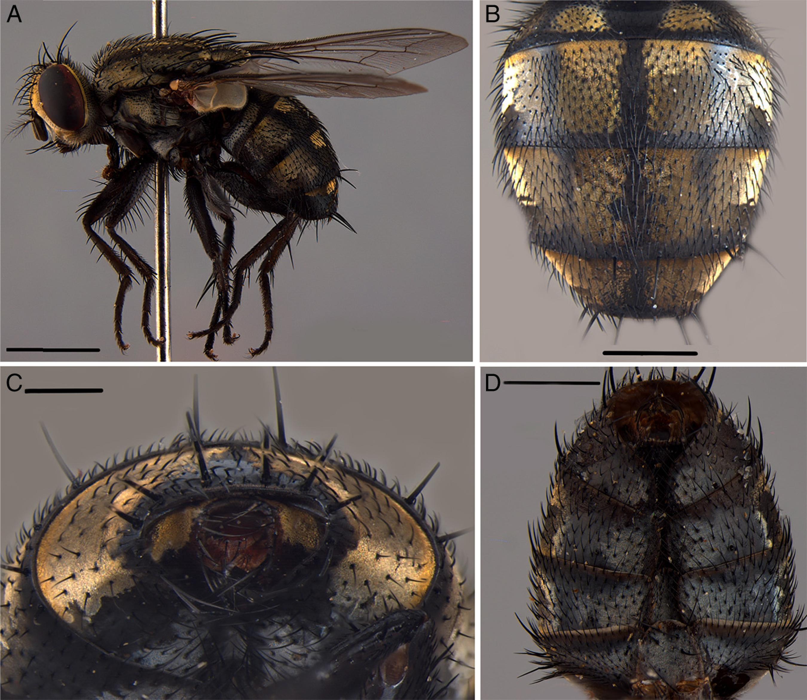
External female morphology of Oxysarcodexia riograndensis. (A) Habitus, lateral view; scale: 2 mm; (B) abdomen, dorsal view; scale: 1 mm; (C) abdominal terminal segments, ventral view; scale: 0.5 mm; (D) abdomen, ventral view; scale: 1 mm.
External female morphology of Peckia(Pattonella) intermutans. (A) Habitus, lateral view; scale: 1 mm; (B) abdomen, dorsal view; scale: 2 mm; (C) abdominal terminal segments, ventral view; scale: 1 mm; (D) abdomen, ventral view; scale: 2 mm.
External female morphology of Peckia(Pattonella) resona. (A) Habitus, lateral view; scale: 2 mm; (B) abdomen, dorsal view; scale: 2 mm; (C) abdominal terminal segments, ventral view; scale: 1 mm; (D) abdomen, ventral view; scale: 2 mm.
External female morphology of Peckia(Euboettcheria) australis. (A) Habitus, lateral view; scales: 2 mm; (B) abdomen, dorsal view; scale: 2 mm; (C) abdominal terminal segments, ventral view; scale: 0.5 mm; (D) abdomen, ventral view; scale: 1 mm.
External female morphology of Peckia(Euboettcheria) florencioi. (A) Habitus, lateral view; scale: 2 mm; (B) abdomen, dorsal view; scale: 1 mm; (C) abdominal terminal segments, ventral view; scale: 0.5 mm; (D) abdomen, ventral view, scale: 1 mm.
External female morphology of Peckia(Sarcodexia) lambens. (A) Habitus, lateral view; scale: 2 mm; (B) abdomen, dorsal view; scale: 1 mm; (C) abdominal terminal segments, ventral view; scale: 0.5 mm; (D) abdomen, ventral view; scale: 1 mm.
External female morphology of Microcerella halli. (A) Habitus, lateral view; scale: 2 mm; (B) abdomen, dorsal view; scale: 2 mm; (C) abdominal terminal segments, ventral view; scale: 1 mm; (D) abdomen, ventral view; scale: 1 mm.
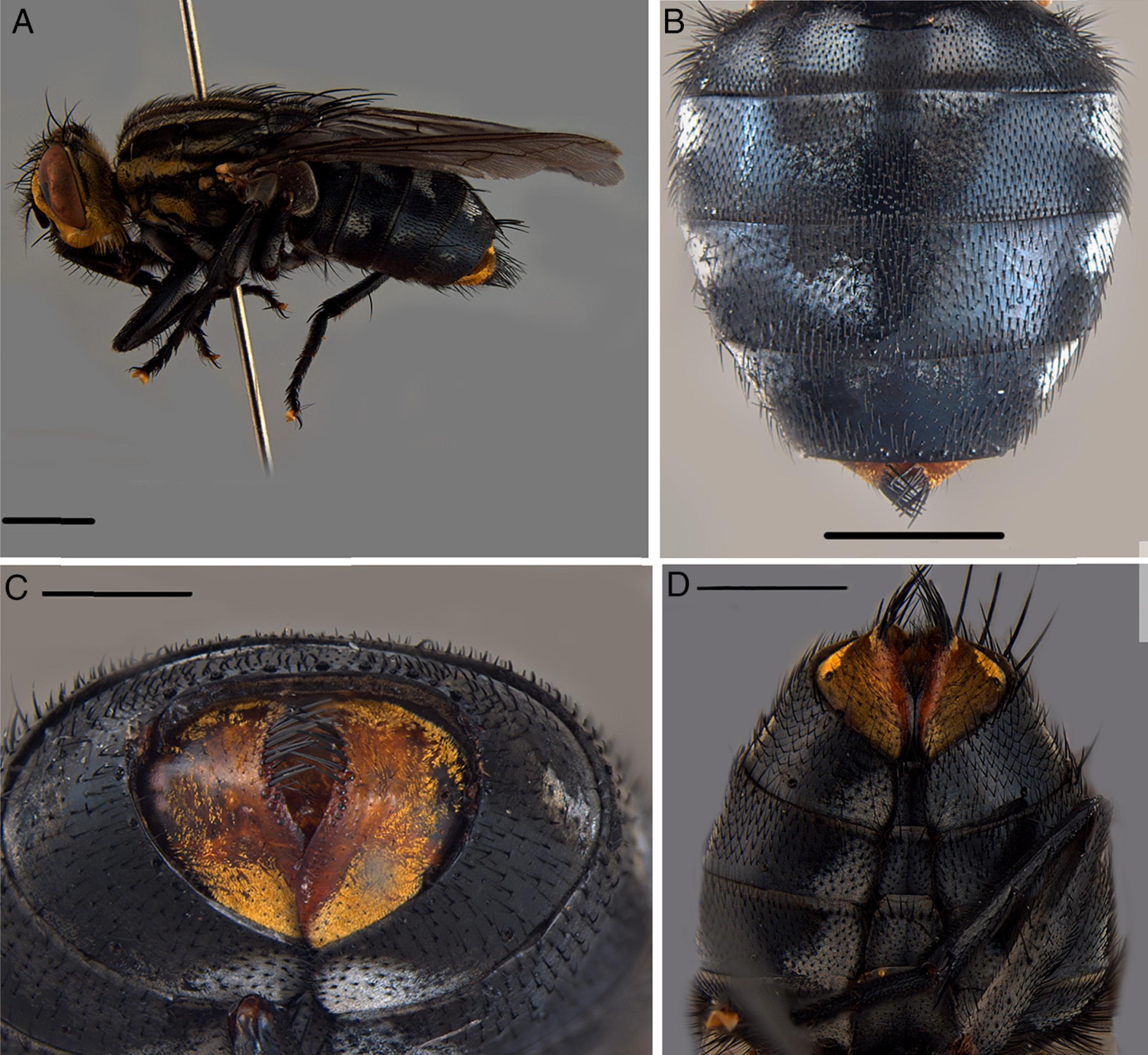
External female morphology of Sarcophaga(Bercaea) africa. (A) Habitus, lateral view; scale: 2 mm; (B) abdomen, dorsal view; scale: 2 mm; (C) abdominal terminal segments, ventral view; scale: 0.5 mm; (D) abdomen, ventral view; scale: 1 mm.
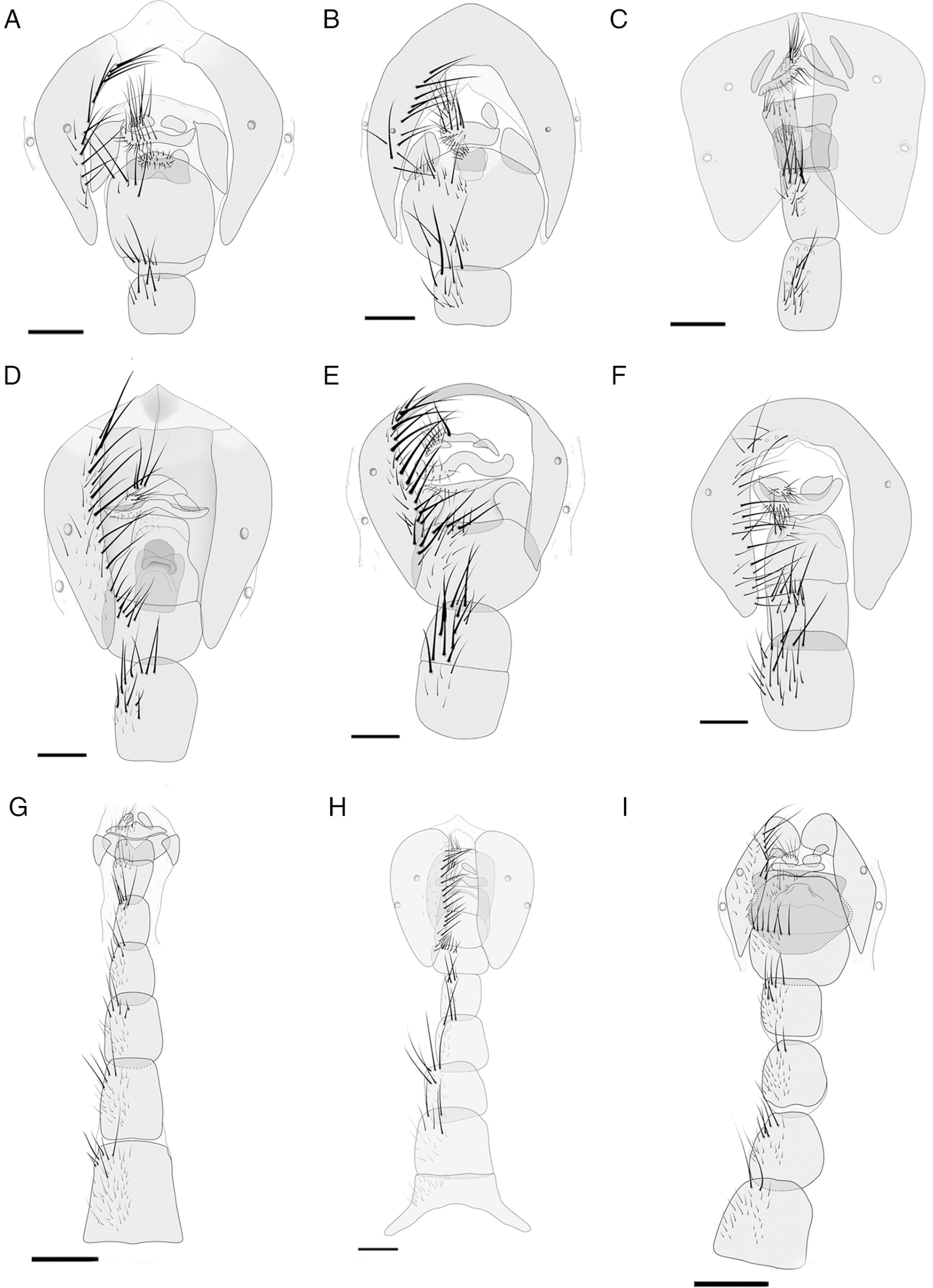
Female terminalia. (A) Oxysarcodexia paulistanensis(sternites 1–4 omitted); (B) Oxysarcodexia riograndensis(sternites 1–4 omitted); (C) Peckia(Pattonella) resona (sternites 1–4 omitted); (D) Peckia (Euboettcheria) florencioi (sternites 1–4 omitted); (E) Peckia (Sarcodexia) lambens (sternites 1–4 omitted); (F) Microcerella halli (sternites 1–4 omitted); (G) Peckia (Pattonella) intermutans (tergite 6 and sternite 1 omitted); (H) Peckia (Euboettcheria) australis; (I) Sarcophaga(Bercaea) africa (sternite 1 omitted). Scales: 1 mm.
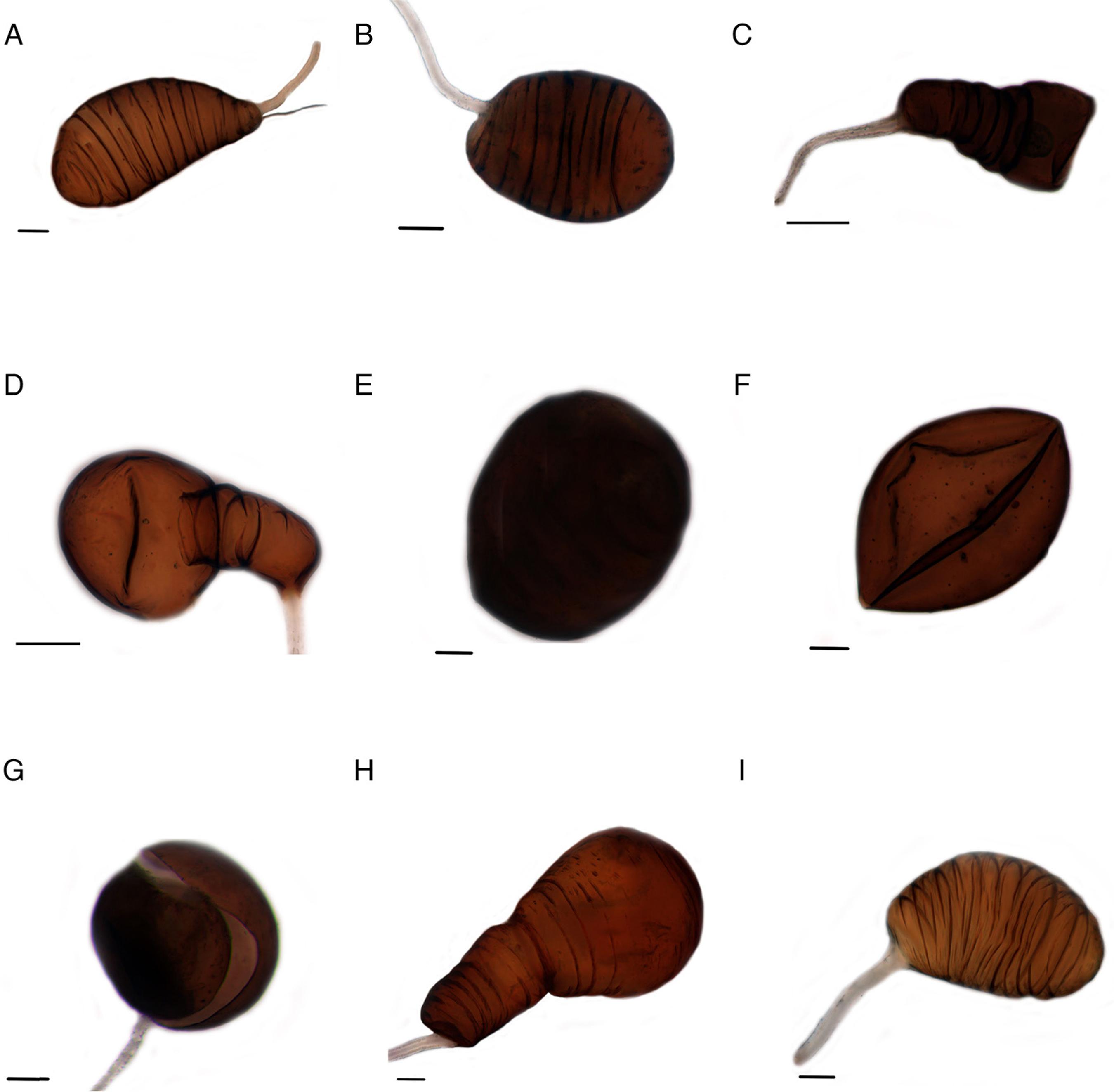
Spermathecae. (A) Oxysarcodexia paulistanensis, lateral view; scale: 0.05 mm; (B) Oxysarcodexia riograndensis, lateral view; scale: 0.05 mm; (C) Peckia(Pattonella) intermutans, lateral view; scale: 0.05 mm; (D) Peckia(Pattonella) resona, lateral view; scale: 0.05 mm; (E) Peckia (Euboettcheria) australis, lateral view; scale: 0.05 mm; (F) Peckia (Euboettcheria) florencioi, ventral view; scale: 0.05 mm; (G) Peckia (Sarcodexia) lambens, ventral view; scale: 0.05 mm; (H) Microcerella halli, lateral view; scale: 0.1 mm; (I) Sarcophaga (Bercaea) africa, lateral view; scale: 0.05 mm.
Key to female flesh flies with forensic importance in Southern Brazil.
-
1. Tergite 6 undivided. . . . . . . . . . . . . . . . . . . . . . . . . . . . . . . . . . . . . . . . . . . . . . . . . . . . . . . . . . . . . . .2
-
1'. Tergite 6 divided into two plates. . . . . . . . . . . . . . . . . . . . . . . . . . . . . . . . . . . . . . . . . . . . . . . .4
-
2. Mid tibia with long median anterior seta that extends beyond the apex of tibia; spermatheca rounded; epiproct present. . . . . . . . . . . . . . . . . . . . . . . . . . .P. (S.) lambens
-
2'. Mid tibia without a long median anterior seta that extends beyond the apex of tibia; spermatheca not rounded, with a different shape as above; epiproct absent. . . . . . . . . . . . . . . . . . . . . . . . . . . . . . . . . . . . . . . . . . . . . . . . . . . . . . . . . . . . . . . . . . . . . . . . . . .3
-
3. Tergite 8 well sclerotized, vaginal plate conspicuous . . . . . . . . . . . . O. riograndensis
-
3'. Tergite 8 absent, vaginal plate absent or completely membranous . . . . . . M. halli
-
4. Tergite 6 as two separated plates dorsally folded. . . . . . . . . . . . . . . . . . . . .S. (B.) africa
-
4'. Tergite 6 as two plates separated by a membrane or by a sclerotized area, notfolded dorsally . . . . . . . . . . . . . . . . . . . . . . . . . . . . . . . . . . . . . . . . . . . . . . . . . . . . . . . . . . . . 5
-
5. Vaginal plate well sclerotized, almost the same size as hypoproct, rectangular, with concave posterior margin and central area with a depression; tergite 6 as two plates separated by a sclerotized area . . . . . . . . . . . . . . . . . . . . . O. paulistanensis
-
5'. Vaginal plate absent or, if present, not as described above; tergite 6 as two plates separated by a membrane . . . . . . . . . . . . . . . . . . . . . . . . . . . . . . . . . . . . . . . . . . . . . 6
-
6. Spermatheca spherical, without striations and segmental constrictions; tergite 8 absent . . . . . . . . . . . . . . . . . . . . . . . . . . . . . . . . . . . . . . . . . . . . . . . . . . . . . . . . . . . . . . . . . . . . . 7
-
6'. Spermatheca with segmental constrictions, divided into proximal and distalpart; tergite 8 present . . . . . . . . . . . . . . . . . . . . . . . . . . . . . . . . . . . . . . . . . . . . . . . . . . . . . . 8
-
7. Vaginal plate absent . . . . . . . . . . . . . . . . . . . . . . . . . . . Peckia (Euboettcheria) australis
-
7'. Vaginal plate present, with a median finger-like projection . . . . P. (E.) florencioi
-
8. Tergite 8 wider than long; tergite 5 with two lateral golden spots . . . . . . . . . P. (P.)intermutans
-
8'. Tergite 8 longer than wide; tergite 5 with no lateral golden spots. . . . . . . . .P. (P.) resona
Oxysarcodexia paulistanensis (Mattos, 1919) (Figs. 2, 11A, and 12A)
Description – differs from male in the following: Two proclinate orbital setae, the superior one with half length of the inferior; inner vertical setae differentiated from postocellar setae. Tergite 5 with a dorsolateral light golden spot. Tergite 6 divided, the median region connecting the two plates are sclerotized; spiracle 6 in membrane and 7 within the sclerites. Six to eight strong marginal setae accompanied by thin setae. Tergite 7 absent. Tergite 8 as two lateral bare plates, relatively pigmented, centrally extended and tapered at the top and bottom, joined by a membrane. Epiproct absent. Sternites 2–6 rectangular with rounded corners with strong setae in the posterior margin and weak setae in the median part; sternite 6 shorter and wider on comparing to sternite 5; sternite 7 wider than 6 with 3 strong setae in each lateral and some setulae; sternites 6, 7 and 8 fused; sternite 8 broadly membranous with an small marginal sclerotized area with setulae. Vaginal plate present, well sclerotized, almost the same size as hypoproct, rectangular, with concave posterior margin and central area with a depression. Spermatheca elongated and slightly oval with transversal striations in all extension.
Distribution: Argentina (Buenos Aires, Córdoba, Entre Ríos), Brazil (Distrito Federal, Minas Gerais, Paraná, Rio de Janeiro, Rio Grande do Sul, São Paulo), Chile (Santiago).
Material examined: eight females from colonies initiated by specimens collected in Brazil, Paraná, Curitiba, ii. 2011. K. Vairo col.
Oxysarcodexia riograndensis (Lopes, 1946) (Figs. 3, 11B, and 12B)
Description – differs from male in the following: Two proclinate orbital setae, the superior one with similar size as frontals and the inferior one two times the size as the superior; inner vertical setae differentiated from the postocellar setae. Tergite 5 with a dorsolateral golden light spot. Tergite 6 undivided; spiracle 6 in membrane and spiracle 7 within the sclerite, with 6–9 strong marginal setae. Tergite 7 absent. Tergite 8 as two lateral sclerotized bare plates, two times the cercus size. Epiproct absent. Sternites 1–5 dark-brown, darker compared to the others; sternites 2 and 5 with square shape, posterior corners rounded, strong setae in the posterior margin and some setulae in the median part; sternite 5 shorter than 6; sternites 6, 7 and 8 fused; sternite 6 wider than 5 with one row of setae, 3 strong setae on each side and with many setulae in central part; sternite 7 almost 1.5 times the size of sternite 5, posterior margin concave, marginal setae being three strong lateral ones and other small weak setae; sternite 8 membranous with median area rounded and pigmented, margin with some setulae. Vaginal plate sub-rectangular, posterior margin slightly concave. Spermathecae slightly elongated with transversal striations in all extension.
Distribution: Argentina (Jujuy), Brazil (Paraná, Rio de Janeiro, Rio Grande do Sul).
Material examined: six females from colonies initiated by specimens collected in Brazil, Paraná, Curitiba, ii. 2011. K. Vairo col.
Peckia (Pattonella) intermutans(Walker, 1861) (Figs. 4, 11G, and 12C)
Description – differs from male in the following: two well-developed proclinate orbital setae; inner vertical setae differentiated from postocellar setae. Tergite 5 with one lateral golden spot and a light golden coloration at posterior margin in dorsal view. Tergite 6 divided into two big plates separated by a narrow membrane; spiracles 6 and 7 within the sclerite; 10–12 strong setae on posterior margin. Tergite 7 absent. Tergite 8 as two small bare plates, slightly larger than cercus. Epiproct absent. Sternites 2–5 square shaped with strong setae on posterior margin; sternites 6, 7 and 8 separated; sternite 6 square shaped, a bit smaller than sternite 5, with numerous strong marginal and premarginal setae; sternite 7 square with setae more concentrated on posterior margin, with a strong pair on each side; sternite 8 membranous, not well pigmented, about half of length of sternite 7, with 5 long setae. Vaginal plate membranous, slightly pigmented; anterior margin rounded and posterior margin with a median depression. Spermatheca elongated with a segmental constriction separating a narrower proximal part and a not striated distal part.
Distribution: Brazil (Amazonas, Ceará, Distrito Federal, Goiás, Mato Grosso, Minas Gerais, Pará, Rio de Janeiro, Paraná, Santa Catarina, São Paulo), Costa Rica, Ecuador, Guatemala, Guiana, Honduras, Mexico (Jalisco), Panama, Paraguay, Peru, St. Lúcia, Trinidad and Tobago (Tobago, Trinidad).
Material examined: nine females from colonies initiated by specimens collected in Brazil, São Paulo, Mogi Guaçu, iv. 2011. M. Grella col.
Peckia (Pattonella) resona (Lopes, 1935) (Figs. 5, 11C, and 12D)
Description – differs from male in the following: two proclinate well-developed orbital setae, both twice the size of frontal setae; inner vertical setae distinguish from the postocellar setae. Tergite 5 with an anterior silver spot in dorsal view. Tergite 6 divided into two big plates separated by a narrow membrane; spiracles 6 and 7 within the sclerite; 12 strong marginal setae concentrated in the median region. Tergite 7 absent. Tergite 8 as two small and narrow bare plates, a bit bigger than cercus. Epiproct absent. Sternites 2–6 squared shaped with strong and long setae on the posterior margin; sternites 6, 7 and 8 individualized; sternite 6 square, a bit smaller than sternite 5, with strong and long setae concentrated on the posterior third; sternite 7 with the half length of sternite 6, with long setae on the posterior half and strong posterior marginal setae; sternite 8 membranous; sparsely pigmented, with a similar length of sternite 7, with long and thin setae on posterior margin. Vaginal plate absent or probably completely membranous and not apparent. Spermatheca elongated with a segmental constriction separating a narrower proximal part, and a rounded not striated distal part.
Distribution: Argentina (Corrientes), Brazil (Rio de Janeiro, Rio Grande do Sul, Santa Catarina, Paraná, Minas Gerais, São Paulo).
Material examined: two females from colonies initiated by specimens collected in Brazil, Paraná, Curitiba, v. 2012. K. Vairo col.
Peckia (Euboettcheria) australis(Townsend, 1927) (Figs. 6, 11H, 12E)
Description – differs from male in the following: two proclinate orbital setae well developed, superior with half of the length of inferior; inner vertical setae differentiated of postocellar setae. Tergite 5 with a light golden microtomentum. Tergite 6 divided into two plates connected by a broad membrane; spiracle 6 in membrane and spiracle 7 within the sclerite, near the margin; 15–17 strong and long marginal setae. Tergites 7 and 8 not absent. Epiproct entire, narrow, with numerous setae on median region. Sternites 2–5 squared shaped with strong marginal setae; sternites 6 separated, 7 and 8 fused; sternite 6 larger than 5, but shorter in length, with strong marginal setae; sternite 7 with a depressed central area, sternite 8 represented by a narrow posterior membranous area with setulae, separated of the sternite 7 by a semicircular, swollen, and setose area. Vaginal plate absent. Spermatheca spherical not striated.
Distribution: Argentina (Misiones), Brazil (Mato Grosso, Rio Grande do Sul, Santa Catarina, Paraná, São Paulo), Paraguay.
Material examined: eight females from colonies initiated by specimens collected in Brazil, Paraná, Curitiba, vii. 2011. K. Vairo col.
Peckia (Euboettcheria) florencioi(Prado and Fonseca, 1932) (Figs. 7, 11D, and 12F)
Description – differs from male in the following: two proclinate orbital setae well developed; inner vertical setae differentiated of postocellar setae. Tergite 5 with light golden microtomentum in dorsal view. Tergite 6 divided into two plates with a broad connecting membrane; spiracle 6 in membrane and spiracle 7 within the sclerite near the margin; 12–15 strong and long marginal setae. Tergites 7 and 8 not absent. Epiproct entire, short, median region unpigmented, with strong and long setae. Sternites 6, 7 and 8 fused; sternite 7 with the same width as sternite 6, anteriorly rounded, without setae; sternite 8 narrower than sternite 7, posterior margin slightly swollen with sparse setulae. Vaginal plate present, well sclerotized, with a digitiform discal apophysis projecting inwards. Spermatheca spherical not striated, with a postero-ventral unsclerotized area.
Distribution: Argentina (Misiones, San Luis), Brazil (Mato Grosso, Rio Grande do Sul, Santa Catarina, Paraná, São Paulo).
Material examined: eight females from colonies initiated by specimens collected in Brazil, Paraná, Curitiba, vi. 2012. K. Vairo col.
Peckia (Sarcodexia) lambens(Wiedemann, 1830) (Figs. 8, 11E, and 12G)
Description – differs from male in the following: posterior femur without a patch of black short setae in the apical third of the anterior surface (male femoral organ). Tergite 5 with golden microtomentum in lateral and dorsal view. Tergite 6 undivided; spiracle 6 in membrane and 7 within the sclerite; 14–16 marginal strong setae accompanied by some setulae. Tergites 7 and 8 absent. Epiproct entire, with some fine setulae along the margin and one conspicuous strong setae on each side. Hypoproct broad with a conspicuous hollow at the medium part. Sternite 2 with 1.5 times the size of sternites 3 and 4; sternite 5 subrectangular with rounded corners and several developed setae; sternite 6 two times the sternite 5 width, with strong marginal setae and sparse discal setulae; sternites 7 and 8 narrower than sternite 6, both linked to the sternite 6 by a lateral conspicuous membranes; sternite 7 with no setae and sternite 8 broadly membranous, represented by a swollen and setulose marginal area. Vaginal plate absent. Spermatheca circular not striated with a posteroventral unsclerotized area.
Distribution: Argentina (Misiones, Tucumán), Bahamas (Grand Bahamas, New Providence), Bolivia, Brazil (Amazonas, Ceará, Mato Grosso, Rio de Janeiro, Santa Catarina, São Paulo, Paraná), Chile (Tarapacá), Colombia, Costa Rica, Cuba, El Salvador, Guyana, Haiti, Jamaica, Mexico (Jalisco, Nuevo Leon, Tamaulipas), Panamá, Paraguay, Peru, Puerto Rico, St. Vincent, Trinidad & Tobago (Tobago).
Material examined: seven females from colonies initiated by specimens collected in Brazil, Paraná, Curitiba, iv. 2011. K. Vairo col.
Microcerella halli (Engel, 1931) (Figs. 9, 11F, and 12H)
Description – differs from male in the following: two proclinate orbital setae well developed; no row of small and strong setae on anteroventral part of trochanter 3; tibia 2 with tree anterior setae and presence of a reddish sensorial area on posterior part of femur. Tergite 5 black with silver microtomentum. Tergite 6 undivided; reddish brown to orange, contrasting with the dark tergite 5; spiracle 6 in membrane and spiracle 7 within the sclerite; 20–24 strong marginal setae accompanied of small ones. Tergite 7, tergite 8 and epiproct absent. Sternites 1–5 reddish brown, darker than the others; sternites 2–6 squared shaped with a row of strong setae on posterior margin; sternites 6, 7 and 8 fused; sternite 6 wider and shorter than the sternite 5; sternite 7 quadrangular; central surface slightly depressed relative to the posterior margin, without setae; sternite 8 swollen, widely membranous except for the sclerotized posterior margin, posterior angles expanded with three apical setae each. Vaginal plate absent or probably completely membranous and not apparent. Spermatheca divided into two parts by a constriction, a narrow and cylindrical proximal part and a rounded distal one, less striated than the proximal and 2.0 times its width.
Distribution: Argentina (no further data), Bolivia, Brazil (Ceará, Minas Gerais, São Paulo, Paraná, Rio Grande do Sul).
Material examined: 10 females from colonies initiated by specimens collected in Brazil, Paraná, Curitiba, vi. 2011. K. Vairo col.
Sarcophaga (Bercaea) africa(Wiedemann, 1824) (Figs. 10, 11I, and 12I)
Description – differs from male in the following: two proclinate orbital setae well developed; inner vertical setae differentiated of postocellar setae. Tergite 5 with golden microtomentum more conspicuous in lateral view. Tergite 6 divided into two plates well separated and dorsally folded; spiracle 6 in membrane and spiracle 7 within the sclerite; 15–16 strong and long marginal setae. Tergites 7 and 8 absent. Epiproct represented by two small dorsal plates without setae. Sternites 2–4 squared shaped with posterior margin rounded; two strong setae in each angle of posterior margin; sternite 5 quadrangular with strong marginal angular setae. Sternites 6, 7 and 8 fused; Sternite 6 almost two times wider than sternite 5, with a medially interrupted row of setae on posterior margin; sternite 7 with a noticeably elevated central area; sternite 8 like a narrow and swollen range fused with the posterior margin of sternite 7, with two lateral groups of setae, two strongest setae and many setulae. Vaginal plate well sclerotized, darker than the sternites, and very long, from the hypoproct to the middle of sternite 6 with a median suture. Spermatheca oval and slightly elongated with transversal striations in all surfaces.
Distribution: Argentina (Buenos Aires), Brazil (Rio de Janeiro, Paraná, Rio Grande do Sul), Costa Rica, Cuba, Mexico, Paraguay.
Material examined: 10 females from colonies initiated by specimens collected in Brazil, Paraná, Curitiba, viii. 2012. K. Vairo col.
Discussion
Undoubtedly the main female diagnostic characters are in terminalia. However, in a forensic context, where, in most of the cases, fresh material is collected at the death scene, the external color could help to identify some species. Color of the gena, postgena, and of the spots in tergites and sternites, for instance, can be very effective in identifying some species, like S.(B.) africa and M. halli.
On the other hand, other external characters may also be useful, like the presence of long setae in tibia and the size of orbital and postocellar setae. In some cases, these external characters are the main differences between males and females of some species, requiring attention in the identification because of this dimorphism.
The characters from the terminalia, such as the microtomentum of tergite 5, can distinguish species even in the same subgenera, as showed in Pattonella. The tergite 6 could be divided or undivided. We considered as divided tergite 6 when it has even a narrow or large, pigmented or not, membrane connecting the two plates, as occurs in O. paulistanensis. Oxysarcodexia has the three already described states of tergite 6, entire, divided and membranous (Tibana and de Mello-Patiu, 1985Tibana, R., de Mello-Patiu, C.A., 1985. O sintergito 6 + 7 nas fêmeas de Oxysarcodexia Townsend, 1917 (Diptera, Sarcophagidae). Rev. Bras. Biol. 45, 439-445.). An undivided tergite 6, but with different degrees of reduction, also occurs in Nephochaetopteryx (de Mello-Patiu and Santos, 2001de Mello-Patiu, C.A., Santos, J.M., 2001. Nephochaetopteryx Townsend, 1934: descriptions and comparative morphological notes on the female terminalia (Diptera: Sarcophagidae). Stud. Dipterol. 8, 303-315.). In this work, species with tergite 6 undivided were O. riograndensis, P.(S.) lambens, and M. halli.
Concerning the spiracles 6 and 7, all studied species, except those of Peckia subgenus Pattonella, have the spiracle 6 inside the membrane and the spiracle 7 within the sclerite (tergite 6). Shewell (1987)Shewell, G.E., 1987. Sarcophagidae. In: McAlpine, J.F., Peterson, B.V., Shewell, G.E., Teskey, H.J., Vockeroth, J.R., Wood, D.M. (Eds.), Manual of Neartic Diptera, vol. 2. Monograph 108, Research Branch, Agriculture Canada, Otawa, pp. 1159–1186. considered that the tergite 7 in Sarcophagidae is frequently absent and the tergite 8 is nearly always present, but usually reduced to bare lateral plates. In this work, we used the same interpretation and named as tergite 8 the bare plates in lateral position to the sternites 7 and 8. This sclerite was visible only in Oxysarcodexiaand in Peckia (Pattonella) and its presence and shape showed to be an important character to discriminate some females of forensic species in Southern Brazil.
The epiproct, if present, can appear divided and undivided (Camargo, 2014Camargo, S., (Dissertação de mestrado) 2014. Descrição e notas taxonômicas comparativas das terminálias femininas de espécies de Peckia Robineau-Desvoidy, 1830 (Diptera, Sarcophagidae) da Amazônia Brasileira. Universidade Federal do Pará. Museu Paraense Emílio Goeldi.). In this work only P.(S.) lambens, P.(E.) australis, P.(E.) florencioi, and S.(B.) africa have epiproct, entire in the first three species and divided in the last one. The undivided epiproct seems to be the most common state of this character in Peckia, but this condition differs in some species of the same genera or subgenera such as P.(E.) collusor and P.(E.) epimelia (Camargo, 2014Camargo, S., (Dissertação de mestrado) 2014. Descrição e notas taxonômicas comparativas das terminálias femininas de espécies de Peckia Robineau-Desvoidy, 1830 (Diptera, Sarcophagidae) da Amazônia Brasileira. Universidade Federal do Pará. Museu Paraense Emílio Goeldi.).
Although the shape of sternites can be a useful character in a general context, important information to distinguish sarcophagid females comes from the presence (or absence) of fusion of the sternites 6, 7 and 8. These sternites may be considered fused when the posterior margin of preceding sternite is contiguous, main laterally, with the anterior margin of the subsequent one, without a well-marked suture. In O. paulistanensis, O. riograndensis, and S. (B.) africa these sternites are fused; in P.(E.) australis, P. (E.) florencioi and M. halli only sternites 7 and 8 are fused; and in P. (P.) resona, P. (P.) intermutans and P. (S.) lambens all sternites are individualized.
Another key character to identify females of these flesh fly species is the presence and shape of vaginal plate. For some genera like Oxysarcodexia and Nephochaetopteryx the vaginal plate is one of the most important characters to segregate species because it has conspicuous interspecific differences (Tibana and Mello, 1983Tibana, R., Mello, C.A., 1983. Estudo sobre as fêmeas de Oxysarcodexia do grupo Peltata (Diptera: Sarcophagidae). Rev. Bras. Biol. 43, 241-250.; de Mello-Patiu and Santos, 2001de Mello-Patiu, C.A., Santos, J.M., 2001. Nephochaetopteryx Townsend, 1934: descriptions and comparative morphological notes on the female terminalia (Diptera: Sarcophagidae). Stud. Dipterol. 8, 303-315.). As previously stated, we found that the shape of vaginal plate is a major character to properly identify O. paulistanensis, O. riograndensis, P. (E.) florencioi, P. (P.) resona and S. (B.) africa.
The morphology of the spermathecae also can help the differentiation of genera and subgenera. In Oxysarcodexia the shape is more elongate (pyriform) and the striations are conspicuous, similar as in Sarcophaga. In P. (S.) lambens and P. (E.) florencioi while also rounded it has an opening in ventral view, a characteristic that we are describing for the first time. In Peckia (Pattonella) and Microcerella, the spermathecae are quite different, as it is divided into well-defined distal and proximal portions, possessing some constrictions along.
Although Sarcophagidae, in general, and its females, in particular, are considered hard to identify, the key and the descriptions provided makes this task possible to both, taxonomists and non-taxonomists. So, we expect that forensic entomologists can identify the necrophagous female flesh flies in Southern Brazil in a short time and with low cost, broadening the number of species that can be used in crime scene investigations.
Acknowledgments
We thank TaxonLine – Rede Paranaense de Coleções Biológicas – for the photographs in this work. Funding was provided by Conselho Nacional de Desenvolvimento Científico e Tecnológico (CNPq): Ph.D. scholarship 141487/2011-9 (K.P.V.); research grant 302584/2012-9 (C.A.M.P.) and 307947/2009-2 (M.O.M.) and Fundação Araucaria research grant 686/2014 (M.O.M.).
References
- Barbosa, R.R., de Mello-Patiu, C.A., Mello, R.P., Queiroz, M.M.C., 2009. New records of calyptrate dipterans (Fanniidae, Muscidae and Sarcophagidae) associated with the decomposition of domestic pigs in Brazil. Mem. Inst. Oswaldo Cruz 104, 923-926.
- Barros, R.M., de Mello-Patiu, C.A., Pujol-Luz, J.R., 2008. Sarcophagidae (Insecta: Diptera) associados à decomposição de carcaças de Sus scrofa em área de cerrado do Distrito Federal, Brasil. Rev. Bras. Entomol. 52, 606-609.
- Buenaventura, E., Pape, T., 2013. Revision of the New World genus Peckia Robineau-Desvoidy (Diptera: Sarcophagidae). Zootaxa 3622, 1-87.
- Camargo, S., (Dissertação de mestrado) 2014. Descrição e notas taxonômicas comparativas das terminálias femininas de espécies de Peckia Robineau-Desvoidy, 1830 (Diptera, Sarcophagidae) da Amazônia Brasileira. Universidade Federal do Pará. Museu Paraense Emílio Goeldi.
- Carvalho, L.M.L., Linhares, A.X., 2001. Seasonality of insect succession and pig carcass decomposition in a natural Forest area in Southeastern Brazil. J. Forensic Sci. 46, 604-608.
- Corrêa, R.C., Moura, M.O., Almeida, L.M., 2014. Coleoptera associated with buried carrion: potential forensic importance and seasonal composition. J. Med. Entomol. 51, 1057-1066.
- de Carvalho, C.J.B., Mello-Patiu, C.A., 2008. Key to the adults of the most common forensic species of Diptera in South America. Rev. Bras. Entomol. 52, 390-406.
- Lopes, H.S., 1939. Contribuição ao conhecimento do gênero Helicobia Coquillett (Dipt. Sarcophagidae). Rev. Entomol. 10, 497-517.
- Lopes, H.S., 1941. Sôbre o aparelho Genital Feminino dos Sarcophagidae e sua importância na classificação (Diptera). Rev. Bras. Biol. 1, 215-221.
- Lopes, H.S., 1958. Considerações sobre as espécies de Peckia Desvoidy, 1830 e de gêneros afins (Diptera, Sarcophagidae). An. Acad. Bras. Ciênc. 30, 211-243.
- de Mello-Patiu, C.A., Santos, J.M., 2001. Nephochaetopteryx Townsend, 1934: descriptions and comparative morphological notes on the female terminalia (Diptera: Sarcophagidae). Stud. Dipterol. 8, 303-315.
- Moura, M.O., de Carvalho, C.J.B., Monteiro-Filho, E.L.A., 1997. A preliminary analysis of insects of medico-legal importance in Curitiba, State of Paraná. Mem. Inst. Oswaldo Cruz 92, 269-274.
- Moura, M.O., de Carvalho, C.J.B., Monteiro-Filho, E.L.A., 1998. Carrion attendant arthropods in southern Brazil. Cienc. Cult. 50, 377-381.
- Moura, M.O., de Carvalho, C.J.B., Monteiro-Filho, E.L.A., 2005. Estrutura de Comunidades necrófagas: efeito da partilha de recursos na diversidade. Rev. Bras. Zool. 22, 1134-1140.
- Mulieri, P.R., Mariluis, J.C., Patitucci, L.D., 2010. Review of the Sarcophaginae (Diptera: Sarcophagidae) of Buenos Aires Province (Argentina), with a key and description of a new species. Zootaxa 2575, 1-37.
- Oliveira, T.C., Vasconcelos, S.D., 2010. Insects (Diptera) associated with cadavers at the Institute of Legal Medicine in Pernambuco, Brazil: implications for forensic entomology. Forensic Sci. Int. 198, 97-102.
- Pape, T., 1996. A catalogue of Sarcophagidae of the world (Insecta: Diptera). Mem. Entomol. 8, Associated Publishers.
- Rosa, T.A., Babata, M.L.Y., de Souza, C.M., de Souza, D., de Mello-Patiu, C.A., Mendes, J., 2009. Dípteros de Interesse Forense em Dois Perfis de Vegetação de Cerrado em Uberlândia, MG. Neotrop. Entomol. 38, 859-866.
- Salviano, R.J.B., 1996. Sucessão de Diptera Caliptrata em carcaça de Sus scrofa L. Dissertação de Mestrado. Universidade Federal Rural do Rio de Janeiro, Rio de Janeiro.
- Shewell, G.E., 1987. Sarcophagidae. In: McAlpine, J.F., Peterson, B.V., Shewell, G.E., Teskey, H.J., Vockeroth, J.R., Wood, D.M. (Eds.), Manual of Neartic Diptera, vol. 2. Monograph 108, Research Branch, Agriculture Canada, Otawa, pp. 1159–1186.
- Souza, J.R.P., Esposito, M.C., Carvalho Filho, F.S., 2011. Composition, abundance and richness of Sarcophagidae (Diptera:Oestroidea) in forests and forest gaps with different vegetation cover. Neotrop. Entomol. 40, 20-27.
- Tibana, R., de Mello-Patiu, C.A., 1985. O sintergito 6 + 7 nas fêmeas de Oxysarcodexia Townsend, 1917 (Diptera, Sarcophagidae). Rev. Bras. Biol. 45, 439-445.
- Tibana, R., Mello, C.A., 1983. Estudo sobre as fêmeas de Oxysarcodexia do grupo Peltata (Diptera: Sarcophagidae). Rev. Bras. Biol. 43, 241-250.
- Tomberlin, J.K., Mohr, R., Benhow, M.E., Tarone, A.M., VanLaerhoven, S., 2011. A roadmap for bridging basic and applied research in forensic entomology. Annu. Rev. Entomol. 56, 401-421.
- Vairo, K.P., de Mello-Patiu, C.A., de Carvalho, C.J.B., 2011. Pictorial identification key for species of Sarcophagidae (Diptera) of potential forensic importance in southern Brazil. Rev. Bras. Entomol. 55, 333-347.
- Vairo, K.P., Ururahy-Rodrigues, A., Moura, M.O., de Mello-Patiu, C.A., 2014. Sarcophagidae (Diptera) with forensic potential in Amazonas: a pictorial key. Trop. Zool. 27, 140-152.
- Vairo, K.P., Corrêa, R.C., Lecheta, M.C., Caneparo, M.F., Mise, K.M., de Carvalho, C.J.B., Almeida, L.M., Moura, M.O., 2015. Forensic use of a subtropical blowfly: the first case indicating minimum post-mortem interval (mPMI) in Southern Brazil and first record of Sarconesia chlorogaster from a human corpse. J. Forensic Sci. 1, 257-260.
Publication Dates
-
Publication in this collection
Jul-Sep 2015
History
-
Received
01 Feb 2015 -
Accepted
04 May 2015