
Abstract
Coconut palm (Cocos nucifera L.) is one of the most important tropical species used by man, known as “tree of life”. In recent decades, with the expansion of coconut growing areas, limitations such as the occurrence of pests, uniformity of crops, adaptation to different ecosystems and others have affected the production and longevity of coconut trees. This review describes the major advances in propagation techniques of coconut palm, conventionally, by seeds and through plant tissue culture techniques from the mid-twentieth century onwards, as well as phytosanitary aspects that should be considered to mitigate the spread of pests and diseases.
Index terms
Cocos nucifera; seedling; micropropagation and diseases
Resumo
O coqueiro (Cocos nucifera L.) é uma das mais importantes espécies tropicais utilizadas pelo homem, conhecida como “árvore da vida”. Nas últimas décadas, com a expansão das áreas de cultivo, limitações como a ocorrência de pragas, uniformidade de culturas, adaptação a diferentes ecossistemas e outras têm afetado a produção e a longevidade dos plantios. Esta revisão descreverá os principais avanços nas técnicas de propagação do coqueiro, convencionalmente, por sementes e por meio de técnicas de cultura de tecidos de plantas a partir de meados do século XX, bem como aspectos fitossanitários que devem ser considerados para mitigar a disseminação de pragas e doenças.
Termos para indexação
Cocos nucifera; mudas; micropropagação e doenças
Introduction
Coconut palm (Cocos nucifera L.) is considered one of the most important tropical species used by man, known as the “tree of life”, as it allows the elaboration of more than 100 products and byproducts, being found in all tropical regions of the globe.
According to reports by botanist Huge Harries, the coconut palm was originated in the ancient supercontinent of Gondwana. Coconut fruits floated in the prehistoric Tethys Seas with natural distribution on the coasts and islands of the Indian Ocean, atolls of the mid-Pacific ocean, India, China and Eastern Africa (BOURDEIX et al., 2005 BOURDEIX, R.; KONAN, J.L.; N’CHO, Y.O. Coconut: a guide to traditional and improved varieties. Barcelona: Editions Diversiflora, 2005. 104 p. ). In the 16th century, after the discovery of the Cape of Good Hope, Portuguese and Spanish distributed coconut seedlings in Western Africa and then in the Americas and the entire tropical region of the globe. In the Caribbean, the first introduction from Cape Verde dates back to 1549, with planting in Puerto Rico. Between 1571 and 1816, coconut fruits from Philippines were introduced in Mexico and across the Pacific coast of America down to Peru (BOURDEIX et al., 2005 BOURDEIX, R.; KONAN, J.L.; N’CHO, Y.O. Coconut: a guide to traditional and improved varieties. Barcelona: Editions Diversiflora, 2005. 104 p. ). In the 1950s, it arrived in Brazil through the State of Bahia, with fruits from the Island of Cape Verde (ARAGÃO et al., 2007 ARAGÃO, W.M. Cultivares de coqueiros. In: FONTES, H.R.; FERREIRA, J.M.S.; SIQUEIRA, L.A. A cultura do coqueiro. Aracaju: Embrapa Tabuleiros Costeiros, 2007. (Sistemas de Produção, 1) ). This species is widespread in almost all continents, being found between parallels 23°N and 23°S in more than 200 countries (FOALE and HARRIES, 2009 FOALE, M.; HARRIES, H. Farm and Forestry Production and Marketing Profile for Coconut (Cocos nucifera). In: ELEVITCH, C. R. (ed.). Specialty crops for pacific island agroforestry. Holualoa: Permanent Agriculture Resources. 2009. Disponível em: http://agroforestry.net/scps. Acesso em: 4 de setembro de 2017. ).
Data provided by FAO estimate coconut cultivation area of 12.3 million hectares in 2017, reaching production of 60.7 million tons of coconut fruits in 92 countries, with Philippines, Indonesia and India accounting for 72.7% of area. Brazil ranks sixth, with planted area of 215,683 hectares and production of around 2.4 million tons of fruit per year (FAO, 2017 FAO- Food and Agriculture Organization of the United Nations. FAOSTAT Beta. 2017. Disponível em: http://fenix.fao.org/faostat/beta/en/#data/QC. Acesso em: 17 jul. 2017.
http://fenix.fao.org/faostat/beta/en/#da...
). The Northeastern region accounts for 74% of the national production, which is concentrated in the states of Bahia, Sergipe, Rio Grande do Norte and Pernambuco (IBGE, 2017 IBGE - Instituto Brasileiro de Geografia e Estatística. Produção e área plantada de lavouras permanentes. Sistema IBGE de recuperação automática, 2015. Disponível em: http://www.sidra.ibge.gov.br/bda/tabela/protabl.asp?c=1613ez=teo=11ei=P. Acesso em: 04 out. 2016.
http://www.sidra.ibge.gov.br/bda/tabela/...
).
Commercial exploitation in most countries that cultivate this palm is focused on the production of fruits to obtain oil and dry coconut dehydrated from copra. In Brazil, it is cultivated with the purpose of producing fruits destined to the production of grated coconut and coconut milk, in addition to coconut water (MARTINS; JESUS JUNIOR, 2013 MARTINS, C.R.; JESUS JUNIOR, L.A. Produção e comercialização de coco no Brasil frente ao comércio internacional: panorama 2014. Aracaju: Embrapa Tabuleiros Costeiros, 2013. 51p. (Documentos, 184) ).
Considering its botanical characteristics, coconut palm is divided into two groups: tall and dwarf. Tall group varieties are more heterogeneous due to crosspollination and destined to the production of copra, while more homogeneous dwarf varieties, due to their lower allogamy, are destined to the production of coconut water (BOURDEIX et al., 2005 BOURDEIX, R.; KONAN, J.L.; N’CHO, Y.O. Coconut: a guide to traditional and improved varieties. Barcelona: Editions Diversiflora, 2005. 104 p. ). The main varieties used in the world, around 34, are divided into these two groups, in addition to hybrids, resulting from crosses between the dwarf and tall varieties, which have dual purpose, for both fresh consumption and industrial processing.
The production of coconut hybrids has boosted the installation of seedling production areas in several regions of the world. In India, for example, there are around 200 ha with 23,000 mother plants for the production of more than 2 million hybrid coconut seedlings (FAO, 2017 FAO- Food and Agriculture Organization of the United Nations. FAOSTAT Beta. 2017. Disponível em: http://fenix.fao.org/faostat/beta/en/#data/QC. Acesso em: 17 jul. 2017.
http://fenix.fao.org/faostat/beta/en/#da...
).
Despite the restrictions in some countries on the entry of coconut seeds due to the occurrence of quarantine pests, sexual propagation by seeds continues to be the main form of multiplication of this species.
In Brazil, there are 10 registered coconut genotypes, six from the dwarf group, highlighting the Brazilian green dwarf coconut, widely cultivated in the country, one from the tall group and three hybrids developed by from the Brazilian Agricultural Research Company (EMBRAPA) BRS 001, BRS 002 and BRS 003 (MINISTÉRIO DA AGRICULTURA, 2017 MINISTÉRIO DA AGRICULTURA. CultivarWEB. Registro nacional de cultivares. Brasília, DF, 2016. Disponível em: http://extranet.agricultura.gov.br/php/snpc/cultivarweb/cultivares_registradas.php. Acesso em: 04 out. 2016.
http://extranet.agricultura.gov.br/php/s...
).
In the last decades, with the expansion of coconut areas, limitations inherent to the occurrence of pests, mainly lethal yellowing and cadang-cadang diseases, unevenness of crops, adaptation to different ecosystems, among others, have affected the production and longevity of coconut trees. Coconut propagation by seeds has not met the demands for replanting of areas affected by diseases (SOLÍS-RAMOS et al., 2012 SOLÍS-RAMOS, L.Y.; ANDRADE-TORRES, A.; CARBONELL, L.A.S.; SALÍN, C.M. O.; DE LA SERNA, E.C. Somatic embryogenesis in recalcitrant plants. In: KEN-ICHI SATO, K-I. Embryogenesis. Rijeka: Intech, p.598-618, 2012. ; HARRISON; OROPEZA, 2011 HARRISON, N.A.; OROPEZA, C. Coconut lethal yellowing. In: HARRISON, N.A.; RAO, G.P.; MARCONE, C. Characterization, diagnosis, and management of phytoplasmas. Houston: Studium Press LLC. 2008, p.219-248. ).
Considering several production factors, the genetic and phytosanitary quality of seedlings has been imperative for the sustainability of the coconut agribusiness.
The present review focuses on the major advances in propagation techniques of coconut palm by seeds and plant tissue culture from the mid-twentieth century onwards, as well as phytosanitary aspects that should be considered to mitigate the spread of pests and diseases.
Characterization of coconut seeds-The seed, surrounded by the endocarp, consists of a thin layer of brown color - the integument - located between the endocarp and the solid albumen (Figure 1).
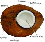
The latter is a fleshy, white and very oily layer, forming a large cavity containing an opalescent liquid or liquid albumen. In the distal part of the seed, where the fruit attaches to the bunch, there are three germination pores and the embryo is under one of them, surrounded by the solid albumen.
Branched vascular tubes are observed on the inner wall of the endocarp, which are responsible for conducting elaborated sap through the peduncle, which feeds the albumen. Moisture also penetrates by these tubes during seed germination (BONDAR, 1955 BONDAR, G. A cultura do coqueiro (Cocos nucifera) no Brasil. Salvador: Tipografia Naval, 1955. 91p. ).
At the beginning of fruit development until the fifth or seventh month after fertilization of the female flower, the central cavity and the liquid endosperm develop. From this age, there are depositions on the walls of this cavity, governed by biochemical processes that incorporate the liquid endosperm to the solid endosperm. The solid endosperm (albumen) initially forms in the polar region, opposite to the point of union of the fruit to the bunch, progressively extending throughout the cavity, reaching the greatest thickness at 12 months of age, when the fruit completes maturation. At this stage, the water volume of the fruit is lower than at six months of maturation and the bark acquires dark coloration.
After fruit maturation, seed germination begins, when the embryo undergoes dilation, producing a whitish mass (haustorium), which around five months occupies the entire internal seed cavity (FRÉMOND et al., 1975 FRÉMOND, Y.; ZILLER, R.; NUCÉ DE LAMOTHE, M. El cocotero: técnicas agrícolas y producciones tropicales. Barcelona: Editorial Blume, 1975. 236p. ), digesting the albumen to nourish the developing seedling.
Evolution of coconut seedling production systems by seed-The planting of the tall variety has been characterized over the years by the use of poor-quality seedlings, without selection criteria such as genetic material, phytosanitary status and vigor. Under this condition, plantations were mostly developed with etiolated seedlings, often derived from fallen seeds germinated in the field or, in many cases, stored in the shade where seeds were collected. This situation can still be observed in some cases, which in a way, causes an increase in the replanting index, especially when seedlings find adverse conditions in the field, such as the occurrence of water deficit, inadequate preparation, nonuse of planting pits and absence of cultural treatment.
Along these lines, plantation heterogeneity and reduction of precocity of plant production is promoted, consequently generating losses to the farmers.
From the 1980s, with the introduction of intervarietal hybrids (tall x dwarf genotypes) and later with the use of the dwarf coconut variety, seedling production systems were developed by adopting two stages: seed bed and nursery. In these systems, seeds are distributed next to each other, covered with soil up to 2/3 of its length, which allows seed placement either in the horizontal or vertical position to germinate; the latter being the most used. When the seedlings reach an average of 0.20 m in height, they are transplanted from the seed bed to the nursery stage, which can be installed by the bare-root system, where seedlings are directly planted on the soil or into polyethylene bags with 0.2 mm in thickness and dimensions of 40 x 40 cm to 60 x 60 cm. Nursery planting should be performed in an equilateral triangle shape with sides of either 60 or 80 cm, corresponding to 31,944 and 17,968 seedlings ha-1, respectively. Considering an average time of four months for seeds to germinate and seedlings to reach an average of 0.20 m in height and six to nine months in nursery, seedlings can be obtained with approximately one year of age, presenting five to seven live leaves, 1.0 to 1.2 m in height and 15 to 18 cm in (FONTES et al., 1998 FONTES, H.R.; CINTRA, F.L.D.; CARVALHO FILHO, O.M. Implantação e manejo da cultura do coqueiro. In: FERREIRA, M.J.S.; WARWICK, D.R.N.; SIQUEIRA, L.A. (Eds) A cultura do coqueiro no Brasil. Brasília, DF: Embrapa-SPI; Aracaju: Embrapa-CPATC, 1998. p.99-128. ).
Studies were carried out by Embrapa Tabuleiros Costeiros comparing coconut development one year after planting from seedlings produced in bare-root system with 4 and 7 months of nursery in presence and absence of irrigation. In conclusion, no differences in the development of non-irrigated seedlings at the two ages tested were observed, thus demonstrating greater capacity of development of younger seedlings because they have lower leaf area for transpiration and larger reserve in the endosperm.
With the expansion of dwarf variety cultivation areas and consequent need for selected seedlings to meet the coconut water market demand, a significant increase in the use of seedlings directly transplanted from the seed bed place directly to the field was observed. Although this type of seedling has been widely used by farmers, problems related to lower selection rigor due to the transplantation of very young seedlings into the field have been identified.
Based on these observations and previous results, and considering the advantages of using younger seedlings, an alternative production system with a lower sowing density was developed by Fontes and Leal (1998) FONTES, H.R.; LEAL, M. de L. da S. Utilização de sistema alternativo na produção de mudas de coqueiros híbridos (Cocos nucifera L). Revista Brasileira de Fruticultura, Jaboticabal, v.20, n.3, p.290-296, 1998. .
This system allows the transplantation of seedlings directly from the seed bed to the field without going through the nursery stage, maintaining the selection criteria based on germination speed and seedling vigor. In this system, seedlings with six and eight months of age present three to four live leaves (Figure 2).

Seedlings produced at the initial (A) and transplanting growth stages at the nursery (B). Photos: Humberto R. Fontes
In studies evaluating the traditional and alternative nursery systems, with three sowing densities of 15; 20 and 30 seeds m-2 (Figure 3), Fontes and Leal (1998) FONTES, H.R.; LEAL, M. de L. da S. Utilização de sistema alternativo na produção de mudas de coqueiros híbridos (Cocos nucifera L). Revista Brasileira de Fruticultura, Jaboticabal, v.20, n.3, p.290-296, 1998. observed superiority of lower planting densities in relation to the traditional treatment.

Sowing densities of 30, 20 and 15 seeds m-2, respectively adopted in the coconut nurseries. Photo: Humberto R. Fontes.
In addition to the lower production and transportation costs and the advantages related to the reduction of field losses, seedlings produced only in the seed bed require half of the formation time, thus reducing plant stress with the elimination of the transplantation stage to the nursery.
A proper planning for nursery time and field planting is required otherwise shading caused by the proximity of plants can cause the etiolation of seedlings if their ideal planting stage is exceeded. An alternative to prevent quality loss of seedlings maintained for longer times in this nursery system is to transfer them from the soil to polyethylene bags with spacing varying from 0.80 to 1.00 m. This practice allows the maintenance of seedlings under suitable conditions for planting in the next crop calendar.
The coconut nursery should be installed in deep soils with sandy loam texture, well drained, thus avoiding the accumulation of surface water and consequently the onset of diseases. There is also a need for good quality water for irrigation.
Application of tissue culture techniques in coconut propagation-Biotechnology has developed impact tools in the area of breeding and genetic resources. Tissue culture techniques have been widely applied in breeding programs, conservation, germplasm exchange, to increase the genetic variability for selection purposes, in introgression of genes of interest, and in the cloning of genotypes (GRATTAPAGLIA; MACHADO, 1998 GRATTAPAGLIA, D.; MACHADO, M.A. Micropropagação. In: TORRES, A.C.; CALDAS, L.S.; BUSO, J.A. (Ed.). Cultura de tecidos e transformação genética de plantas. Brasília, DF: EMBRAPA, 1998. v. 1, p.183-260. ).
The major advances to date in the application of plant tissue culture techniques in coconut propagation are described in Table 1.
Major advances in the application of plant tissue culture techniques (TCP) in coconut propagation from 1950.
In vitro culture of zygotic embryos-In vitro coconut propagation research began more than 40 years ago when in 1954, Cutter Júnior and Wilson studied the effect of coconut milk during the growth and development of zygotic embryos. In the 1960s, De Guzman and Del Rosario established the first zygotic embryo culture protocol for coconut Makapuno cultivar.
The definition of the basal medium of Malayan dwarf coconut culture by Eeuwens in 1976 boosted research for this culture. The Y3 culture medium was superior to other media in macro elements, especially nitrogen (ammonia), potassium and phosphorus, as well as microelements such as iron, iodine and molybdenum (EEUWENS, 1976 EEUWENS, C.J. Mineral requirements for growth and callus initiation of tissue explants excised from mature coconut palms (Cocos nucifera) and cultured in vitro. Physiologia Plantarum, Lund, v.36, p.23- 28, 1976. ). By the 1980s, the first studies with coconut cloning based on somatic embryogenesis were published.
Coconut germplasm exchange by seeds has been gradually replaced in recent years by the culture of zygotic embryos due to several factors, such as the high cost of transportation due to the size of seeds, the risk of germination during transportation, and the potential introduction of pests and diseases.
Simple and efficient embryo culture techniques have been established by several research groups in various countries to collect, exchange and conserve germplasm in the mid- and long-term (ENGELMANN et al., 2002 ENGELMANN, F.; BATUGAL, P.A. Background on the development and implementation of the coconut embryo in vitro culture project. In: ENGELMANN, F.; BATUGAL, P.A.; OLIVER, J. Coconut embryo in vitro culture. Malaysia: IPGRI-APO, 2002. v.2, p.1-4. ). The main protocols for in vitro cultivation of zygotic coconut embryos were obtained from research institutes in several countries, such as PCA and UPLB (Philippines), CPCRI (India) and ORSTOM/IRD (France) protocols (ENGELMANN et al., 2002 ENGELMANN, F.; BATUGAL, P.A. Background on the development and implementation of the coconut embryo in vitro culture project. In: ENGELMANN, F.; BATUGAL, P.A.; OLIVER, J. Coconut embryo in vitro culture. Malaysia: IPGRI-APO, 2002. v.2, p.1-4. ). Other protocols have been established with variation in culture media in view of different responses depending on genotype. The most commonly used culture medium for coconut is Y3, with variations in Fe2SO4.7H2O, Na2EDTA concentrations, growth regulators, carbon sources, activated carbon and other additives in the different stages of in vitro culture, as a function of protocol and genotype.
In Brazil, some studies have been published; however, the application of these techniques requires the establishment of efficient protocols for in vitro embryo germination and development, and acclimatization of regenerated plantlets for proper plant adaptation and performance in field conditions. The zygotic embryo culture protocol and acclimatization of Brazilian green dwarf coconut seedlings (BGD) was developed by Lédo et al. (2007) LÉDO, A.S.; GOMES, K.K.P.; BARBOZA, S.B.S.C.; VIEIRA, G.S.S.; ARAGÃO, W.M. de; TUPINAMBÁ, E.A. Cultivo in vitro de embriões zigóticos e aclimatação de plântulas de coqueiro anão. Pesquisa Agropecuária Brasileira, Brasília, DF, v.42, p.147-154, 2007. , as illustrated in Figure 4. Culture media were defined for the germination of embryos, development of shoots and root, in addition to the selection of substrate composed of washed sand and dry coconut shell powder, in the proportion of 1:1, for the acclimatization of seedlings.
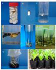
Production of Brazilian green dwarf coconut seedlings from zygotic embryos. (A) endosperm discs; (B) zygotic embryo; (C) zygotic embryo established in liquid Y3 culture medium; (D) shoots and root emergence; (E) seedling developed at 70 days after culture establishment; (F) seedlings developed at 150 days after culture establishment; (G) seedling transferred at 210 days after culture establishment for root induction; (H) seedlings in the acclimatization phase under screen at 270 days after culture establishment. Source: adapted from Lédo et al. (2007)
However, in the study of propagation of different coconut accessions by in vitro zygotic embryo culture, Lédo et al. (2011) LÉDO, A. da S.; RAMOS, S.R.R.; DINIZ, L.E.C.; KONAN, K.J.L. Performance of coconut embryo culture accessions introduced at international coconut genebank for latin america and the Caribbean (ICG-LAC). Acta Horticulturae, The Hague, v.918, p.161-166, 2011. observed differences in the germination percentages and seedling development among accessions.
From the protocol established for the BGD genotype in 2007, Embrapa Tabuleiros Costeiros has been focusing on optimization and adaptation studies for other coconut cultivars.
Iron, considered an essential element in energy transformations, can influence seedling development. For BGD coconut, the presence of 27.8 mg L-1 of Fe2SO4.7H2O provides efficient germination of zygotic embryos and formation of normal seedlings. In studies with Yellow Malayan Dwarf (YMD) and Brazilian tall coconut palm (BT), Fe2SO4.7H2O concentration of 13.9 mg L-1 in the Y3 germination medium was sufficient to favor the formation of shoots and roots (BARIN, LÉDO, 2011 BARIN, L.B.; LÉDO, A. da S. Avaliação de técnicas biotecnológicas para propagação sexuada e conservação in vitro de coqueiro. In: SEMINÁRIO DE INICIAÇÃO CIENTÍFICA E PÓS-GRADUAÇÃO DA EMBRAPA TABULEIROS COSTEIROS, 1., 2011, Aracaju. Anais... Aracaju: Embrapa Tabuleiros Costeiros, 2011. p.44-48. ). The addition of gibberellic acid in the range from 0.5 to 2 μM in the culture medium induces higher germination percentage and better shoot development for BT and BGD varieties (BARIN; LÉDO, 2011 BARIN, L.B.; LÉDO, A. da S. Avaliação de técnicas biotecnológicas para propagação sexuada e conservação in vitro de coqueiro. In: SEMINÁRIO DE INICIAÇÃO CIENTÍFICA E PÓS-GRADUAÇÃO DA EMBRAPA TABULEIROS COSTEIROS, 1., 2011, Aracaju. Anais... Aracaju: Embrapa Tabuleiros Costeiros, 2011. p.44-48. ; SANTOS et al., 2012 SANTOS, J.E. ; LEDO, A.S.; MACHADO, C.A.; ARAUJO, A.G. Estabelecimento de protocolos para cultura in vitro de novas cultivares de coqueiro. In: SEMINÁRIO DE INICIAÇÃO CIENTÍFICA E PÓS-GRADUAÇÃO DA EMBRAPA TABULEIROS COSTEIROS, 2., 2012, Aracaju. Anais... Aracaju: Embrapa Tabuleiros Costeiros, 2012, p.44-48. ).
Additional studies were performed on factors affecting in vitro zygotic coconut embryo cultures. The induction of photoautotrophic growth in YMD coconut seedlings after 4-5 months of in vitro cultivation by reducing the sucrose concentration (43.8 mm or less), associated with CO2 enrichment (1,600 μmol mol-1CO2, in the light phase and 350 μmol mol-1CO2 in the dark phase) for two months before acclimatization resulted in efficient production and cost reduction (SAMOSIR and ADKINS, 2014 SAMOSIR, Y.M.S; ADKINS, S.W. Improving acclimatization through the photoautotrophic culture of coconut (Cocos nucifera) seedlings: an in vitro system for the efficient exchange of germplasm. In Vitro Cellular Development Biology-Plant, Berlin, v.50, p.493-501, 2014. ). The addition of fatty acids, such as lauric acid in the culture medium at 60-75 days of in vitro culture of zygotic YMD coconut embryos accelerated seedling growth and development (LOPEZ-VILLALOBOS et al., 2011 LOPEZ-VILLALOBOS A.; DODDS, P.F.; HORNUNG, R. Lauric acid improves the growth of zygotic coconut (Cocos nucifera L.) embryos in vitro. Plant Cell, Tissue and Organ Culture, Dordrecht, v.106, p.317-327, 2011. ).
In addition to adjustments in the in vitro stages, acclimatization is essential to the success of in vitro techniques to reduce potential losses and increase survival. Few studies have been conducted to improve acclimatization techniques and yields. Comparing three ex vitro acclimatization techniques of Laguna tall coconut seedlings, Magdalita et al. (2010) MAGDALITA, P.M.; DAMASCO, O.P.; ADKINS, S.W. Effects of medium replenishment and acclimatization techniques on growth and survival of embryo cultured coconut seedlings. Philippine Science Letters, Manila, v. 3, n.2, p.1-9, 2010. observed that ex vitro survival was significantly affected when seedlings with two to three leaves acclimated in plastic tent (81.7%) and wet chamber with wooden box (82.2%) showed greater survival when compared to nursery under mist (62.5%).
An excellent example of the application of the zygotic embryo culture technique for the large-scale production of coconut palm seedlings is the Makapuno Laboratory Network in the Philippines (Figure 5). From a protocol established in the 1960s by Guzman due to the low seed germination of this variety in the field, a largescale multiplication program was established in 1992 and is currently composed of six satellite laboratories. The Makapuno cultivar embryo culture protocol was improved in 2003, aiming to reduce costs by reducing the sucrose and activated carbon concentration and the non-addition of myo-inositol and the plant growth regulators naphthalene acetic acid and benzylaminopurine (AREZA-UBALDO et al., 2003 AREZA-UBALDO, M.B.B.; RILLO, E.P.; CUETO, C.A. Application of the improved embryo culture protocol for commercial production of Makapuno seedlings. Philippine Journal of Science, Manila, v.132, n.1, p. 1-11, 2003. ). These results reinforce that the establishment of a standard protocol for the in vitro culture of zygotic embryos of different genotypes cannot be applied to coconut, and constant adjustments are necessary.
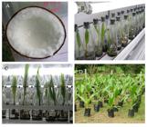
In vitro production and acclimatization of Makapuno coconut seedlings in Zamboanga, Philippines. A - detail of the gelatinous endosperm of the mature fruit; B and C- in vitro development of embryos; D - acclimatization in open-air nursery. Photos: Ana Lédo
The embryo culture technique has also been used for the exchange of genetic resources to minimize the risks of dissemination of pests. Introduction by tissue culture is relatively safe; however, elimination of phytopathogens and maintenance of cultures free from contamination by bacteria, fungi and micro-arthropods have been highlighted (CASSELLS, 2012 CASSELLS, A.C. Pathogen and biological contamination management in plant tissue culture: phytopathogens, vitro pathogens, and vitro pests. Methods in Molecular Biology, New York, v.877, p.57-80, 2012. ). Recent guidelines from Bioversity International and FAO recommend the exchange of endosperm discs containing the zygotic embryo or in vitro zygotic embryos for the safe movement of coconut germplasm (CUETO et al., 2012 CUETO, C.A., JOHNSON, V.B.; ENGELMANN, F.; KEMBU, A.; KONAN, J.L.; KAN, M.K.; RIVERA, R.L.; VIDHANAARACHCHI, V.; BOURDEIX, R.; WEISE, S.F. Technical guidelines for the safe movement and duplication of Coconut (Cocos nucifera L.) germoplasm using embryo culture transfer protocols. Montpellier: COGENT Bioversity International, 2012. 74p. ).
MICROPROPAGATION BY SOMATIC EMBRYOGENESIS-The term micropropagation refers to techniques for in vitro vegetative propagation of plants using small explants as stem tips, nodal segments and zygotic embryos, according to the Glossary of Plant Tissue Culture (CARVALHO et al., 2011 CARVALHO, A. C. P. P. de; TORRES, A. C.; BRAGA, E. J. B.; LEMOS, E. P. de; SOUZA, F. V.D.; PETERS, J. A.; WILLADINO, L.; CÂMARA, T.R. Glossário de cultura de tecidos de plantas. Plant Cell Culture and Micropropagation , Lavras, v.7,n.1, p.30-60, 2011. ). One of the processes of plant micropropagation through somatic embryogenesis involves the in vitro formation of bipolar structures (somatic embryos) from somatic cells under specific culture conditions.
The first studies on coconut somatic embryogenesis were performed in the 1980s using zygotic embryos, immature leaves and inflorescences as explants. These results indicated that the process was feasible, but the production of somatic embryos was only confirmed in 1994 by JL Verdeil’s team (Table 1) using histology techniques that evidenced the formation of calluses and differentiation into somatic embryos, with their subsequent conversion into plants from immature inflorescences (VERDEIL et al., 1994 VERDEIL, J-L.; HUET, C.; GROSDEMANGE, F.; BUFFARD-MOREL, J. Plant regeneration from cultured immature inflorescences of coconut (Cocos nucifera L.): evidence for somatic embryogenesis Plant Cell Reports, Berlin, v.13, p.218-221, 1994. ).
Due to the high efficiency of plumules in the in vitro propagation process by somatic embryogenesis, this tissue has been used to propagate superior dwarf coconut genotypes, which present high self-fertilization rates. Promising results were obtained for green Malayan dwarf coconut (PÉREZ-NUÑEZ et al., 2006 PÉREZ-NUÑEZ, M. T.; CHAN, J. L.; SÁENZ, L.; GONZÁLEZ, T.; VERDEIL, J-L.; OROPEZA, C. Improved somatic embryogenesis from cocos nucifera L. plumule explants. In Vitro Cellular Development Biology-Plant, Berlin, v.42, p.37-43, 2006. ; SÁENZ et al., 2010 SÁENZ, L.; HERRERA-HERRERA, G.; UICAB-BALLOTE, F.; CHAN, J.L.; OROPEZA, C. Influence of form of activated charcoal on embryogenic callus formation in coconut (Cocos nucifera). Plant Cell, Tissue and Organ Culture, Dordrecht, v.100, p.301-308, 2010. ; SÁENZ et al., 2018). Through histological investigation of tissue viability, Pérez-Nuñez et al. (2006) PÉREZ-NUÑEZ, M. T.; CHAN, J. L.; SÁENZ, L.; GONZÁLEZ, T.; VERDEIL, J-L.; OROPEZA, C. Improved somatic embryogenesis from cocos nucifera L. plumule explants. In Vitro Cellular Development Biology-Plant, Berlin, v.42, p.37-43, 2006. found that the insertion of repetitive callogenesis cycles and secondary somatic embryogenesis allowed increasing the technique efficiency, enabling the commercial propagation of certain genotypes from plumules.
However, to ensure clonal propagation, especially in tall coconut genotype and their hybrids with crossfertilization, somatic embryogenesis must occur from other tissues.
Successful cloning protocols were obtained from explants originated from inflorescences (VERDEIL et al., 1994 VERDEIL, J-L.; HUET, C.; GROSDEMANGE, F.; BUFFARD-MOREL, J. Plant regeneration from cultured immature inflorescences of coconut (Cocos nucifera L.): evidence for somatic embryogenesis Plant Cell Reports, Berlin, v.13, p.218-221, 1994. ), from ovaries and anthers (PERERA et al., 2008 PERERA, P.I.P.; PERERA, L.; HOCHER, V.; VERDEIL, J-L.; YAKANDAWALA, D.M. D.; WEERAKOON, L.K. Effect of growth regulators on microspore embryogenesis in coconut anthers. Plant Cell Reports, Berlin, v.27, n.11, p.1697-1703, 2008. , 2010 PERERA, P.; HOCHER, V.; WEERAKOON, L.; YAKANDAWALA, D.; FERNANDO, S.; VERDEIL, J-L. Early inflorescence and floral development in Cocos nucifera L.(Arecaceae: Arecoideae). South African Journal of Botany, Amsterdam,v.76, p.482-492, 2010. ; SANDOVAL-CANCINO et al., 2014; BANDUPRIYA, 2017), respectively, the latter producing haploid plants of great importance for breeding programs.
Despite the great advance of the technique obtained by Pérez-Núñez et al. (2006) PÉREZ-NUÑEZ, M. T.; CHAN, J. L.; SÁENZ, L.; GONZÁLEZ, T.; VERDEIL, J-L.; OROPEZA, C. Improved somatic embryogenesis from cocos nucifera L. plumule explants. In Vitro Cellular Development Biology-Plant, Berlin, v.42, p.37-43, 2006. through secondary embryogenesis in three cycles that increased efficiency by 50,000 times, most studies still present unsatisfactory results for large-scale seedling production, and new studies are being developed in several countries to improve the technique efficiency.
According to results presented by researchers from eight countries at the IV Workshop on Somatic Embryogenesis for Rapid Coconut Multiplication at the International Coconut Biodiversity for Prosperity Conference held in India in 2010, some factors were identified as technical difficulties, which require further investigation. Among these difficulties are genotypedependence, with variations of plant-to-plant results; effects of seasonality on the collection of explants, source of activated carbon in the culture medium; and the formation of fused embryo and pre-mature roots, making somatic embryos more difficult to germinate. According to BANDUPRIYA (2017) there are a lot of constrains that have been addressed for developing a successful protocol for coconut micropropagation, with considerable progress achieved to date. In any plant tissue culture protocol, selection of a suitable starting plant material (explant) is a major factor which governs successful plant regeneration.
Some advances have been obtained with studies on alterations of the culture medium with activated charcoal, gibberellic acid, lauric acid, among others, in addition to gene expression in somatic embryogenesis. Sáenz et al. (2010) SÁENZ, L.; HERRERA-HERRERA, G.; UICAB-BALLOTE, F.; CHAN, J.L.; OROPEZA, C. Influence of form of activated charcoal on embryogenic callus formation in coconut (Cocos nucifera). Plant Cell, Tissue and Organ Culture, Dordrecht, v.100, p.301-308, 2010. studied different brands of activated charcoal and particle size in the induction of somatic embryogenesis of mature Green Malayan Dwarf coconut plumules (GMD), and found that particles smaller than 0.38 μM induced higher percentage of embryogenic calluses (70%). The addition of 0.5 μM gibberellic acid in the culture medium increased the number of embryogenic calluses, of somatic embryos per callus, and the germination of embryos from Malayan dwarf variety plumules (MONTERO-CORTES et al., 2010 b MONTERO-CORTES, M.; SAENZ, L.; CORDOVA, I.; QUIROZ, A.; VERDEIL, J. L.; OROPEZA, C. GA(3) stimulates the formation and germination of somatic embryos and the expression of a KNOTTED- like homeobox gene of Cocos nucifera L. Plant Cell Reports, Berlin, v.29, p.1049-1059, 2010b. ).
The application of histological studies on in vitro coconut culture began in 1990 by Verdeil at CIRAD, France, and since then, the technique associated with histochemistry has been improved and contributed to elucidate the process of somatic embryogenesis.
Studies on the expression of CnSERK, CnCDKA and KNOTTED genes in the process of somatic embryogenesis of Malayan dwarf variety was investigated by PÉREZ-NUNEZ et al. (2009) PÉREZ-NUÑEZ, M.T.; SOUZA, R.; SÁENZ, L.; CHAN, J.L.; ZUNIGA-AGUILAR, J.J.; OROPEZA, C. Detection of a SERK-like gene in coconut and analysis of its expression during the formation of embryogenic callus and somatic embryos. Plant Cell Reports, Berlin, v.8, p.11-19, 2009. and (MONTEROCORTES et al. 2010 a MONTERO-CORTES, M.; RODRIGUEZ-PAREDES, F.; BURGEFF, C.; PEREZ-NUNEZ T.; CORDOVA, I.; OROPEZA, C.; VERDEIL, J.L.; SAENZ, L. Characterization of a cyclin-dependent kinase (CDKA) gene expressed during somatic embryogenesis of coconut palm. Plant Cell, Tissue and Organ Culture, Dordrecht, v.102, p. 251-258, 2010a. ;MONTEROCORTES et al. 2010b MONTERO-CORTES, M.; SAENZ, L.; CORDOVA, I.; QUIROZ, A.; VERDEIL, J. L.; OROPEZA, C. GA(3) stimulates the formation and germination of somatic embryos and the expression of a KNOTTED- like homeobox gene of Cocos nucifera L. Plant Cell Reports, Berlin, v.29, p.1049-1059, 2010b. ). The expression of the CnCDKA gene was higher in the embryogenic callus formation phase, coinciding with its in situ location with meristematic cell centers from which somatic embryos developed. Gene expression, progressively reduced with the embryo development. However, according to Nguyen et al. (2015) NGUYEN, Q.T.; BANDUPRIYA, H.D.D.; LÓPEZ-VILLALOBOS, A.; SISUNANDAR, S.; FOALE, M.; ADKINS, S.W. Tissue culture and associated biotechnological interventions for the improvement of coconut (Cocos nucifera L.): a review. Planta, Berlin, v.242, n.5, p.1059-1076, 2015. , further studies on the molecular control of the expression of somatic embryogenesis in coconut trees are needed to improve yield. The same authors point out the low efficiency of the cloning method for the coconut tree due to the slow in vitro development and low ex vitro vigor when compared to other species.
Along these lines, further studies should be conducted so that the mass propagation of coconut tree becomes a reality, as in the case of palm oil, where biofactories in some regions of the world already produce seedlings on a large-scale. Recent studies have indicated that the use of temporary immersion bioreactors for somatic embryogenesis of palm oil (Elaeis guineenses) has increased the multiplication capacity (MARBUN et al., 2015 MARBUN, C.L.M.; TORUAN-MATHIUS, N.; UTOMO, C.; LIWANG, T. Micropropagation of embryogenic callus of oil palm (Elaeis guineensis jacq.) using temporary immersion system. Procedia Chemistry, Amsterdam, v.14, p.122-129, 2015. ).
Bioreactor technology involves in vitro culture of cells, tissues and organs in liquid medium and in an automated system. This system allows the reduction of labor and production costs and is faster and more efficient for the in vitro propagation of plants in largescale, compared to conventional system with semi-solid agar medium (ZIV, 2005 ZIV, M. Simple bioreactors for mass propagation of plants. Plant Cell, Tissue and Organ Culture, Dordrecht, v.81, p.277-285, 2005. ). The temporary immersion bioreactor system involves controlling the frequency of immersion of explants in liquid medium, with control of the number of immersions per day and the length of each immersion. These systems have been efficiently used in in vitro multiplication of several species. Etienne and Berthouly (2002) ETIENNE, H.; BERTHOULY, M. Temporary immersion systems in plant micropropagation. Plant Cell, Tissue and Organ Culture, Dordrecht, v.69, p.215-231, 2002. described several types of temporary immersion bioreactor systems. These systems have been successful in the processes of in vitro multiplication of buds, microcuttings, microtubers and somatic embryos.
In addition to reducing labor and production costs, other advantages of bioreactors include less space required for production, better plant quality and vigor, increased productivity and overall efficiency (ETIENNE; BERTHOULY, 2002 ETIENNE, H.; BERTHOULY, M. Temporary immersion systems in plant micropropagation. Plant Cell, Tissue and Organ Culture, Dordrecht, v.69, p.215-231, 2002. ).
Advances on in vitro coconut production using somatic embryogenesis techniques may be associated with temporary immersion bioreactor technology, thus providing an efficient system for large-scale clonal coconut propagation. In addition, the inclusion of lightemitting diodes (LED) technology allows an increase in vitro productivity. LED-type lights emit selective illumination at wavelengths suitable for photosynthesis, being 660 nm by means of red light and 450 nm of blue light. The Tropical Research and Education Center (TREC) of the University of Florida recently installed a plant production system using state-of-the-art temporary immersion bioreactors. The system is automated with LED light (Figure 6) and has shown positive results so far in the production of banana, sugarcane, bromeliads and orchids.

A-Temporal immersion bioreactors installed at TREC, University of Florida, under LED-type light; B - System control board (Photos: Wagner Vendrame).
In 2017, TREC started studies to improve protocol for coconut production aiming in vitro mass multiplication of the variety of Fijian dwarf coconut, whose genotype has demonstrated tolerance to lethal yellowing. This project has great economic importance for coconut production in the US, as well as for other countries in the Caribbean, because coconut tree is one of the most popular palm trees for use in landscaping and a symbol of the tropics (VENDRAME, 2017, personal communication).
Phytosanitary aspects of coconut propagation-The coconut palm in its propagation phase is susceptible to the occurrence of diseases and pests that can affect seedling development and reduce its commercial value. Insects, mites and pathogens can be transmitted through transit of coconut propagating material. The transmission may also occur through dispersion of present pests or absent quarantine pests, which can be transported and introduced by different ways in areas free of pests. Of the diseases and pests that threaten coconut cultivation and the safe exchange of propagating material among countries, the most noteworthy are: lethal yellowing, cadang-cadang and the Coleoptera Rhynchophorus ferrugineus (Linnaeus) as absent quarantine pests; red palm mite Raoiella indica a present quarantine pest; and the coconut mite Aceria guerreronis, the coconut transparent mealybug Aspidiotus destructor and the whitefly Aleurodicus pseudugesii, pests with great potential for dispersion and largely distributed in several growing regions worldwide.
Lethal yellowing is a devastating disease caused by different groups of phytoplasmas that affect around 40 palm tree species and is now considered the main coconut disease in the world (OROPEZA et al., 2011 OROPEZA, C.; CORDOVA, I.; CHUMBA, A.; NARVAEZ, M.; SAENZ, L.; ASHBURNER, R.; HARRISON, N. Phytoplasma distribution in coconut palms affected by lethal yellowing disease. Annals of Applied Biology, Wellesbourne, v.159, p.109-117, 2011. ; MYRIE et al., 2012 MYRIE, W.A.; DOUGLAS, L.; HARRISON, N.A.; MCLAUGHLIN, W.; JAMES, M. First report of lethal yellowing disease associated with subgroup 16SrIV, a phytoplasma on St. Kitts in the Lesser Antilles. New Disease Reports, London, v.26, p.25. 2012. ). Currently, lethal yellowing is distributed in countries in North America, the Caribbean, and on the western and eastern coasts of Africa.
The transit of coconut germplasm between countries constitutes a threat of introduction of this disease. Phytoplasma DNA of the lethal yellowing disease were detected in embryos from diseased coconut fruits by the DNA hybridization technique (CORDOVA, 1994) and also by PCR analysis (CORDOVA et al., 2003) cited by OROPEZZA et al. (2005). In another study, phytoplasma DNA was detected in the stem, peduncles, female flowers and embryos of fruits harvested from infected coconut trees. The germination capacity of infected fruits and seedling formation were not affected; however, no phytoplasma DNA was detected in the resulting seedlings.
Also, phytoplasma DNA was not detected in seedlings obtained through in vitro embryo culture extracted from symptomatic plants. Therefore, although phytoplasma DNA can be detected in embryos, there is no evidence that the pathogen is transmitted through seeds to the seedling. However, it is important to emphasize that the presence of phytoplasma DNA in coconut embryo tissues suggests a transmission potential that still needs to be proven (NIPAH et al., 2007 NIPAH, J.O.; JONES, P.; DICKINSON, M.J. Detection of lethal yellowing phytoplasma in embryos from coconut palms infected with Cape St Paul wilt disease in Ghana. Plant Pathology, Bari, v.56, n.5, p.777-784, 2007. ).
The cadang-cadang disease is also lethal to the coconut tree and has the coconut cadang-cadang-viroid (CCCVd) as etiologic agent. CCCVd associated with coconut is found in the central region of Philippines and is slowly and continuously dispersing to the north and south of the site of its first report in 1930, in the province of Albay (HANOLD; RANDLES, 1991a HANOLD, D.; RANDLES, J. W. Coconut cadang-cadang disease and its viroid agent. Plant Disease, Davis, v. 75, p. 330-335, 1991a. ). CCCVd can be detected in embryos and in in vitro cultured seedlings from naturally infected plants and in coconut shells by molecular DNA hybridization. This viroid is transmissible via seeds, but with low rate, around one to 300 (HANOLD; RANDLES, 1991b HANOLD, D.; RANDLES, J.W. Detection of coconut cadang-cadang viroid-like sequences in oil and coconut palm and other monocotyledons in the South-west Pacific. Annals of Applied Biology, Wellesbourne, v.118, p.139-151, 1991b. ; PACUMBABA et al., 1994 PACUMBABA, E.P.; ZELAZNY, B.; ORENSE, J.C.; RILLO, E.P. Evidence for pollen and seed transmission of the coconut cadang-cadang viroid in Cocos nucifera. Journal of Phytopathology, Berlin, v.142, n.1, p.37-42, 1994. ) and more rarely by the pollen (PACUMBABA et al., 1994 PACUMBABA, E.P.; ZELAZNY, B.; ORENSE, J.C.; RILLO, E.P. Evidence for pollen and seed transmission of the coconut cadang-cadang viroid in Cocos nucifera. Journal of Phytopathology, Berlin, v.142, n.1, p.37-42, 1994. ).
Regarding pests, R. ferrugineus (Olivier) is considered potentially dangerous for the American continent today. This species is originated from Southeastern Asia, but has recently been introduced in the Caribbean, Curacao and Aruba, and the United States (RODA et al., 2011; FIABOE et al., 2012; GIBLINDAVIS et al., 2013, cited by FERREIRA et al., 2014 FERREIRA, J.M.S; TEODORO, A.V.; NEGRISOLI JUNIOR, A.S.; GUZZO, E.C. Manejo integrado da broca-do-olho-do-coqueiro Rhynchophorus palmarum L. (Coleoptera: Curculionidae). Aracaju: Embrapa Tabuleiros Costeiros, 2014, 8p. (Comunicado Técnico, 141) ) and it is worth mentioning that the island of Aruba is only 26 km away from the coast of Venezuela. R. ferrugineus is the only species of this genus that has spread to several countries, probably due to its association with date palm (Phoenix dactilifera). Since it is one of the few palm trees propagated by tillers, the transit of seedlings to other countries can transport the beetle as eggs, larvae or pupae (HOWARD, 2001 HOWARD, F.W. Sap-feeders on palms. In: HOWARD, F.W.; MOORE, D.; GIBLIN-DAVIS, R.M.; ABAD, R.G. Insects on palms. New York: CABI Publishing. 2001. p.109-232. ). Another species that presents a great dispersion potential through the propagating material is the red palm mite R. indica Hirst 1924. This species was recently introduced in Brazil through the state of Roraima from Venezuela and is the most outstanding example, among the countless pest species that attack the coconut tree. It was first reported in India in 1924, with dispersion over time throughout the Middle East, Africa, Central America, North America (MENDONÇA et al., 2005 MENDONÇA, R.S.; NAVIA, D.; FLECHTMANN, C.H.W. Raoiella indica Hirst (Prostigmata: Tenuipalpidae), o ácaro vermelho das palmeiras – uma ameaça para as Américas. Brasília: Embrapa Recursos Genéticos e Biotecnologia, 2005. 40p. (Documentos, 146) ).
Until now, Brazil was the last record of this dispersive route of the red palm mite in the American continent (NÁVIA 2011 NAVIA, D.; MARSARO JR, A. L.; SILVA, F. R.; GONDIM JR, M. G. C.; MORAES, G.J. First report of the red palm mite, Raoiella indica Hirst (Acari: Tenuipalpidae) in Brazil. Neotropical Entomology, v. 40, p. 409-411, 2011. ; RODRIGUES; ANTONY, 2011 RODRIGUES, J. C. V.; ANTONY, L. M. K. First report of Raoiella indica (Acari: Tenuipalpidae) in Amazonas state, Brazil. Florida Entomologist, v.94, p.1073-1074, 2011. ; SILVA et al., 2016 SILVA, S. S.; SANTOS, P. M.; SANTOS, M. C.; VIEIRA, I. G.; SARAIVA, W. V. A.; FARIAS, A. P.; SILVA, E. A.; PINHEIRO NETO, M.; TEODORO, A.V. Primeiro registro do ácaro-vermelho-das-palmeiras Raoiella indica em Sergipe. In: XXVI Congresso Brasileiro de Entomologia e IX Congresso Latino-Americano de Entomologia, 2016, Maceió. XXVI Congresso Brasileiro de Entomologia. Maceió. 2016. ).
The coconut mite A. guerreronis was first reported in 1965, in Rio de Janeiro, Brazil, and described at the same year by Keifer, in samples from Mexico. A decade later, it was found on the Eastern Coast of West Africa where producers and residents of some countries have claimed to be aware, for many decades, of similar damages in coconut trees before being scientifically described (HOWARD, 2001 HOWARD, F.W. Sap-feeders on palms. In: HOWARD, F.W.; MOORE, D.; GIBLIN-DAVIS, R.M.; ABAD, R.G. Insects on palms. New York: CABI Publishing. 2001. p.109-232. ).
The coconut mealybug A. destructor, a species native from the tropics of the eastern hemisphere was first described by Signoret in 1869 in coconut samples from Réunion Islands in the Indian Ocean and its presence was detected in the Caribbean islands around 1902.
Throughout the 20th century, dispersion was observed in the tropical regions of the eastern and western hemispheres (HOWARD, 2001 HOWARD, F.W. Sap-feeders on palms. In: HOWARD, F.W.; MOORE, D.; GIBLIN-DAVIS, R.M.; ABAD, R.G. Insects on palms. New York: CABI Publishing. 2001. p.109-232. ).
Whiteflies have been reported in several countries associated with coconut palm, but without causing severe damage. However, in Brazil, of a total of 17 species associated with this palm tree (OMENA et al., 2012 OMENA, R.P.M.; GUZZO, E.C.; FERREIRA, J.M.S.; MENDONÇA F.A.C.; LIMA A.F.; RACCA-FILHO F.; SANTANA, A.E.G. First report on the whitefly, Aleurodicus pseudugesii on the coconut palm, Cocos nucifera in Brazil. Journal of Insect Science, Oxford, v.12, p.1-6, 2012. ), A. pseudugesii has been the most aggressive species causing great damage to coconut crops (FERREIRA et al., 2011 FERREIRA, J.M.S.; LINS, P.M.P.; OMENA, R.P.M.; LIMA, A.F.; RACCA FILHO, F. Mosca branca: uma ameaça à produção do coqueiro no Brasil. Aracaju: Embrapa Tabuleiros Costeiros, 2011. 5p. (Circular Técnica, 62) ). This species was first described in coconut palm in 2008 in specimens collected in Ecuador and Peru (MARTIN, 2008). However, outbreaks of A. pseudugesii were recorded in Brazil in 2006 (Bahia state) and in 2007 (Ceará state). In 2011, the pest had already reached all coconut producing areas of the Brazilian national territory (FERREIRA et al., 2011 FERREIRA, J.M.S.; LINS, P.M.P.; OMENA, R.P.M.; LIMA, A.F.; RACCA FILHO, F. Mosca branca: uma ameaça à produção do coqueiro no Brasil. Aracaju: Embrapa Tabuleiros Costeiros, 2011. 5p. (Circular Técnica, 62) ; OMENA et al., 2012 OMENA, R.P.M.; GUZZO, E.C.; FERREIRA, J.M.S.; MENDONÇA F.A.C.; LIMA A.F.; RACCA-FILHO F.; SANTANA, A.E.G. First report on the whitefly, Aleurodicus pseudugesii on the coconut palm, Cocos nucifera in Brazil. Journal of Insect Science, Oxford, v.12, p.1-6, 2012. ).
The increased trade of seedlings between regions, countries or continents has contributed to accelerate the dispersal and establishment of pests and pathogens to locations favorable to their multiplication and survival.
Conclusion
The transmission of potential pests and pathogens shall be largely considered in the transit of coconut propagation material, since it might disseminate these agents among regions and countries. Care should be taken regarding the internal and external commercialization or the safe exchange of coconut seedlings or any other coconut plant live tissues of coconut tree which can represents risks for the coconut crop. There is a need for standardization of an efficient somatic embryogenesis protocol for fast, large-scale micropropagation via bioreactors and safety considering the phytosanitary aspects of the cultivars of interest.
- ARAGÃO, W.M. Cultivares de coqueiros. In: FONTES, H.R.; FERREIRA, J.M.S.; SIQUEIRA, L.A. A cultura do coqueiro. Aracaju: Embrapa Tabuleiros Costeiros, 2007. (Sistemas de Produção, 1)
- AREZA-UBALDO, M.B.B.; RILLO, E.P.; CUETO, C.A. Application of the improved embryo culture protocol for commercial production of Makapuno seedlings. Philippine Journal of Science, Manila, v.132, n.1, p. 1-11, 2003.
- BARIN, L.B.; LÉDO, A. da S. Avaliação de técnicas biotecnológicas para propagação sexuada e conservação in vitro de coqueiro. In: SEMINÁRIO DE INICIAÇÃO CIENTÍFICA E PÓS-GRADUAÇÃO DA EMBRAPA TABULEIROS COSTEIROS, 1., 2011, Aracaju. Anais.. Aracaju: Embrapa Tabuleiros Costeiros, 2011. p.44-48.
- BONDAR, G. A cultura do coqueiro (Cocos nucifera) no Brasil. Salvador: Tipografia Naval, 1955. 91p.
- BOURDEIX, R.; KONAN, J.L.; N’CHO, Y.O. Coconut: a guide to traditional and improved varieties. Barcelona: Editions Diversiflora, 2005. 104 p.
- CARVALHO, A. C. P. P. de; TORRES, A. C.; BRAGA, E. J. B.; LEMOS, E. P. de; SOUZA, F. V.D.; PETERS, J. A.; WILLADINO, L.; CÂMARA, T.R. Glossário de cultura de tecidos de plantas. Plant Cell Culture and Micropropagation , Lavras, v.7,n.1, p.30-60, 2011.
- CASSELLS, A.C. Pathogen and biological contamination management in plant tissue culture: phytopathogens, vitro pathogens, and vitro pests. Methods in Molecular Biology, New York, v.877, p.57-80, 2012.
- CUETO, C.A., JOHNSON, V.B.; ENGELMANN, F.; KEMBU, A.; KONAN, J.L.; KAN, M.K.; RIVERA, R.L.; VIDHANAARACHCHI, V.; BOURDEIX, R.; WEISE, S.F. Technical guidelines for the safe movement and duplication of Coconut (Cocos nucifera L.) germoplasm using embryo culture transfer protocols. Montpellier: COGENT Bioversity International, 2012. 74p.
- EEUWENS, C.J. Mineral requirements for growth and callus initiation of tissue explants excised from mature coconut palms (Cocos nucifera) and cultured in vitro. Physiologia Plantarum, Lund, v.36, p.23- 28, 1976.
- ENGELMANN, F.; BATUGAL, P.A. Background on the development and implementation of the coconut embryo in vitro culture project. In: ENGELMANN, F.; BATUGAL, P.A.; OLIVER, J. Coconut embryo in vitro culture. Malaysia: IPGRI-APO, 2002. v.2, p.1-4.
- ETIENNE, H.; BERTHOULY, M. Temporary immersion systems in plant micropropagation. Plant Cell, Tissue and Organ Culture, Dordrecht, v.69, p.215-231, 2002.
- FAO- Food and Agriculture Organization of the United Nations. Deejay farms India coconut breeding and the benefits of hybrid coconut farms. 2014. Disponível em: http://www.fao.org/fileadmin/templates/rap/files/meetings/2013/131030-breedings.pdf Acesso em: 25 out. 2016.
» http://www.fao.org/fileadmin/templates/rap/files/meetings/2013/131030-breedings.pdf - FAO- Food and Agriculture Organization of the United Nations. FAOSTAT Beta. 2017. Disponível em: http://fenix.fao.org/faostat/beta/en/#data/QC Acesso em: 17 jul. 2017.
» http://fenix.fao.org/faostat/beta/en/#data/QC - FERREIRA, J.M.S.; LINS, P.M.P.; OMENA, R.P.M.; LIMA, A.F.; RACCA FILHO, F. Mosca branca: uma ameaça à produção do coqueiro no Brasil. Aracaju: Embrapa Tabuleiros Costeiros, 2011. 5p. (Circular Técnica, 62)
- FERREIRA, J.M.S; TEODORO, A.V.; NEGRISOLI JUNIOR, A.S.; GUZZO, E.C. Manejo integrado da broca-do-olho-do-coqueiro Rhynchophorus palmarum L. (Coleoptera: Curculionidae). Aracaju: Embrapa Tabuleiros Costeiros, 2014, 8p. (Comunicado Técnico, 141)
- FOALE, M.; HARRIES, H. Farm and Forestry Production and Marketing Profile for Coconut (Cocos nucifera). In: ELEVITCH, C. R. (ed.). Specialty crops for pacific island agroforestry. Holualoa: Permanent Agriculture Resources. 2009. Disponível em: http://agroforestry.net/scps. Acesso em: 4 de setembro de 2017.
- FONTES, H.R.; CINTRA, F.L.D.; CARVALHO FILHO, O.M. Implantação e manejo da cultura do coqueiro. In: FERREIRA, M.J.S.; WARWICK, D.R.N.; SIQUEIRA, L.A. (Eds) A cultura do coqueiro no Brasil. Brasília, DF: Embrapa-SPI; Aracaju: Embrapa-CPATC, 1998. p.99-128.
- FONTES, H.R.; LEAL, M. de L. da S. Utilização de sistema alternativo na produção de mudas de coqueiros híbridos (Cocos nucifera L). Revista Brasileira de Fruticultura, Jaboticabal, v.20, n.3, p.290-296, 1998.
- FRÉMOND, Y.; ZILLER, R.; NUCÉ DE LAMOTHE, M. El cocotero: técnicas agrícolas y producciones tropicales. Barcelona: Editorial Blume, 1975. 236p.
- GRATTAPAGLIA, D.; MACHADO, M.A. Micropropagação. In: TORRES, A.C.; CALDAS, L.S.; BUSO, J.A. (Ed.). Cultura de tecidos e transformação genética de plantas. Brasília, DF: EMBRAPA, 1998. v. 1, p.183-260.
- HANOLD, D.; RANDLES, J. W. Coconut cadang-cadang disease and its viroid agent. Plant Disease, Davis, v. 75, p. 330-335, 1991a.
- HANOLD, D.; RANDLES, J.W. Detection of coconut cadang-cadang viroid-like sequences in oil and coconut palm and other monocotyledons in the South-west Pacific. Annals of Applied Biology, Wellesbourne, v.118, p.139-151, 1991b.
- HARRISON, N.A.; OROPEZA, C. Coconut lethal yellowing. In: HARRISON, N.A.; RAO, G.P.; MARCONE, C. Characterization, diagnosis, and management of phytoplasmas. Houston: Studium Press LLC. 2008, p.219-248.
- HOWARD, F.W. Sap-feeders on palms. In: HOWARD, F.W.; MOORE, D.; GIBLIN-DAVIS, R.M.; ABAD, R.G. Insects on palms. New York: CABI Publishing. 2001. p.109-232.
- IBGE - Instituto Brasileiro de Geografia e Estatística. Produção e área plantada de lavouras permanentes. Sistema IBGE de recuperação automática, 2015. Disponível em: http://www.sidra.ibge.gov.br/bda/tabela/protabl.asp?c=1613ez=teo=11ei=P. Acesso em: 04 out. 2016.
» http://www.sidra.ibge.gov.br/bda/tabela/protabl.asp?c=1613ez=teo=11ei=P. - LÉDO, A. da S.; RAMOS, S.R.R.; DINIZ, L.E.C.; KONAN, K.J.L. Performance of coconut embryo culture accessions introduced at international coconut genebank for latin america and the Caribbean (ICG-LAC). Acta Horticulturae, The Hague, v.918, p.161-166, 2011.
- LÉDO, A.S.; GOMES, K.K.P.; BARBOZA, S.B.S.C.; VIEIRA, G.S.S.; ARAGÃO, W.M. de; TUPINAMBÁ, E.A. Cultivo in vitro de embriões zigóticos e aclimatação de plântulas de coqueiro anão. Pesquisa Agropecuária Brasileira, Brasília, DF, v.42, p.147-154, 2007.
- LOPEZ-VILLALOBOS A.; DODDS, P.F.; HORNUNG, R. Lauric acid improves the growth of zygotic coconut (Cocos nucifera L.) embryos in vitro. Plant Cell, Tissue and Organ Culture, Dordrecht, v.106, p.317-327, 2011.
- MAGDALITA, P.M.; DAMASCO, O.P.; ADKINS, S.W. Effects of medium replenishment and acclimatization techniques on growth and survival of embryo cultured coconut seedlings. Philippine Science Letters, Manila, v. 3, n.2, p.1-9, 2010.
- MARBUN, C.L.M.; TORUAN-MATHIUS, N.; UTOMO, C.; LIWANG, T. Micropropagation of embryogenic callus of oil palm (Elaeis guineensis jacq.) using temporary immersion system. Procedia Chemistry, Amsterdam, v.14, p.122-129, 2015.
- MARTINS, C.R.; JESUS JUNIOR, L.A. Produção e comercialização de coco no Brasil frente ao comércio internacional: panorama 2014. Aracaju: Embrapa Tabuleiros Costeiros, 2013. 51p. (Documentos, 184)
- MENDONÇA, R.S.; NAVIA, D.; FLECHTMANN, C.H.W. Raoiella indica Hirst (Prostigmata: Tenuipalpidae), o ácaro vermelho das palmeiras – uma ameaça para as Américas. Brasília: Embrapa Recursos Genéticos e Biotecnologia, 2005. 40p. (Documentos, 146)
- MINISTÉRIO DA AGRICULTURA. CultivarWEB. Registro nacional de cultivares. Brasília, DF, 2016. Disponível em: http://extranet.agricultura.gov.br/php/snpc/cultivarweb/cultivares_registradas.php Acesso em: 04 out. 2016.
» http://extranet.agricultura.gov.br/php/snpc/cultivarweb/cultivares_registradas.php - MONTERO-CORTES, M.; SAENZ, L.; CORDOVA, I.; QUIROZ, A.; VERDEIL, J. L.; OROPEZA, C. GA(3) stimulates the formation and germination of somatic embryos and the expression of a KNOTTED- like homeobox gene of Cocos nucifera L. Plant Cell Reports, Berlin, v.29, p.1049-1059, 2010b.
- MONTERO-CORTES, M.; RODRIGUEZ-PAREDES, F.; BURGEFF, C.; PEREZ-NUNEZ T.; CORDOVA, I.; OROPEZA, C.; VERDEIL, J.L.; SAENZ, L. Characterization of a cyclin-dependent kinase (CDKA) gene expressed during somatic embryogenesis of coconut palm. Plant Cell, Tissue and Organ Culture, Dordrecht, v.102, p. 251-258, 2010a.
- MYRIE, W.A.; DOUGLAS, L.; HARRISON, N.A.; MCLAUGHLIN, W.; JAMES, M. First report of lethal yellowing disease associated with subgroup 16SrIV, a phytoplasma on St. Kitts in the Lesser Antilles. New Disease Reports, London, v.26, p.25. 2012.
- NAVIA, D.; MARSARO JR, A. L.; SILVA, F. R.; GONDIM JR, M. G. C.; MORAES, G.J. First report of the red palm mite, Raoiella indica Hirst (Acari: Tenuipalpidae) in Brazil. Neotropical Entomology, v. 40, p. 409-411, 2011.
- NGUYEN, Q.T.; BANDUPRIYA, H.D.D.; LÓPEZ-VILLALOBOS, A.; SISUNANDAR, S.; FOALE, M.; ADKINS, S.W. Tissue culture and associated biotechnological interventions for the improvement of coconut (Cocos nucifera L.): a review. Planta, Berlin, v.242, n.5, p.1059-1076, 2015.
- NIPAH, J.O.; JONES, P.; DICKINSON, M.J. Detection of lethal yellowing phytoplasma in embryos from coconut palms infected with Cape St Paul wilt disease in Ghana. Plant Pathology, Bari, v.56, n.5, p.777-784, 2007.
- OMENA, R.P.M.; GUZZO, E.C.; FERREIRA, J.M.S.; MENDONÇA F.A.C.; LIMA A.F.; RACCA-FILHO F.; SANTANA, A.E.G. First report on the whitefly, Aleurodicus pseudugesii on the coconut palm, Cocos nucifera in Brazil. Journal of Insect Science, Oxford, v.12, p.1-6, 2012.
- OROPEZA, C.; CORDOVA, I.; CHUMBA, A.; NARVAEZ, M.; SAENZ, L.; ASHBURNER, R.; HARRISON, N. Phytoplasma distribution in coconut palms affected by lethal yellowing disease. Annals of Applied Biology, Wellesbourne, v.159, p.109-117, 2011.
- OROPEZA, C.; CORDOVA, I.; CHUMBA, A.; NARVAEZ, M.; SAENZ, L.; ASHBURNER, R.; HARRISON, N. Phytoplasma distribution in coconut palms affected by lethal yellowing disease. Annals of Applied Biology, Wellesbourne, v.159, p.109- 117, 2011.
- PACUMBABA, E.P.; ZELAZNY, B.; ORENSE, J.C.; RILLO, E.P. Evidence for pollen and seed transmission of the coconut cadang-cadang viroid in Cocos nucifera. Journal of Phytopathology, Berlin, v.142, n.1, p.37-42, 1994.
- PERERA, P.; HOCHER, V.; WEERAKOON, L.; YAKANDAWALA, D.; FERNANDO, S.; VERDEIL, J-L. Early inflorescence and floral development in Cocos nucifera L.(Arecaceae: Arecoideae). South African Journal of Botany, Amsterdam,v.76, p.482-492, 2010.
- PERERA, P.I.P.; PERERA, L.; HOCHER, V.; VERDEIL, J-L.; YAKANDAWALA, D.M. D.; WEERAKOON, L.K. Effect of growth regulators on microspore embryogenesis in coconut anthers. Plant Cell Reports, Berlin, v.27, n.11, p.1697-1703, 2008.
- PÉREZ-NUÑEZ, M. T.; CHAN, J. L.; SÁENZ, L.; GONZÁLEZ, T.; VERDEIL, J-L.; OROPEZA, C. Improved somatic embryogenesis from cocos nucifera L. plumule explants. In Vitro Cellular Development Biology-Plant, Berlin, v.42, p.37-43, 2006.
- PÉREZ-NUÑEZ, M.T.; SOUZA, R.; SÁENZ, L.; CHAN, J.L.; ZUNIGA-AGUILAR, J.J.; OROPEZA, C. Detection of a SERK-like gene in coconut and analysis of its expression during the formation of embryogenic callus and somatic embryos. Plant Cell Reports, Berlin, v.8, p.11-19, 2009.
- RODRIGUES, J. C. V.; ANTONY, L. M. K. First report of Raoiella indica (Acari: Tenuipalpidae) in Amazonas state, Brazil. Florida Entomologist, v.94, p.1073-1074, 2011.
- SÁENZ, L.; HERRERA-HERRERA, G.; UICAB-BALLOTE, F.; CHAN, J.L.; OROPEZA, C. Influence of form of activated charcoal on embryogenic callus formation in coconut (Cocos nucifera). Plant Cell, Tissue and Organ Culture, Dordrecht, v.100, p.301-308, 2010.
- SAMOSIR, Y.M.S; ADKINS, S.W. Improving acclimatization through the photoautotrophic culture of coconut (Cocos nucifera) seedlings: an in vitro system for the efficient exchange of germplasm. In Vitro Cellular Development Biology-Plant, Berlin, v.50, p.493-501, 2014.
- SANTOS, J.E. ; LEDO, A.S.; MACHADO, C.A.; ARAUJO, A.G. Estabelecimento de protocolos para cultura in vitro de novas cultivares de coqueiro. In: SEMINÁRIO DE INICIAÇÃO CIENTÍFICA E PÓS-GRADUAÇÃO DA EMBRAPA TABULEIROS COSTEIROS, 2., 2012, Aracaju. Anais... Aracaju: Embrapa Tabuleiros Costeiros, 2012, p.44-48.
- SILVA, S. S.; SANTOS, P. M.; SANTOS, M. C.; VIEIRA, I. G.; SARAIVA, W. V. A.; FARIAS, A. P.; SILVA, E. A.; PINHEIRO NETO, M.; TEODORO, A.V. Primeiro registro do ácaro-vermelho-das-palmeiras Raoiella indica em Sergipe. In: XXVI Congresso Brasileiro de Entomologia e IX Congresso Latino-Americano de Entomologia, 2016, Maceió. XXVI Congresso Brasileiro de Entomologia. Maceió. 2016.
- SOLÍS-RAMOS, L.Y.; ANDRADE-TORRES, A.; CARBONELL, L.A.S.; SALÍN, C.M. O.; DE LA SERNA, E.C. Somatic embryogenesis in recalcitrant plants. In: KEN-ICHI SATO, K-I. Embryogenesis. Rijeka: Intech, p.598-618, 2012.
- VERDEIL, J-L.; HUET, C.; GROSDEMANGE, F.; BUFFARD-MOREL, J. Plant regeneration from cultured immature inflorescences of coconut (Cocos nucifera L.): evidence for somatic embryogenesis Plant Cell Reports, Berlin, v.13, p.218-221, 1994.
- ZIV, M. Simple bioreactors for mass propagation of plants. Plant Cell, Tissue and Organ Culture, Dordrecht, v.81, p.277-285, 2005.
Publication Dates
-
Publication in this collection
29 Apr 2019 -
Date of issue
2019
History
-
Received
08 June 2018 -
Accepted
21 Sept 2018


