Abstracts
OBJECTIVE: To determine the influence of intraabdominal pressure in the ultrasonographic evaluation of the urethrovesical junction (UVJ) and proximal urethra (PU) in patients with stress urinary incontinence (SUI). MATERIALS AND METHODS: A prospective, cross-sectional study was conducted at the Urinary Incontinence Research Unit of "Universidade Federal de Pernambuco", Brazil, from January 2002 to January 2005. Perineal ultrasounds of the UVJ and PU were performed in thirty-six women complaining of SUI with the bladder virtually empty (< 50 ml); simultaneous measurement of the intraabdominal pressure was also performed. An ultrasound machine with a computer chip and a high-resolution photographic camera connected to a 7 MHz vaginal probe was used for the evaluation. In order to measure the intraabdominal pressure, an urodynamic equipment with a 10fr rectal catheter connected to a pressure sensitive balloon was used. RESULTS: The age of the patients ranged from 25 to 69 years (mean 46.4 ± 10.2 years). On Valsava maneuver, the intra-abdominal pressure ranged from 7 to 193 cmH2O (mean: 99.3 ± 51.8 cmH2O; median: 99.5 cmH2O). Eight out of 31 (25.8%) patients with UVJ hypermobility had an intraabdominal pressure lower than 60 cmH2O. There was no statistically significant association between intraabdominal pressure and the ultrasound parameters studied. CONCLUSION: There is a specific urethral pressure index for each woman with SUI. However, there is no significant association between increase in intraabdominal pressure and increase of UVJ and UP hypermobility in women with clinical SUI.
Stress urinary incontinence; Abdominal pressure; Perineal ultrasound; Urethrovesical junction
OBJETIVO: Determinar a influência da aferição da pressão intra-abdominal na avaliação ultra-sonográfica da junção uretrovesical (JUV) e da uretra proximal (UP) em pacientes com incontinência urinária de esforço (IUE). MATERIAIS E MÉTODOS: Estudo prospectivo de corte transversal realizado na Unidade de Pesquisa em Incontinência Urinária da Universidade Federal de Pernambuco, de janeiro de 2002 a janeiro de 2005. Trinta e seis pacientes com queixas de IUE foram submetidas a ultra-sonografia perineal para avaliação da JUV e da UP com a bexiga praticamente vazia (< 50 ml), com aferição simultânea de pressão intra-abdominal. Para as avaliações, foi utilizado aparelho de ultra-som com transdutor vaginal de 7 MHz e seletor eletrônico de mensuração de imagem real, equipado com computador e câmera fotográfica de resolução instantânea. Para a medida da pressão intra-abdominal, foi utilizado aparelho de urodinâmica com cateter de 10 fr retal acoplado a um balão de sensor para medida da pressão intra-abdominal. RESULTADOS: As pacientes tinham idade entre 25 e 69 anos (média de 46,4 ± 10,2 anos). À manobra de Valsalva, a pressão intra-abdominal variou entre 7 cmH2O e 193 cmH2O (média de 99,3 ± 51,8 cmH2O; mediana de 99,5 cmH2O). Oito das 31 (25,8%) pacientes com hipermobilidade da JUV apresentaram pressão intra-abdominal inferior a 60 cmH2O. Não foi detectada relação estatisticamente significante entre a variação de pressão intra-abdominal e os parâmetros ultra-sonográficos em questão. CONCLUSÃO: Há um índice específico de pressão de deslocamento uretral para cada mulher com IUE. Porém, não há associação significativa entre o aumento de pressão intra-abdominal e aumento de mobilidade da JUV e UP em mulheres com quadro clínico de IUE.
Incontinência urinária de esforço; Pressão intra-abdominal; Ultra-sonografia perineal; Junção uretrovesical
ORIGINAL ARTICLE
Intra-abdominal pressure measurement during ultrasound assessment of female patients with stress urinary incontinence* * Study developed at the Urinary Incontinence Research Unit of the Universidade Federal de Pernambuco, Recife, PE.
Frederico Teixeira BrandtI; Leonor Viana NóbregaII; Carla Daisy Costa AlbuquerqueIII; Felipe Rinald Barbosa LorenzatoIV; Glaucia Fonseca de AlmeidaV; Darla Siqueira Tenório LimaVI; Mariana Vila Nova de OliveiraVII
IAdjunct Professor of Urology at the Universidade Federal de Pernambuco
IIMaster of Urology by the Universidade Federal de Pernambuco
IIIAdjunct Professor of Obstetrics at the Universidade Federal de Pernambuco
IVCoordinator for the Discipline of Women Health, Master Course of Maternal-Infant Health at the Instituto Materno Infantil de Pernambuco
VAssistant Professor of Gynecology and Obstetrics at the Universidade Federal de Pernambuco
VIStudent of Medicine at the Universidade Federal de Pernambuco
VIIStudent of Medicine at the Universidade Federal de Pernambuco
Mailing address Mailing address: Prof. Dr. Frederico Teixeira Brandt Avenida 17 de Agosto, 2475, ap. 2801 Recife, PE, Brasil 52060-590 E-mail: fcbrandt@elogica.com.br
ABSTRACT
OBJECTIVE: Determination of the intra-abdominal pressure influence on the ultrasonographic evaluation of the urethrovesical junction (UVJ) and proximal urethra (PU) in patients with stress urinary incontinence (SUI).
MATERIALS AND METHODS: A prospective, cross-sectional study was undertaken at the Urinary Incontinence Research Unit of the "Universidade Federal de Pernambuco", Brazil, from January 2002 to January 2005. Thirty-six women complaining of SUI underwent perineal ultrasound assessment of the UVJ and PU having the bladder practically empty (< 50 ml) with simultaneous measurement of the intra-abdominal pressure. An Aloka ultrasound equipment with a computer chip plus a high-resolution photographic camera connected to a 7 MHz vaginal probe was used for the evaluation. In order to measure the intra-abdominal pressure, an urodynamic equipment (MPX616 by Dynamed) with a 10 fr rectal catheter connected to a pressure sensitive balloon was used.
RESULTS: The average population age studied was 46.4 ± 10.2 years. On maximum straining (Valsalva maneuver) the intra-abdominal pressure varied from 7 to 193 cmH2O (mean 99.3 ± 51.8 cmH2O) and the average was 99.5 cmH2O. Eight of 31 (25.8%) patients with UVJ hypermobility had an intra-abdominal pressure lower than 60 cmH2O. There was no significant association between the intra-abdominal pressure and the parameters studied.
CONCLUSION: Each woman affected by SUI produces her own urethral pressure-descent index. However, there is no significant association between the increase in intra-abdominal pressure and the changes in UVJ and UP characteristics in women presenting SUI complaints.
Keywords: Stress urinary incontinence; Abdominal pressure; Perineal ultrasound; Urethrovesical junction.
INTRODUCTION
A multicentric study in France has estimated a 37% prevalence of urinary incontinence in women with ages between 30 and 86 years(1).
Urinary incontinence affects about 10 million women in the United States, in different age ranges and social classes, leading to an estimate government expenditure of approximately 20 billion dollars per year, mainly as a result of the systematic employment of the urodynamic study for investigation of urinary incontinence(2).
In the last decades, ultrasonography has been fairly used for investigation of the urethrovesical junction (UVJ) and of the proximal urethra (PU) as a simple, low-cost, innocuous and easily reproducible method. Since 2001, some authors have associated the UVJ ultrasound with the color Doppler, for obtaining images and pressure measurements of the urinary tract blood flow(38).
Brandt et al. defend the concept of excluding from the UVJ and PU ultrasound study those factors that might cause false motility of such structures. Therefore, the proximal urethra mobility is assessed in a primary way, without the interference of eventual detrusor contraction, secondarily determining the UVJ and PU mobility(3).
Presently, the urodynamic study remains as the gold standard, presenting as a basic element the measurement of the intra-abdominal pressure in the moment of urinary loss, with an estimate cut-point of 60 cm H2O(3).
The authors indirectly relate the loss pressure to the urethrovesical junction and the proximal urethra motility and infer about the urethral resistance. Classically, one has reported that the full bladder and the increased intra-abdominal pressure are determinant of the stress urinary incontinence (SUI) and for this reason the examination is performed with a full bladder with the purpose of measuring the intra-abdominal pressure at the moment of the stress urinary loss(915).
To date, no reference has been made about the intra-abdominal pressure measurement in the moment of the UVJ and PU ultrasound evaluation. Therefore, we might be in face of a new and important concept when studying the mathematical and clinical relation between intra-abdominal pressure and UVJ and PU mobility.
We assume that the intra-abdominal pressure measurement during the evaluation of the UVJ and PU motility will be a further step toward the elucidation of the urinary incontinence physiopathology and possibly will provide economic and functional rationalization of diagnosis and treatment of patients affected by SUI.
This study involving women affected by SUI aims at establishing the necessity of intra-abdominal pressure measurement during the UVJ and PU ultrasound evaluation.
MATERIALS AND METHODS
The present study research protocol was reviewed and approved by the Committee of Ethics in Research involving human beings at the Universidade Federal de Pernambuco (UFPE), before any woman was invited to participate in the study. From January/2002 to January/2005, women complaining of SUI referred to the Unit of Research in Urinary Incontinence at the UFPE were invited to participate in the present study.
The inclusion criteria adopted were: age between 25 and 70 years, presence of symptoms or signs of SUI, absence of surgical history involving bladder, urethra or vagina. Absence of grade 2 or greater urethrocystocele.
The exclusion criteria were the following: diagnosis of neurogenic bladder defined before or during the research, complaint suggestive of urinary infection on the day of the ultrasound examination.
Sample characterization
Thirty-six women complaining of SUI were investigated. Age range was 2569 years, mean age of 46.4 ± 10.2 years and median 46.5 years. Seventy-five percent of women were 40 years old or more.
The equipment used for the performance of examinations was an Aloka SSD500, connected to a 7 MHz vaginal transducer and an electronic selector for real image measurement, equipped with computer and photographic camera with instantaneous resolution capability.
Patients were asked to spontaneously empty the bladder a little before undergoing the test and, after, remaining in a dorsal lithotomy position, with legs bend, as previously described by Brandt et al.(3,4).
After rectal catheterization for measurement of intra-abdominal pressure by the urodynamics device, the transducer previously covered by a condom and lubricated with water-soluble gel was placed touching the vulva, in a localization that allowed the sonographist to identify the urethra, the bladder, the vesical cervix and the pubic shymphysis % structures presenting characteristic echotextures.
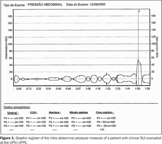
For the measurement of the intra-abdominal pressure, a Dynamed Uromaster MPX616 urodynamics device was used, measuring only the intra-abdominal pressure during the Valsalva maneuver, by means of a 10 fr rectal balloon catheter (Figure 1). The authors of the study undertook all the exams performed urogynecological, measurement of intra-abdominal pressure and ultrasound.
During the examination, the intra-abdominal pressure was measured in the moment of the Valsalva maneuver, by means of a rectal transducer, generating a graphic register (Figure 1).
RESULTS
During the Valsalva maneuver, the intra-abdominal pressure ranged between 7 cmH2O and 193 cmH2O, an average of 99.3 ± 51.8 cmH2O with pattern-deviation and a 99.5 cmH2O median. Eight of the 31 (25.8%) patients with UVJ hypermobility presented intra-abdominal pressure lower than 60 cmH2O.
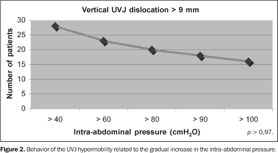
Figure 2 demonstrates that the higher the intra-abdominal pressure during UVJ perineal ultrasound evaluation, the lower number of women who presented vertical dislocation above 9 mm (hypermobility).
As shown in Table 1, the Kruskal-Wallis test, corresponding to the variance analysis for unpaired groups and determining the relation between variation of the intra-abdominal pressure and of each ultrasonographic parameter, has not detected statistical significance in these relations.
In a non-stratified way, one demonstrates a general trend toward UVJ vertical hypermobility with an intra-abdominal pressure higher than 60 cm de H2O, as per Table 2.
DISCUSSION
The SUI is highly relevant not only as a disease, but principally due to the social repercussions and the consequences for the quality of patients' life. For this reason, the SUI is considered as a problem of public health(1620).
Due to the relevance of SUI, huge public and private resources are employed for diagnosis and treatment of patients affected, mainly by means of the systematic employment of urodynamics.
In the last decade we have insisted on the necessity of rationalizing the diagnostic investigation of patients affected by SUI, above all, employing the UVJ and PU ultrasound, since it is a simple, low-cost, physiological and easily reproducible study.
The purpose of this study is not presenting a formal opposition to the indication of the complete or simplified urodynamic evaluation in the investigation of patients with SUI. On the contrary, our purpose is the enhancement of the urodynamic evaluation status as a gold standard in the investigation of patients with SUI. But, it is necessary to accept the existence of different and contradictory interpretations regarding this matter.
On the other hand, for analysis purposes, it is worthwhile mentioning that, historically, urodynamics has presented several shortcomings which were recognized and demystified along time, for example, the exaggerated significance assigned to the urethral pressure values.
Presently, the main aspect of the urodynamics is essentially the loss pressure. This measure is used to indirectly define the urethra mobility type. On the other hand, ultrasonography, as a method that does not induce detrusor contraction, directly evaluates the vertical and horizontal mobility of the UVJ and PU, besides being a real time imaging and more comfortable study(3,4).
Brandt(3) considers that urodynamics does not produce a concrete diagnosis of the SUI. As a result, there is no study using this method, showing a standard compatible with the pre and post-operatory condition of patients affected by SUI. However, there is a consensus that this test diagnoses associated conditions significant for definition of the treatment(915). Ultrasonography also does not diagnose the SUI, but clearly defines the parameters related to the UVJ and PU mobility both before and after SUI treatment(3,4).
Frequently one mentions that there is a necessity of determining if patients with SUI also have detrusor contraction diagnosed by urodynamics(915). But, we have disagreed with this interpretation because: 1) about 50% of patients with SUI have involuntary detrusor contraction as a result of the urethra hypermobility and that disappears with an adequate surgical treatment; 2) Additionally, a patient presenting clinical SUI associated with involuntary detrusor contraction and primary urethra hypermobility has indication of surgical correction of the abnormal mobility, even if the patient remains postoperatively with involuntary detrusor contraction. In this hypothesis, the involuntary detrusor contraction should be treated independently from the SUI complaint(3,4).
The patients inclusion was based on conceptual symptoms according to Abrams et al., considering that the urinary incontinence investigation and consequent determination of presence of UVJ hypermobility would be the criterion for diagnosis and therapeutic decision(19).
There is an old discussion, more and more interesting, related to the relevance of the UVJ and PU for the SUI multifactorial etiopathogeny, including whether such mobility is directly connected to the variable gradient of the intra-abdominal pressure(17,18). There is a consensus that each patient produces a different Valsalva pressure in the moment of examination, so it is our understanding that this research is important and valid for the search of a simultaneous measurement of the increase in the intra-abdominal pressure and evaluation of the UVJ and PU mobility.
Based on the literature, we understand that there are variable parameters of intra-abdominal pressure, vesical pressure and urethral mobility for each patient affected by SUI and in each moment of the examination(915).
This study initial hypothesis would be to demonstrate that, based on the simultaneous intra-abdominal pressure measurement with the UVJ and PU ultrasound, it would be possible to establish a pressoric constant for the urethral dislocation. Each patient would have a real urethral dislocation pressure that, in an association with the clinical history of SUI, could objectively guide the therapeutic conduct. However, this hypothesis is discarded, considering that our data demonstrate that there is no significant influence of the pressure over the parameters studied.
The causal factor of the changes in the measures between the rest and stress situations was the increase in the intra-abdominal pressure obtained by means of the Valsalva maneuver. This does not mean that the SUI in these patients has been caused exclusively by an increase in the intra-abdominal pressure, even because this was not the objective of this study as well as there is a concept that the SUI has multifactorial mechanisms(17,18).
However, as each patient was her own standard, the test was performed with the bladder almost emptied and the only factor changed was the intra-abdominal pressure with the Valsalva maneuver. So, one may assume the existence of a causal relationship between UVJ and PU mobility and the increase in the intra-abdominal pressure, at least with relatively low pressures between 40 e 60 cmH2O. Therefore, eight (25.8%) of the 31 patients presenting UVJ vertical hypermobility had an intra-abdominal pressure lower than 60 cmH2O (Table 2), whilst 23 (74.2%) presented hypermobility with an intra-abdominal pressure higher than 60 cmH2O, similarly to the concept of urethral loss pressure(9,10). Based on this information, one may suggest that a great abdominal effort, like coughing in the moment of the test, is dispensable; it is only necessary that the patient performs the usual Valsalva maneuver.
One may say that the intra-abdominal pressure is not an isolate parameter for changing ultrasonographic measures, in compliance with findings of other authors who report that the intra-abdominal pressure acts as one but not the only causal factors of the SUI(11,17,20).
Our ultrasonographic study is similar to that of Madjar et al.(9) who, analyzing the correlation between variation of intra-abdominal pressure and the loss pressure under stress, by means of urodynamic study, has concluded that the basal value of the intra-abdominal pressure presents a high variation among patients, depending on the pondo-statural composition and the patient's habituation to the high intra-abdominal pressure. On the other hand, it reinforces this research theory that pursues the concept of urethral dislocation pressure % a specific index for each patient that may be useful as an additional parameter in the therapeutical guidance of the SUI, independently from the multifactorial cause hypothesis.
This study demonstrates that there is not an exclusive relation between the increase in the intra-abdominal pressure and the UVJ and PU dislocations, and that there is a specific index of urethral dislocation pressure for each patient, indicating the necessity of extending the present investigation in order to adequately define the preponderant factors in the SUI etiopathogenesis. It also demonstrates that, from a practical viewpoint, each patient affected by SUI presents a higher index of dislocation with an intra-abdominal pressure lower than 40 cmH2O, compatible with the Valsalva maneuver; so it is unnecessary to measure the intra-abdominal pressure in the routine ultrasound studies for investigation of UVJ and PU.
CONCLUSION
There is a specific index of urethral dislocation pressure for each patient. However, there is no significant association between the increase in the intra-abdominal pressure and the increase in the UVJ and PU dislocation in patients presenting a clinical picture of SUI.
REFERENCES
Received June 2, 2005
Accepted after revision June 26, 2005.
- 1. Minaire P, Jacquetin B. La prévalence de l'incontinence urinaire féminine en médicine générale. J Gynecol Obstet Biol Reprod 1992;21:731738.
- 2. Johnson VY. Bladder neck suspension nursing care: preop, postop, and beyond. Perspectives in Nursing Strategies 2002;8:5357.
- 3. Brandt FT. Estudo dos parâmetros ultra-sonográficos da uretra proximal e da mobilidade da junção uretrovesical em mulheres, visando estabelecer a importância clínica. (Tese de Livre-Docência). São Paulo: USP, 1999.
- 4. Brandt FT, Albuquerque CDC, Arraes AF, Albuquerque GF, Barbosa CD, Araújo CM. Influência do volume vesical na avaliação ultra-sonográfica da junção uretrovesical e uretra proximal. Radiol Bras 2005;38:3336.
- 5. Sandvik H. Health information and interaction on the internet: a survey of female urinary incontinence. BMJ 1999;319:2932.
- 6. Brandt FT, Albuquerque CDC, Lorenzato FR, Amaral FJ. Perineal assessment of urethrovesical junction mobility in young continent females. Int Urogynecol J Pelvic Floor Dysfunct 2000;11:1822.
- 7. Dietz HP, Clarke B. The influence of posture on perineal ultrasound imaging parameters. Int Urogynecol J Pelvic Floor Dysfunct 2001;12:104106.
- 8. Dietz HP, Clarke B. Translabial color Doppler urodynamics. Int Urogynecol J Pelvic Floor Dysfunct 2001;12:304307.
- 9. Koelbl H, Bernaschek G, Deutinger J. Assessment of female urinary incontinence by introital sonography. J Clin Ultrasound 1990;18:370374.
- 10. Madjar S, Balzarro M, Appell RA, Tchegen MB, Nelson D. Baseline abdominal pressure and Valsalva leak point pressures-correlation with clinical and urodynamic data. Neurourol Urodyn 2003;22: 26.
- 11. Howard D, Miller JM, Delancey JO, Ashton-Miller JA. Differential effects of cough, Valsalva, and continence status on vesical neck movement. Obstet Gynecol 2000;95:535540.
- 12. Fusco MA, Martin RS, Chang MC. Estimation of intra-abdominal pressure by bladder pressure measurement: validity and methodology. J Trauma 2001;50:297302.
- 13. McIntosh SL, Griffiths CJ, Drinnan MJ, Robson WA, Ramsden PD, Pickard RS. Noninvasive measurement of bladder pressure. Does mechanical interruption of the urinary stream inhibit detrusor contraction? J Urol 2003;169:10031006.
- 14. Sultana CJ. Urethral closure pressure and leak-point pressure in incontinent women. Obstet Gynecol 1995;86:839842.
- 15. Swift SE, Ostergard DR. A comparison of stress leak-point pressure and maximal urethral closure pressure in patients with genuine stress incontinence. Obstet Gynecol 1995;85(5 Pt 1):704708.
- 16. Gray M. Stress urinary incontinence in women. J Am Acad Nurse Pract 2004;16:188197.
- 17. Ulmsten U. Some reflections and hypotheses on the pathophysiology of female urinary incontinence. Acta Obstet Gynecol Scand Suppl 1997;166:38.
- 18. Mouritsen L, Lose G, Ulmsten U, et al. Consensus of basic assessment of female incontinence. Acta Obstet Gynecol Scand Suppl 1997;166:5960.
- 19. Abrams P, Cardozo L, Fall M, et al. The standardisation of terminology of lower urinary tract function: report from the Standardisation Sub-Committee of the International Continence Society. Neurourol Urodyn 2002;21:16761678.
- 20. Culligan PJ, Heit M. Urinary incontinence in women: evaluation and management. Am Fam Physician 2000;62:243344, 2447, 2452.
Publication Dates
-
Publication in this collection
25 May 2006 -
Date of issue
Apr 2006
History
-
Accepted
29 June 2005 -
Received
02 June 2005


