
Abstract
Ultrasonography is an instrument that is present in the maternal-fetal assessment throughout pregnancy and with widely documented benefits, but its use in intrapartum is becoming increasingly relevant. From the assessment of labor progression to the assessment of placental disorders, ultrasound can be used to correlate with physiological findings and physical examination, as its benefit in the delivery room cannot yet be proven. There are still few professionals with adequate training for its use in the delivery room and for the correct interpretation of data. Thus, this article aims to present a review of the entire applicability of ultrasound in the delivery room, considering the main stages of labor. There is still limited research in evidence-based medicine of its various possible uses in intrapartum, but it is expected that further studies can bring improvements in the quality of maternal and neonatal health during labor.
Keywords
ultrasound; delivery room; labor; placental disorders
Resumo
A ultrassonografia é um instrumento que está presente na avaliação materno-fetal durante toda a gestação e com benefícios largamente documentados, porém sua utilização no intraparto vem sendo cada vez mais pertinente. Desde a avaliação de progressão de trabalho de parto a avaliação das desordens placentárias, a ultrassonografia pode ser empregada correlacionando com os achados fisiológicos e do exame físico, pois o seu benefício na sala de parto ainda não pode ser comprovado. Há ainda poucos profissionais com treinamento adequado para seu uso na sala de parto e para interpretação correta dos dados. Dessa forma, este artigo tem como finalidade apresentar uma revisão de toda a aplicabilidade do ultrassom na sala de parto, considerando as principais etapas do trabalho de parto. Ainda são limitadas as pesquisas em medicina baseada em evidências sobre os diversos usos possíveis no intraparto, mas espera-se que novos estudos possam trazer melhorias na qualidade da saúde materno-neonatal durante o trabalho de parto.
Palavras-chave
ultrassom; sala de parto; parto; desordens placentárias
Introduction
The use of intrapartum ultrasound has been widely reported as an additional tool for predicting the evolution of successful labor.11 Ghi T, Eggebø T, Lees C, et al. ISUOG Practice Guidelines: intrapartum ultrasound. Ultrasound Obstet Gynecol. 2018;52(01):128–139. Doi: 10.1002/uog.19072
https://doi.org/10.1002/uog.19072...
The sonographic evaluation was not shown to be superior to the vaginal examination (VE), but complementary, as the first is better for the evaluation of head station, position, and caput succedaneum, while cervix dilatation in the active stage of labor (> 4 cm) is better assessed by VE.22 Usman S, Wilkinson M, Barton H, Lees CC. The feasibility and accuracy of ultrasound assessment in the labor room. J Matern Fetal Neonatal Med. 2019;32(20):3442–3451. Doi: 10.1080/14767058.2018.1465553
https://doi.org/10.1080/14767058.2018.14...
Sonographic assistance during the first and second stages of labor has the potential to improve labor outcomes, although its real benefits have not yet been proven in large randomized trials.33 Kahrs BH, Eggebø TM. Intrapartum ultrasound in women with prolonged first stage of labor. Am J Obstet Gynecol MFM. 2021;3(6S):100427. Doi: 10.1016/j.ajogmf.2021.100427
https://doi.org/10.1016/j.ajogmf.2021.10...
4 Gimovsky AC. Intrapartum ultrasound for the diagnosis of cephalic malpositions and malpresentations. Am J Obstet Gynecol MFM. 2021;3(6S):100438. Doi: 10.1016/j.ajogmf.2021.100438
https://doi.org/10.1016/j.ajogmf.2021.10...
5 Chan VYT, Lau WL. Intrapartum ultrasound and the choice between assisted vaginal and cesarean delivery. Am J Obstet Gynecol MFM. 2021;3(6S):100439. Doi: 10.1016/j.ajogmf.2021.100439
https://doi.org/10.1016/j.ajogmf.2021.10...
-66 Ghi T. Intrapartum ultrasound and evidence-based medicine: a necessary but challenging marriage. Am J Obstet Gynecol MFM. 2021;3(6S):100428. Doi: 10.1016/j.ajogmf.2021.100428
https://doi.org/10.1016/j.ajogmf.2021.10...
In contrast, the intrapartum Doppler assessment has shown no benefit in perinatal outcomes.77 Dall'Asta A, Kumar S. Prelabor and intrapartum Doppler ultrasound to predict fetal compromise. Am J Obstet Gynecol MFM. 2021;3(6S):100479. Doi: 10.1016/j.ajogmf.2021.100479
https://doi.org/10.1016/j.ajogmf.2021.10...
Differentiated normal and abnormal sonographic postpartum findings can also be an extra implement for the patients' well-being when the clinical evaluations are doubtful.88 Steinkeler J, Coldwell BJ, Warner MA. Ultrasound of the postpartum uterus. Ultrasound Q. 2012;28(02):97–103. Doi: 10.1097/RUQ.0b013e31824e6b7d
https://doi.org/10.1097/RUQ.0b013e31824e...
Despite great acceptability by patients,99 Solaiman SA, Atwa KA, Gad AA. Al- Shatouri M. Transperineal ultrasound of fetal head progression in prolonged labor: women's acceptance and ability to predict the mode of delivery. Egypt J Radiol Nucl Med. 2020;51:94. Doi: 10.1186/s43055-020-00215-0
https://doi.org/10.1186/s43055-020-00215...
specially during stressful situations such as prolonged labor (more than 12 hours from the beginning of active phase of the first stage) and unplanned operative delivery,1010 Rizzo G, Aloisio F, Bacigalupi A, et al. Women's compliance with ultrasound in labor: a prospective observational study. J Matern Fetal Neonatal Med. 2021;34(09):1454–1458. Doi: 10.1080/14767058.2019.1638903
https://doi.org/10.1080/14767058.2019.16...
the use of intrapartum ultrasound requires a steep learning curve for good reproducibility; thus, younger obstetricians prefer to rely on clinical and digital examinations,1111 Gilboa Y, Perlman S, Karp H, Rabinovitch R, Achiron R. What do obstetricians really think about ultrasound in the delivery room? Isr Med Assoc J. 2017;19(04):234–236,1212 Plurien A, Berveiller P, Guerby P, et al. Ultrasound in delivery room: Does it have a place for the younger generation? J Gynecol Obstet Hum Reprod. 2020;49(10):101915. Doi: 10.1016/j.jogoh.2020.101915
https://doi.org/10.1016/j.jogoh.2020.101...
even though ultrasound has been proven to be more reliable than VE.
The aim of this article is to present a revision of all ultrasound applicability in the delivery room, considering the main stages of labor.
Placenta and Cord Anomalies
Placenta and cord anomalies are associated with 30% of intrauterine death risk factors and a high risk of cerebral palsy.1313 Hasegawa J. Ultrasound screening of umbilical cord abnormalities and delivery management. Placenta. 2018;62:66–78. Doi: 10.1016/j.placenta.2017.12.003
https://doi.org/10.1016/j.placenta.2017....
Therefore, they are a great cause of concern during prenatal and intrapartum period. The best time to diagnose placental implantations abnormalities is during the second trimester of pregnancy, ideally with a gestational age between 18 and 26 weeks,1414 Melcer Y, Maymon R, Jauniaux E. Vasa previa: prenatal diagnosis and management. Curr Opin Obstet Gynecol. 2018;30(06):385–391. Doi: 10.1097/GCO.0000000000000478
https://doi.org/10.1097/GCO.000000000000...
when is still possible to program the optimum time to perform cesarean section (c-section)—usually around 36 gestational weeks—modifying the neonatal and obstetric outcomes.
The umbilical cord is protected from trauma and compression through the presence of the Warthon jelly and spiraling of blood vessels.1515 Degani S, Lewinsky RM, Berger H, Spiegel D. Sonographic estimation of umbilical coiling index and correlation with Doppler flow characteristics. Obstet Gynecol. 1995;86(06):990–993. Doi: 10.1016/0029-7844(95)00307-d
https://doi.org/10.1016/0029-7844(95)003...
Literature has shown that both hypocoiled cords (spiral index below the 10th percentile) and hypercoiled cords (spiral index above the 90th percentile) are associated with unfavorable neonatal outcomes,1616 Hayes DJL, Warland J, Parast MM, et al. Umbilical cord characteristics and their association with adverse pregnancy outcomes: A systematic review and meta-analysis. PLoS One. 2020;15(09):e0239630. Doi: 10.1371/journal.pone.0239630
https://doi.org/10.1371/journal.pone.023...
17 Pergialiotis V, Kotrogianni P, Koutaki D, Christopoulos-Timogiannakis E, Papantoniou N, Daskalakis G. Umbilical cord coiling index for the prediction of adverse pregnancy outcomes: a meta-analysis and sequential analysis. J Matern Fetal Neonatal Med. 2019;33(23):1–8. Doi: 10.1080/14767058.2019.1594187
https://doi.org/10.1080/14767058.2019.15...
-1818 Mittal A, Nanda S, Sen J. Antenatal umbilical coiling index as a predictor of perinatal outcome. Arch Gynecol Obstet. 2015;291(04):763–768. Doi: 10.1007/s00404-014-3456-5
https://doi.org/10.1007/s00404-014-3456-...
such as higher rates of fetal growth restriction, fetal death, intrapartum fetal heart decelerations, karyotype abnormalities,1919 Moshiri M, Zaidi SF, Robinson TJ, et al. Comprehensive imaging review of abnormalities of the umbilical cord. Radiographics. 2014;34(01):179–196. Doi: 10.1148/rg.341125127
https://doi.org/10.1148/rg.341125127...
low birth weight (< 2,500 g), and Appearance, Pulse, Grimace, Activity, and Respiration (APGAR) score < 7 on the 1st and 5th minutes of life.1515 Degani S, Lewinsky RM, Berger H, Spiegel D. Sonographic estimation of umbilical coiling index and correlation with Doppler flow characteristics. Obstet Gynecol. 1995;86(06):990–993. Doi: 10.1016/0029-7844(95)00307-d
https://doi.org/10.1016/0029-7844(95)003...
A prenatal ultrasound assessment of cord coiling is possible; however, no benefit was found in this diagnostic screening since there are no revised means to prevent intrauterine death or a nonreassuring pattern of fetal heart rate in these cases.2020 Hasegawa J. Ultrasound assessment of the umbilical cord. Donald School J Ultrasound Obstet Gynecol. 2014;8(04):382–390. Doi: 10.5005/jp-journals-10009-1378
https://doi.org/10.5005/jp-journals-1000...
While 97% of vasa previa cases are diagnosed during prenatal scanning,1212 Plurien A, Berveiller P, Guerby P, et al. Ultrasound in delivery room: Does it have a place for the younger generation? J Gynecol Obstet Hum Reprod. 2020;49(10):101915. Doi: 10.1016/j.jogoh.2020.101915
https://doi.org/10.1016/j.jogoh.2020.101...
the benefit of performing the intrapartum diagnosis to foresee possible complications such as maternal bleeding, fetal bleeding, and neonatal death is questioned. Due to the low prevalence of this pathology (0.02–0.27% of all pregnancies),1212 Plurien A, Berveiller P, Guerby P, et al. Ultrasound in delivery room: Does it have a place for the younger generation? J Gynecol Obstet Hum Reprod. 2020;49(10):101915. Doi: 10.1016/j.jogoh.2020.101915
https://doi.org/10.1016/j.jogoh.2020.101...
prenatal screening through transvaginal ultrasound becomes unfeasible and is recommended only for women at high risk: in vitro fertilization pregnancies, placenta previa, placenta with accessory lobe, velamentous cord insertion, and multiple gestations.2121 Harding JA, Lewis DF, Major CA, Crade M, Patel J, Nageotte MP. Color flow Doppler–a useful instrument in the diagnosis of vasa previa. Am J Obstet Gynecol. 1990;163(5 Pt 1):1566–1568. Doi: 10.1016/0002-9378(90)90628-k
https://doi.org/10.1016/0002-9378(90)906...
,2222 Ruiter L, Kok N, Limpens J, et al. Incidence of and risk indicators for vasa praevia: a systematic review. BJOG. 2016;123(08):1278–1287. Doi: 10.1111/1471-0528.13829
https://doi.org/10.1111/1471-0528.13829...
Data in the literature are very vague about intrapartum diagnosis of vasa previa using the Doppler ultrasound, with only two case reports.2323 Arts H, van Eyck J. Antenatal diagnosis of vasa previa by transvaginal color Doppler sonography. Ultrasound Obstet Gynecol. 1993;3(04):276–278. Doi: 10.1046/j.1469-0705.1993.03040276.x
https://doi.org/10.1046/j.1469-0705.1993...
,2424 Baschat AA, Gembruch U. Ante- and intrapartum diagnosis of vasa praevia in singleton pregnancies by colour coded Doppler sonography. Eur J Obstet Gynecol Reprod Biol. 1998;79(01):19–25. Doi: 10.1016/s0301-2115(98)00026-8
https://doi.org/10.1016/s0301-2115(98)00...
In both cases, the correct diagnostic enables the performance of c-section before the rupture of the vasa previa, with a favorable outcome for the maternal-fetal binominal. Another condition that can lead to risk of maternal and fetal life due to bleeding is placental abruption, present in 0.4 to 1% of all pregnancies.2525 Tikkanen M. Placental abruption: epidemiology, risk factors and consequences. Acta Obstet Gynecol Scand. 2011;90(02):140–149. Doi: 10.1111/j.1600-0412.2010.01030.x
https://doi.org/10.1111/j.1600-0412.2010...
,2626 Downes KL, Grantz KL, Shenassa ED. Maternal, labor, delivery, and perinatal outcomes associated with placental abruption: a systematic review. Am J Perinatol. 2017;34(10):935–957. Doi: 10.1055/s-0037-1599149
https://doi.org/10.1055/s-0037-1599149...
The sonographic visualization of retroplacental clots is a finding present in only 15 to 25% of cases and does not interfere with the conduct regarding the interruption of pregnancy, both in term and preterm pregnancies, since maternal and fetal conditions are more important for clinical management.2727 Sholl JS. Abruptio placentae: clinical management in nonacute cases. Am J Obstet Gynecol. 1987;156(01):40–51. Doi: 10.1016/0002-9378(87)90200-6
https://doi.org/10.1016/0002-9378(87)902...
,2828 Tikkanen M, Nuutila M, Hiilesmaa V, Paavonen J, Ylikorkala O. Clinical presentation and risk factors of placental abruption. Acta Obstet Gynecol Scand. 2006;85(06):700–705. Doi: 10.1080/00016340500449915
https://doi.org/10.1080/0001634050044991...
The intrapartum ultrasound represents a sensitivity of less than 30% for the diagnosis of placental abruption, and the clinical diagnosis remains the gold standard of this obstetric emergency.2929 Qiu Y, Wu L, Xiao Y, Zhang X. Clinical analysis and classification of placental abruption. J Matern Fetal Neonatal Med. 2021;34(18):2952–2956. Doi: 10.1080/14767058.2019.1675625
https://doi.org/10.1080/14767058.2019.16...
On the other hand, the benefit of intrapartum ultrasound use has been proven in relation to the diagnosis of nuchal cord, with a sensibility of 90.2 to 96.8% when using the Doppler mode.1616 Hayes DJL, Warland J, Parast MM, et al. Umbilical cord characteristics and their association with adverse pregnancy outcomes: A systematic review and meta-analysis. PLoS One. 2020;15(09):e0239630. Doi: 10.1371/journal.pone.0239630
https://doi.org/10.1371/journal.pone.023...
,3030 Jauniaux E, Mawissa C, Peellaerts C, Rodesch F. Nuchal cord in normal third-trimester pregnancy: a color Doppler imaging study. Ultrasound Obstet Gynecol. 1992;2(06):417–419. Doi: 10.1046/j.1469-0705.1992.02060417.x
https://doi.org/10.1046/j.1469-0705.1992...
This finding is present in 22 to 45% of all pregnancies, and it is known that single nuchal cord is not associated with unfavorable perinatal outcomes.1616 Hayes DJL, Warland J, Parast MM, et al. Umbilical cord characteristics and their association with adverse pregnancy outcomes: A systematic review and meta-analysis. PLoS One. 2020;15(09):e0239630. Doi: 10.1371/journal.pone.0239630
https://doi.org/10.1371/journal.pone.023...
,3131 Clapp JF III, Stepanchak W, Hashimoto K, Ehrenberg H, Lopez B. The natural history of antenatal nuchal cords. Am J Obstet Gynecol. 2003;189(02):488–493. Doi: 10.1067/s0002-9378(03)00371-5
https://doi.org/10.1067/s0002-9378(03)00...
32 Mastrobattista JM, Hollier LM, Yeomans ER, et al. Effects of nuchal cord on birthweight and immediate neonatal outcomes. Am J Perinatol. 2005;22(02):83–85. Doi: 10.1055/s-2005-837737
https://doi.org/10.1055/s-2005-837737...
-3333 Lal N, Deka D, Mittal S. Does the nuchal cord persist? An ultrasound and color-Doppler-based prospective study. J Obstet Gynaecol Res. 2008;34(03):314–317. Doi: 10.1111/j.1447-0756.2007.00695.x
https://doi.org/10.1111/j.1447-0756.2007...
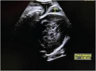
However, multiple nuchal cord is associated with worse outcomes, such as perinatal mortality, APGAR score < 7 on the 1st and 5th minutes of life, fetal distress, and meconium (Figure 1).1616 Hayes DJL, Warland J, Parast MM, et al. Umbilical cord characteristics and their association with adverse pregnancy outcomes: A systematic review and meta-analysis. PLoS One. 2020;15(09):e0239630. Doi: 10.1371/journal.pone.0239630
https://doi.org/10.1371/journal.pone.023...
,3434 Tagliaferri S, Esposito FG, Esposito G, et al. Impact of nuchal cord on antenatal and intrapartum foetal heart rate surveillance and perinatal outcome. J Obstet Gynaecol. 2020;40(03):316–323. Doi: 10.1080/01443615.2019.1621816
https://doi.org/10.1080/01443615.2019.16...
35 Peesay M. Nuchal cord and its implications. Matern Health Neonatol Perinatol. 2017;3:28. Doi: 10.1186/s40748-017-0068-7
https://doi.org/10.1186/s40748-017-0068-...
36 Spellacy WN, Gravem H, Fisch RO. The umbilical cord complications of true knots, nuchal coils, and cords around the body. Report from the collaborative study of cerebral palsy. Am J Obstet Gynecol. 1966;94(08):1136–1142. Doi: 10.1016/0002-9378(66)90777-0
https://doi.org/10.1016/0002-9378(66)907...
-3737 Pergialiotis V, Fanaki M, Bellos I, Tzortzis A, Loutradis D, Daskalakis G. Evaluation of umbilical cord entanglement as a predictive factor of adverse pregnancy outcomes: A meta-analysis. Eur J Obstet Gynecol Reprod Biol. 2019;243:150–157. Doi: 10.1016/j.ejogrb.2019.10.038
https://doi.org/10.1016/j.ejogrb.2019.10...

The intrapartum diagnostic of nuchal cord is a good tool in situations of variable deceleration in cardiotocography during labor, as it helps to recognize the cases in which the cardiotocographic pattern is not reassuring due to fetal distress and the cases when the deceleration is due to the presence of nuchal cord.3434 Tagliaferri S, Esposito FG, Esposito G, et al. Impact of nuchal cord on antenatal and intrapartum foetal heart rate surveillance and perinatal outcome. J Obstet Gynaecol. 2020;40(03):316–323. Doi: 10.1080/01443615.2019.1621816
https://doi.org/10.1080/01443615.2019.16...
Lastly, the umbilical cord prolapse is a rare situation that affects 0.12 to 0.62% of all pregnancies, with a mortality rate of up to 10% due to compression of the umbilical cord.2020 Hasegawa J. Ultrasound assessment of the umbilical cord. Donald School J Ultrasound Obstet Gynecol. 2014;8(04):382–390. Doi: 10.5005/jp-journals-10009-1378
https://doi.org/10.5005/jp-journals-1000...
Some risk factors for this comorbidity are polyhydramnios, prematurity, multiparity, multiple pregnancies, breech presentation, and low birth weight (< 2,500 g).1919 Moshiri M, Zaidi SF, Robinson TJ, et al. Comprehensive imaging review of abnormalities of the umbilical cord. Radiographics. 2014;34(01):179–196. Doi: 10.1148/rg.341125127
https://doi.org/10.1148/rg.341125127...
,2020 Hasegawa J. Ultrasound assessment of the umbilical cord. Donald School J Ultrasound Obstet Gynecol. 2014;8(04):382–390. Doi: 10.5005/jp-journals-10009-1378
https://doi.org/10.5005/jp-journals-1000...
,3838 Behbehani S, Patenaude V, Abenhaim HA. Maternal risk factors and outcomes of umbilical cord prolapse: a population-based study. J Obstet Gynaecol Can. 2016;38(01):23–28. Doi: 10.1016/j.jogc.2015.10.008
https://doi.org/10.1016/j.jogc.2015.10.0...
,3939 Dilbaz B, Ozturkoglu E, Dilbaz S, Ozturk N, Sivaslioglu AA, Haberal A. Risk factors and perinatal outcomes associated with umbilical cord prolapse. Arch Gynecol Obstet. 2006;274(02):104–107. Doi: 10.1007/s00404-006-0142-2
https://doi.org/10.1007/s00404-006-0142-...
The evident umbilical cord prolapse occurs when the umbilical cord passes between the fetal parts after the premature rupture of membranes and the diagnosis is possible through the VE, while the occult umbilical cord prolapse occurs when the membranes are intact but the cord is ahead of fetal presentation, and the diagnosis is made by ultrasound.2020 Hasegawa J. Ultrasound assessment of the umbilical cord. Donald School J Ultrasound Obstet Gynecol. 2014;8(04):382–390. Doi: 10.5005/jp-journals-10009-1378
https://doi.org/10.5005/jp-journals-1000...
The literature has shown low accuracy for the diagnosis of cord prolapse in routine ultrasound,4040 Ezra Y, Strasberg SR, Farine D. Does cord presentation on ultrasound predict cord prolapse? Gynecol Obstet Invest. 2003;56(01):6–9. Doi: 10.1159/000072323
https://doi.org/10.1159/000072323...
but has shown benefit in the use of transvaginal ultrasound to predict occult umbilical in breech presentation,4141 Kinugasa M, Sato T, Tamura M, Suzuki H, Miyazaki Y, Imanaka M. Antepartum detection of cord presentation by transvaginal ultrasonography for term breech presentation: potential prediction and prevention of cord prolapse. J Obstet Gynaecol Res. 2007;33(05):612–618. Doi: 10.1111/j.1447-0756.2007.00620.x
https://doi.org/10.1111/j.1447-0756.2007...
and the results were better when the occult cord prolapse was previously diagnosed when compared with the evident cord prolapse, suggesting that in high-risk situations, ultrasound evaluation could improve the neonatal outcomes.4242 Hasegawa J, Ikeda T, Sekizawa A, Ishiwata I, Kinoshita KJapan Association of Obstetricians and Gynecologists, Tokyo, Japan. Obstetric risk factors for umbilical cord prolapse: a nationwide population-based study in Japan. Arch Gynecol Obstet. 2016;294(03):467–472. Doi: 10.1007/s00404-015-3996-3
https://doi.org/10.1007/s00404-015-3996-...
Fetal Wellbeing During the Labor
The use of Doppler ultrasound during the labor is still limited for research purposes. However, new studies are emerging, and the application of Doppler is increasingly being studied at this time. Sütterlin et al.4343 Sütterlin MW, Seelbach-Göbel B, Oehler MK, Heupel M, Dietl J. Doppler ultrasonographic evidence of intrapartum brain-sparing effect in fetuses with low oxygen saturation according to pulse oximetry. Am J Obstet Gynecol. 1999;181(01):216–220. Doi: 10.1016/s0002-9378(99)70462-x
https://doi.org/10.1016/s0002-9378(99)70...
evaluated 70 pregnant women in early labor between 38 and 41 weeks of gestation, obtaining Doppler waveforms before and during abnormal fetal heart rate patterns. When an oxygen saturation level of < 30% was maintained for more than 2 minutes, the middle cerebral artery Doppler indices were reversed, indicating morbid fetal hypoxia. These results were considered consistent with the concept that the fetus maintains the oxygen supply to the brain by redistributing blood flow during active labor.
Chainarong and Petpichetchian4444 Chainarong N, Petpichetchian C. The relationship between intrapartum cerebroplacental ratio and adverse perinatal outcomes in term fetuses. Eur J Obstet Gynecol Reprod Biol. 2018;228:82–86. Doi: 10.1016/j.ejogrb.2018.06.016
https://doi.org/10.1016/j.ejogrb.2018.06...
evaluated the cerebroplacental ratio (CPR) during the labor, and no association was found between CPR and adverse perinatal outcomes with any CPR cut-off values. This study found that fetuses that ended up in a non-reassuring state, necessitating operative delivery, had significantly lower CPR compared with fetuses that did not. Dall'Asta et al.4545 Dall'Asta A, Ghi T, Rizzo G, et al. Cerebroplacental ratio assessment in early labor in uncomplicated term pregnancy and prediction of adverse perinatal outcome: prospective multicenter study. Ultrasound Obstet Gynecol. 2019;53(04):481–487. Doi: 10.1002/uog.19113
https://doi.org/10.1002/uog.19113...
studied the relationship between CPR measured at the beginning of labor and perinatal and delivery outcomes in a cohort of uncomplicated term pregnancies with a single child. The study's conclusion suggests that reduced CPR by itself, although associated with an increased risk of intrapartum distress, represents a poor predictor of adverse perinatal outcomes. Cochrane review assessed the effectiveness of fetal movement monitoring and Doppler ultrasound for the detection and surveillance of high-risk pregnancies and their effect in preventing stillbirths. The combined results of 16 studies showed that the umbilical arterial Doppler assessment in high-risk pregnancies leads to a 29% reduction in perinatal mortality compared with no Doppler assessment.4646 Alfirevic Z, Stampalija T, Dowswell T. Fetal and umbilical Doppler ultrasound in high-risk pregnancies. Cochrane Database Syst Rev. 2017;6(06):CD007529. Doi: 10.1002/14651858.CD007529.pub4
https://doi.org/10.1002/14651858.CD00752...
Intrapartum ultrasound (including Doppler) allowed for a greater understanding of the complex physiology of childbirth. Although promising, neither maternal nor fetal intrapartum Doppler has played a role in the true management of intrapartum ultrasound to date.4343 Sütterlin MW, Seelbach-Göbel B, Oehler MK, Heupel M, Dietl J. Doppler ultrasonographic evidence of intrapartum brain-sparing effect in fetuses with low oxygen saturation according to pulse oximetry. Am J Obstet Gynecol. 1999;181(01):216–220. Doi: 10.1016/s0002-9378(99)70462-x
https://doi.org/10.1016/s0002-9378(99)70...
Labor Progression Through Ultrasound
While digital VE are uncomfortable and subjective exams,4747 Wiafe YA, Whitehead B, Venables H, Nakua EK. The effectiveness of intrapartum ultrasonography in assessing cervical dilatation, head station and position: A systematic review and meta-analysis. Ultrasound. 2016;24(04):222–232. Doi: 10.1177/1742271X16673124
https://doi.org/10.1177/1742271X16673124...
with an error rate ranging from 26.64848 Akmal S, Kametas N, Tsoi E, Hargreaves C, Nicolaides KH. Comparison of transvaginal digital examination with intrapartum sonography to determine fetal head position before instrumental delivery. Ultrasound Obstet Gynecol. 2003;21(05):437–440. Doi: 10.1002/uog.103
https://doi.org/10.1002/uog.103...
to 33.5%4949 Akmal S, Tsoi E, Kametas N, Howard R, Nicolaides KH. Intrapartum sonography to determine fetal head position. J Matern Fetal Neonatal Med. 2002;12(03):172–177. Doi: 10.1080/jmf.12.3.172.177
https://doi.org/10.1080/jmf.12.3.172.177...
due to interexaminer reproducibility, sonographic measurements are more reliable and could be an additional tool for the evaluation and estimation of a successful labor.5050 Yeo L, Romero R. Sonographic evaluation in the second stage of labor to improve the assessment of labor progress and its outcome. Ultrasound Obstet Gynecol. 2009;33(03):253–258. Doi: 10.1002/uog.6336
https://doi.org/10.1002/uog.6336...
Besides, multiple digital VE are associated with ascending infection to the fetus and the uterus5151 Ahn KH, Oh MJ. Intrapartum ultrasound: A useful method for evaluating labor progress and predicting operative vaginal delivery. Obstet Gynecol Sci. 2014;57(06):427–435. Doi: 10.5468/ogs.2014.57.6.427
https://doi.org/10.5468/ogs.2014.57.6.42...
,5252 Benediktsdottir S, Eggebø TM, Salvesen KÅ Agreement between transperineal ultrasound measurements and digital examinations of cervical dilatation during labor. BMC Pregnancy Childbirth. 2015;15:273. Doi: 10.1186/s12884-015-0704-z
https://doi.org/10.1186/s12884-015-0704-...
and are contraindicated in some situations, such as preterm prelabor rupture of membranes and placenta previa.5353 Usman S, Lees C. Benefits and pitfalls of the use of intrapartum ultrasound. Australas J Ultrasound Med. 2015;18(02):53–59. Doi: 10.1002/j.2205-0140.2015.tb00042.x
https://doi.org/10.1002/j.2205-0140.2015...
It is possible to get valuable information that could not be obtained in a VE, such as angle of progression (AoP) which is the angle between a line in the midline of the pubic symphysis and a line running tangentially from the anterior edge of the symphysis to the fetal skull evaluated through transperineal ultrasound (Figure 2); the head progression distance (HPD) which is the shortest distance between the infrapubic line and the leading edge of the fetal skull, also evaluated through transperineal ultrasound; and the head direction (HD) which is the angle between the infrapubic line, perpendicular to the most caudal part of the pubic symphysis, and a line drawn perpendicular to the widest diameter of fetal head, evaluated through abdominal ultrasound.4747 Wiafe YA, Whitehead B, Venables H, Nakua EK. The effectiveness of intrapartum ultrasonography in assessing cervical dilatation, head station and position: A systematic review and meta-analysis. Ultrasound. 2016;24(04):222–232. Doi: 10.1177/1742271X16673124
https://doi.org/10.1177/1742271X16673124...
,5454 Kameyama S, Sato A, Miura H, et al. Prediction of spontaneous vaginal delivery by transperineal ultrasound performed just after full cervical dilatation is determined. J Med Ultrason. 2016;43(02):243–248 The AoP is the most useful measure to predict the success of vaginal delivery, with the manual parasagittal technique being the most reliable,5555 Frick A, Kostiv V, Vojtassakova D, Akolekar R, Nicolaides KH. Comparison of different methods of measuring angle of progression in prediction of labor outcome. Ultrasound Obstet Gynecol. 2020;55(03):391–400. Doi: 10.1002/uog.21913
https://doi.org/10.1002/uog.21913...
in which the angle is formed between a line drawn along the superior-inferior axis of the pubic bone and a line drawn along the inferior end of the hyperechogenic pelvic bone forming the vertex of the angle with the fetal head.

A systematic review has shown that ultrasound is superior to digital VE for evaluation of fetal head position in the first stage of labor, in addition to the great agreement between the two methods in the assessment of cervical dilatation and a moderate correlation for fetal head station.4848 Akmal S, Kametas N, Tsoi E, Hargreaves C, Nicolaides KH. Comparison of transvaginal digital examination with intrapartum sonography to determine fetal head position before instrumental delivery. Ultrasound Obstet Gynecol. 2003;21(05):437–440. Doi: 10.1002/uog.103
https://doi.org/10.1002/uog.103...
Although the success rate of digital VE increases with the progression of cervical dilatation, approximately a quarter of digital assessments differ by more than 45° when compared with the sonographic evaluation of fetal head position,4848 Akmal S, Kametas N, Tsoi E, Hargreaves C, Nicolaides KH. Comparison of transvaginal digital examination with intrapartum sonography to determine fetal head position before instrumental delivery. Ultrasound Obstet Gynecol. 2003;21(05):437–440. Doi: 10.1002/uog.103
https://doi.org/10.1002/uog.103...
,4949 Akmal S, Tsoi E, Kametas N, Howard R, Nicolaides KH. Intrapartum sonography to determine fetal head position. J Matern Fetal Neonatal Med. 2002;12(03):172–177. Doi: 10.1080/jmf.12.3.172.177
https://doi.org/10.1080/jmf.12.3.172.177...
,5353 Usman S, Lees C. Benefits and pitfalls of the use of intrapartum ultrasound. Australas J Ultrasound Med. 2015;18(02):53–59. Doi: 10.1002/j.2205-0140.2015.tb00042.x
https://doi.org/10.1002/j.2205-0140.2015...
which may lead to unfavorable neonatal outcomes, especially when instrumented deliveries are necessary.5353 Usman S, Lees C. Benefits and pitfalls of the use of intrapartum ultrasound. Australas J Ultrasound Med. 2015;18(02):53–59. Doi: 10.1002/j.2205-0140.2015.tb00042.x
https://doi.org/10.1002/j.2205-0140.2015...
Nevertheless, ultrasound evaluation is associated with higher rates of instrumental vaginal delivery,5656 Popowski T, Porcher R, Fort J, Javoise S, Rozenberg P. Influence of ultrasound determination of fetal head position on mode of delivery: a pragmatic randomized trial. Ultrasound Obstet Gynecol. 2015;46(05):520–525. Doi: 10.1002/uog.14785
https://doi.org/10.1002/uog.14785...
with no difference in maternal and neonatal morbidities when compared with exclusive VE evaluation before operative delivery,5656 Popowski T, Porcher R, Fort J, Javoise S, Rozenberg P. Influence of ultrasound determination of fetal head position on mode of delivery: a pragmatic randomized trial. Ultrasound Obstet Gynecol. 2015;46(05):520–525. Doi: 10.1002/uog.14785
https://doi.org/10.1002/uog.14785...
57 Ramphul M, Ooi PV, Burke G, et al. Instrumental delivery and ultrasound : a multicentre randomised controlled trial of ultrasound assessment of the fetal head position versus standard care as an approach to prevent morbidity at instrumental delivery. BJOG. 2014;121(08):1029–1038. Doi: 10.1111/1471-0528.12810
https://doi.org/10.1111/1471-0528.12810...
-5858 Mappa I, Tartaglia S, Maqina P, et al. Ultrasound vs routine care before instrumental vaginal delivery: A systematic review and meta-analysis. Acta Obstet Gynecol Scand. 2021;100(11):1941-–1948. Doi: 10.1111/aogs.14236
https://doi.org/10.1111/aogs.14236...
nor in relation to c-section rate, even though ultrasound evaluation corresponds to a greater success in the diagnoses of fetal head position and station.5858 Mappa I, Tartaglia S, Maqina P, et al. Ultrasound vs routine care before instrumental vaginal delivery: A systematic review and meta-analysis. Acta Obstet Gynecol Scand. 2021;100(11):1941-–1948. Doi: 10.1111/aogs.14236
https://doi.org/10.1111/aogs.14236...
Kameyama et al.5454 Kameyama S, Sato A, Miura H, et al. Prediction of spontaneous vaginal delivery by transperineal ultrasound performed just after full cervical dilatation is determined. J Med Ultrason. 2016;43(02):243–248 described an optimal cut-off from prediction of spontaneous vaginal delivery of 83° for HPD (positive predictive value, PPV = 92.9%), 56 mm for PD (PPV = 94.4%) and AoP of 146° (PPV = 94.3%) right after full cervical dilatation. Ghi et al.5959 Ghi T, Youssef A, Maroni E, et al. Intrapartum transperineal ultrasound assessment of fetal head progression in active second stage of labor and mode of delivery. Ultrasound Obstet Gynecol. 2013;41(04):430–435. Doi: 10.1002/uog.12379
https://doi.org/10.1002/uog.12379...
have shown that women with spontaneous vaginal delivery had a wider AoP in the begging of second stage of labor (140° ± 20.2°) than the women who had been submitted to operative delivery (122.9° ± 16.7°). Sainz et al.6060 Sainz JA, García-Mejido JA, Aquise A, et al. Intrapartum transperineal ultrasound used to predict cases of complicated operative (vacuum and forceps) deliveries in nulliparous women. Acta Obstet Gynecol Scand. 2017;96(12):1490–1497. Doi: 10.1111/aogs.13230
https://doi.org/10.1111/aogs.13230...
have found that an AoP of 122° ( ± 17.8°) is associated with a complicated operative delivery in nulliparous woman, while an AoP of 149.2° ( ± 15.6°) and a HPD of 50.5 mm are good predictors of uncomplicated deliveries. These facts are consistent with the findings Bultez et al.,6161 Bultez T, Quibel T, Bouhanna P, Popowski T, Resche-Rigon M, Rozenberg P. Angle of fetal head progression measured using transperineal ultrasound as a predictive factor of vacuum extraction failure. Ultrasound Obstet Gynecol. 2016;48(01):86–91. Doi: 10.1002/uog.14951
https://doi.org/10.1002/uog.14951...
in which the median of the AoP of 145° is associated with a successful delivery with vacuum extraction, whereas the median of AoP of 136° corresponds to vacuum extraction failure. On the other hand, Kalache et al.6262 Kalache KD, Dückelmann AM, Michaelis SA, Lange J, Cichon G, Dudenhausen JW. Transperineal ultrasound imaging in prolonged second stage of labor with occipitoanterior presenting fetuses: how well does the ‘angle of progression’ predict the mode of delivery? Ultrasound Obstet Gynecol. 2009;33(03):326–330. Doi: 10.1002/uog.6294
https://doi.org/10.1002/uog.6294...
described an AoP of 120° as leading to the probability of an easy and successful vacuum or spontaneous vaginal delivery in 90% of the cases.
Chan et al.6363 Chan WWY, Chaemsaithong P, Lim WT, et al. Pre-induction transperineal ultrasound assessment of the prediction of labor outcome. Fetal Diagn Ther. 2019;45(04):256–267. Doi: 10.1159/000489122
https://doi.org/10.1159/000489122...
have shown that parasagittal AoP is an independent predictor for c-section and for non-progression before induction of labor: women with manual parasagittal AoP of 102° (93–111°) and automated parasagittal AoP of 108° (99–115°) were more likely to give birth through vaginal delivery, while women with manual parasagittal AoP of 93° (90–102°) and automated parasagittal AoP of 99° (93–104°) were submitted to c-sections, with no difference between nulliparous and multiparous women. Tse et al.6464 Tse WT, Chaemsaithong P, Chan WWY, et al. Labor progress determined by ultrasound is different in women requiring cesarean delivery from those who experience a vaginal delivery following induction of labor. Am J Obstet Gynecol. 2019;221(04):335.e1–335.e18. Doi: 10.1016/j.ajog.2019.05.040
https://doi.org/10.1016/j.ajog.2019.05.0...
have also shown an additional decrease of 5.28° in the parasagittal AoP and an additional increase of 0.27 cm in HPD for a unit increase in fetal head station and cervical dilation in women requiring c-section, while the additional decrease was 1.35° in the parasagittal AoP and the additional increase was 0.12 cm in HPD in women who achieved vaginal delivery.
Birth weight is an important predictor of neonatal morbidity and mortality and has a strong influence on obstetric and neonatal management.6565 Faschingbauer F, Dammer U, Raabe E, et al. Intrapartum sonographic weight estimation. Arch Gynecol Obstet. 2015;292(04):805–811. Doi: 10.1007/s00404-015-3720-3
https://doi.org/10.1007/s00404-015-3720-...
Stubert et al.6666 Stubert J, Peschel A, Bolz M, Glass Ä, Gerber B. Accuracy of immediate antepartum ultrasound estimated fetal weight and its impact on mode of delivery and outcome - a cohort analysis. BMC Pregnancy Childbirth. 2018;18(01):118. Doi: 10.1186/s12884-018-1772-7
https://doi.org/10.1186/s12884-018-1772-...
confirmed that the ultrasound-derived estimated fetal weight during labor at term is an appropriate diagnostic tool, with an average accuracy of 70% within a relative difference of ± 10% to the real birth weight. Furthermore, term-estimated fetal weight has been shown to be unreliable for predicting macrosomia and is therefore not recommended.6666 Stubert J, Peschel A, Bolz M, Glass Ä, Gerber B. Accuracy of immediate antepartum ultrasound estimated fetal weight and its impact on mode of delivery and outcome - a cohort analysis. BMC Pregnancy Childbirth. 2018;18(01):118. Doi: 10.1186/s12884-018-1772-7
https://doi.org/10.1186/s12884-018-1772-...
Considering international guidelines, the cesarean delivery rate should not be higher if fetal weight is estimated immediately before delivery. However, overestimation of fetal weight was associated with an increased risk of c-section.6565 Faschingbauer F, Dammer U, Raabe E, et al. Intrapartum sonographic weight estimation. Arch Gynecol Obstet. 2015;292(04):805–811. Doi: 10.1007/s00404-015-3720-3
https://doi.org/10.1007/s00404-015-3720-...
In this study, the increase in the rate of c-section was not accompanied by a decrease in fetal or maternal morbidity. No differences were observed in shoulder dystocia and in third- and fourth-degree perineal lacerations.6666 Stubert J, Peschel A, Bolz M, Glass Ä, Gerber B. Accuracy of immediate antepartum ultrasound estimated fetal weight and its impact on mode of delivery and outcome - a cohort analysis. BMC Pregnancy Childbirth. 2018;18(01):118. Doi: 10.1186/s12884-018-1772-7
https://doi.org/10.1186/s12884-018-1772-...
Yang et al.6767 Yang JM, Hyett JA, Mcgeechan K, Phipps H, de Vries BS. Is ultrasound measured fetal biometry predictive of intrapartum caesarean section for failure to progress? Aust N Z J Obstet Gynaecol. 2018;58(06):620–628. Doi: 10.1111/ajo.12776
https://doi.org/10.1111/ajo.12776...
found that biparietal diameter, abdominal circumference, and estimated fetal weight at 38 weeks of gestation were associated with c-sections for failure to progress in labor after adjusting for confounders. Routine biometry may help identify patients whose intrapartum c-section risk could be reduced by elective induction at 39 weeks. Faschingbauer et al.6565 Faschingbauer F, Dammer U, Raabe E, et al. Intrapartum sonographic weight estimation. Arch Gynecol Obstet. 2015;292(04):805–811. Doi: 10.1007/s00404-015-3720-3
https://doi.org/10.1007/s00404-015-3720-...
found that the best results regarding intrapartum estimated fetal weight can be obtained with formulas that use biparietal diameter as the only head measurement. Little et al.6868 Little SE, Edlow AG, Thomas AM, Smith NA. Estimated fetal weight by ultrasound: a modifiable risk factor for cesarean delivery? Am J Obstet Gynecol. 2012;207(04):309.e1–309.e6. Doi: 10.1016/j.ajog.2012.06.065
https://doi.org/10.1016/j.ajog.2012.06.0...
suggest that provider knowledge may be associated with a higher rate of c-section; therefore, limiting ultrasound check of fetal weight in the short term may help reduce c-section rate.
A different use of ultrasound during the labor is by creating a sonopartogram, which is a conformation of the conventional partogram, with the use of ultrasound parameters of recording assessments during the labor.6969 Hassan WA, Eggebø T, Ferguson M, et al. The sonopartogram: a novel method for recording progress of labor by ultrasound. Ultrasound Obstet Gynecol. 2014;43(02):189–194. Doi: 10.1002/uog.13212
https://doi.org/10.1002/uog.13212...
,7070 Dira LM, Tudorache S, Antsaklis P, et al. Sonographic Evaluation of the Mechanism of Active Labor (SonoLabor Study): observational study protocol regarding the implementation of the sonopartogram. BMJ Open. 2021;11(09):e047188. Doi: 10.1136/bmjopen-2020-047188
https://doi.org/10.1136/bmjopen-2020-047...
It is possible to evaluate cervical dilatation, fetal head rotation, and fetal head descent, as it is in the conventional partogram, as well as to evaluate caput and molding6969 Hassan WA, Eggebø T, Ferguson M, et al. The sonopartogram: a novel method for recording progress of labor by ultrasound. Ultrasound Obstet Gynecol. 2014;43(02):189–194. Doi: 10.1002/uog.13212
https://doi.org/10.1002/uog.13212...
,7070 Dira LM, Tudorache S, Antsaklis P, et al. Sonographic Evaluation of the Mechanism of Active Labor (SonoLabor Study): observational study protocol regarding the implementation of the sonopartogram. BMJ Open. 2021;11(09):e047188. Doi: 10.1136/bmjopen-2020-047188
https://doi.org/10.1136/bmjopen-2020-047...
(Figure 3). Although a good agreement was shown between VE and ultrasound evaluation regarding cervical dilatation and head rotation during the first period of labor, the evaluation of head descent was better estimated by VE.6060 Sainz JA, García-Mejido JA, Aquise A, et al. Intrapartum transperineal ultrasound used to predict cases of complicated operative (vacuum and forceps) deliveries in nulliparous women. Acta Obstet Gynecol Scand. 2017;96(12):1490–1497. Doi: 10.1111/aogs.13230
https://doi.org/10.1111/aogs.13230...
Another possibility for the use of ultrasound in the delivery room would be the prediction of success for vaginal delivery on patients with leiomyomas located in regions close to cervix; however, we did not find any data about this topic (Figure 4).

Ultrasonography at the Immediate Postpartum
Considering postpartum hemorrhage is the leading cause of maternal death worldwide,7171 Urner F, Zimmermann R, Krafft A. Manual removal of the placenta after vaginal delivery: an unsolved problem in obstetrics. J Pregnancy. 2014;2014:274651. Doi: 10.1155/2014/274651
https://doi.org/10.1155/2014/274651...
it is logical to think that ultrasound in the immediate postpartum could be an extra resource to identify possible cases of hemorrhagic complications, including uterine atony, retained products of conception, uterine arteriovenous malformations, and hematomas due birth canal trauma, as well as a good tool for specific treatments, such as curettage, embolization of uterine arteries, and the use of the Bakri balloon. In the immediate postpartum period, the transabdominal approach is preferable for uterus evaluation rather than transvaginal approach.7272 Mulic-Lutvica A. Postpartum ultrasound. Donald School J Ultrasound Obstet Gynecol. 2012;6(01):76–92
The main cause of postpartum hemorrhage is uterine atony, which can be identified by the loss of at least 500 ml of blood after vaginal delivery or 1000 ml after c-section, associated with the lack of the Pinnard security globe, which represents the uterus involution and can be diagnosed by physical exam, through palpation of the uterine height.7373 Üçyiğit A, Johns J. The postpartum ultrasound scan. Ultrasound. 2016;24(03):163–169. Doi: 10.1177/1742271X16653779
https://doi.org/10.1177/1742271X16653779...
,7474 Plunk M, Lee JH, Kani K, Dighe M. Imaging of postpartum complications: a multimodality review. AJR Am J Roentgenol. 2013;200(02):W143-54. Doi: 10.2214/AJR.12.9637
https://doi.org/10.2214/AJR.12.9637...
Through sonographic exam, the mean uterine length is 16.1 ± 1.7 cm,7575 Sokol ER, Casele H, Haney EI. Ultrasound examination of the postpartum uterus: what is normal? J Matern Fetal Neonatal Med. 2004;15(02):95–99. Doi: 10.1080/14767050310001650798
https://doi.org/10.1080/1476705031000165...
while the maximum anterior-posterior uterine dimeter reported was 9.2 cm7676 Mulic-Lutvica A, Bekuretsion M, Bakos O, Axelsson O. Ultrasonic evaluation of the uterus and uterine cavity after normal, vaginal delivery. Ultrasound Obstet Gynecol. 2001;18(05):491–498. Doi: 10.1046/j.0960-7692.2001.00561.x
https://doi.org/10.1046/j.0960-7692.2001...
(Figure 5). Concerning endometrial evaluation, some studies have shown no correlation between the duration or amount of bleeding and the presence of echogenic material diagnosed by postpartum ultrasound.7575 Sokol ER, Casele H, Haney EI. Ultrasound examination of the postpartum uterus: what is normal? J Matern Fetal Neonatal Med. 2004;15(02):95–99. Doi: 10.1080/14767050310001650798
https://doi.org/10.1080/1476705031000165...
,7777 Deans R, Dietz HP. Ultrasound of the post-partum uterus. Aust N Z J Obstet Gynaecol. 2006;46(04):345–349. Doi: 10.1111/j.1479-828X.2006.00604.x
https://doi.org/10.1111/j.1479-828X.2006...
78 Fuller KP, Feldman DM. Ultrasound evaluation of the postpartum endometrial cavity. J Reprod Med. 2015;60(1-2):3–5-7979 Edwards A, Ellwood DA. Ultrasonographic evaluation of the postpartum uterus. Ultrasound Obstet Gynecol. 2000;16(07):640–643. Doi: 10.1046/j.1469-0705.2000.00234.x
https://doi.org/10.1046/j.1469-0705.2000...
A recent systematic review has found that the upper limit for endometrial thickness (95th centile) measured by abdominal ultrasound within 24 hours postpartum is 22 mm,8080 Ucci MA, Di Mascio D, Bellussi F, Berghella V. Ultrasound evaluation of the uterus in the uncomplicated postpartum period: a systematic review. Am J Obstet Gynecol MFM. 2021;3(03):100318. Doi: 10.1016/j.ajogmf.2021.100318
https://doi.org/10.1016/j.ajogmf.2021.10...
with no statistically significant difference between vaginal delivery or c-section, or between nulliparous and multiparous women.7777 Deans R, Dietz HP. Ultrasound of the post-partum uterus. Aust N Z J Obstet Gynaecol. 2006;46(04):345–349. Doi: 10.1111/j.1479-828X.2006.00604.x
https://doi.org/10.1111/j.1479-828X.2006...
,8080 Ucci MA, Di Mascio D, Bellussi F, Berghella V. Ultrasound evaluation of the uterus in the uncomplicated postpartum period: a systematic review. Am J Obstet Gynecol MFM. 2021;3(03):100318. Doi: 10.1016/j.ajogmf.2021.100318
https://doi.org/10.1016/j.ajogmf.2021.10...

Another possible cause of postpartum bleeding is the presence of retained placental tissue, which can happen in approximately 1% of term deliveries.8181 Weissbach T, Haikin-Herzberger E, Bacci-Hugger K, Shechter-Maor G, Fejgin M, Biron-Shental T. Immediate postpartum ultrasound evaluation for suspected retained placental tissue in patients undergoing manual removal of placenta. Eur J Obstet Gynecol Reprod Biol. 2015;192:37–40. Doi: 10.1016/j.ejogrb.2015.06.004
https://doi.org/10.1016/j.ejogrb.2015.06...
The literature has shown a variable sensitivity (42–94%) and specificity (62–92%) for the use of ultrasound in the uterus evaluation after placental removal.8282 Carlan SJ, Scott WT, Pollack R, Harris K. Appearance of the uterus by ultrasound immediately after placental delivery with pathologic correlation. J Clin Ultrasound. 1997;25(06):301–308. Doi: 10.1002/(sici)1097-0096(199707)25:6<301:aid-jcu3>3.0.co;2-g
https://doi.org/10.1002/(sici)1097-0096(...
The gray scale itself is not the best option as a diagnostic method, as the appearances of retained placental tissue in the immediate postpartum are highly variable and can be represented as echogenic mass, heterogeneous mixed density mass, and normal endometrial cavity. Therefore, it could not be correlated with a need for intervention and might not change patient outcomes.8181 Weissbach T, Haikin-Herzberger E, Bacci-Hugger K, Shechter-Maor G, Fejgin M, Biron-Shental T. Immediate postpartum ultrasound evaluation for suspected retained placental tissue in patients undergoing manual removal of placenta. Eur J Obstet Gynecol Reprod Biol. 2015;192:37–40. Doi: 10.1016/j.ejogrb.2015.06.004
https://doi.org/10.1016/j.ejogrb.2015.06...
,8282 Carlan SJ, Scott WT, Pollack R, Harris K. Appearance of the uterus by ultrasound immediately after placental delivery with pathologic correlation. J Clin Ultrasound. 1997;25(06):301–308. Doi: 10.1002/(sici)1097-0096(199707)25:6<301:aid-jcu3>3.0.co;2-g
https://doi.org/10.1002/(sici)1097-0096(...
The identification of thickened endometrial echo complex > 10 mm, associated with vascular flow detection on color Doppler, is highly suggestive for retained placental tissue.8383 Kamaya A, Ro K, Benedetti NJ, Chang PL, Desser TS. Imaging and diagnosis of postpartum complications: sonography and other imaging modalities. Ultrasound Q. 2009;25(03):151–162. Doi: 10.1097/RUQ.0b013e3181b5451e
https://doi.org/10.1097/RUQ.0b013e3181b5...
However, a hypervascular area can be physiologic in the postpartum period and disappear spontaneously or after removal of placental remnants8484 Van den Bosch T, Van Schoubroeck D, Lu C, De Brabanter J, Van Huffel S, Timmerman D. Color Doppler and gray-scale ultrasound evaluation of the postpartum uterus. Ultrasound Obstet Gynecol. 2002;20(06):586–591. Doi: 10.1046/j.1469-0705.2002.00851.x
https://doi.org/10.1046/j.1469-0705.2002...
,8585 Durfee SM, Frates MC, Luong A, Benson CB. The sonographic and color Doppler features of retained products of conception. J Ultrasound Med. 2005;24(09):1181–1186, quiz 1188–1189. Doi: 10.7863/jum.2005.24.9.1181
https://doi.org/10.7863/jum.2005.24.9.11...
and, therefore, there is no gold standard protocol for diagnosing retained placental tissue through imaging exams.8686 De Winter J, De Raedemaecker H, Muys J, Jacquemyn Y. The value of postpartum ultrasound for the diagnosis of retained products of conception: A systematic review. Facts Views Vis ObGyn. 2017;9(04):207–216
As for the placenta accreta spectrum, the diagnostic should preferably happen prenatally, so the best time and place for delivery can be arranged, as an intraoperative hysterectomy might be necessary.8787 Cahill AG, Beigi R, Heine RP, Silver RM, Wax JRSociety of Gynecologic Oncology American College of Obstetricians and Gynecologists and the Society for Maternal–Fetal Medicine. Placenta accreta spectrum. Am J Obstet Gynecol. 2018;219(06):B2–B16. Doi: 10.1016/j.ajog.2018.09.042
https://doi.org/10.1016/j.ajog.2018.09.0...
However, when there is no prenatal diagnosis and the patient goes through labor, the diagnostic is made during the third stage of labor, which may lead to major bleeding.8787 Cahill AG, Beigi R, Heine RP, Silver RM, Wax JRSociety of Gynecologic Oncology American College of Obstetricians and Gynecologists and the Society for Maternal–Fetal Medicine. Placenta accreta spectrum. Am J Obstet Gynecol. 2018;219(06):B2–B16. Doi: 10.1016/j.ajog.2018.09.042
https://doi.org/10.1016/j.ajog.2018.09.0...
,8888 Krapp M, Baschat AA, Hankeln M, Gembruch U. Gray scale and color Doppler sonography in the third stage of labor for early detection of failed placental separation. Ultrasound Obstet Gynecol. 2000;15(02):138–142. Doi: 10.1046/j.1469-0705.2000.00063.x
https://doi.org/10.1046/j.1469-0705.2000...
The normal placental separation can be sonographically characterized by decreased blood flow while the placenta is detaching from the myometrium, whereas the presence of placenta accreta spectrum can be sonographically characterized by the persistent blood flow between the placenta and the myometrium.8888 Krapp M, Baschat AA, Hankeln M, Gembruch U. Gray scale and color Doppler sonography in the third stage of labor for early detection of failed placental separation. Ultrasound Obstet Gynecol. 2000;15(02):138–142. Doi: 10.1046/j.1469-0705.2000.00063.x
https://doi.org/10.1046/j.1469-0705.2000...
A rare, but serious situation of postpartum hemorrhage, is the uterine rupture. Its prevalence is less than 1% after a vaginal delivery after one c-section, increasing to up to 2% when the vaginal delivery occurs after more than two previous c-sections.8989 Habak PJ, Kole M. Vaginal birth after cesarean delivery. In: StatPearls [Internet]. Treasure Island: StatPearls Publishing; 2021 [cited 2022 Jan 20]. Available from: https://www.ncbi.nlm.nih.gov/books/NBK507844/
https://www.ncbi.nlm.nih.gov/books/NBK50...
It should be suspected in patients with vaginal delivery after c-section or any uterine surgery, presenting postpartum hemorrhage and hypovolemic shock.9090 Oba T, Hasegawa J, Sekizawa A. Postpartum ultrasound: postpartum assessment using ultrasonography. J Matern Fetal Neonatal Med. 2017;30(14):1726–1729. Doi: 10.1080/14767058.2016.1223034
https://doi.org/10.1080/14767058.2016.12...
The diagnostic must be done as soon as possible, and a transabdominal ultrasound would show an echo-free space or mass lesions, possibly corresponding to intraperitoneal bleeding or retroperitoneal hematoma.9090 Oba T, Hasegawa J, Sekizawa A. Postpartum ultrasound: postpartum assessment using ultrasonography. J Matern Fetal Neonatal Med. 2017;30(14):1726–1729. Doi: 10.1080/14767058.2016.1223034
https://doi.org/10.1080/14767058.2016.12...
Lastly, an unusual but possible cause of hemodynamic instability in the delivery room is the presence of a vulvar or paravaginal hematoma.9191 Bellussi F, Cataneo I, Dodaro MG, Youssef A, Salsi G, Pilu G. The use of ultrasound in the evaluation of postpartum paravaginal hematomas. Am J Obstet Gynecol MFM. 2019;1(01):82–88. Doi: 10.1016/j.ajogmf.2019.03.002
https://doi.org/10.1016/j.ajogmf.2019.03...
,9292 Oong GC, Eruo FU. Vulvar hematoma. In: StatPearls [Internet]. Treasure Island: StatPearls Publishing; 2021 [cited 2022 Jan 20]. Available from: https://www.ncbi.nlm.nih.gov/books/NBK560753/
https://www.ncbi.nlm.nih.gov/books/NBK56...
This complication might happen specially after direct injury of the perineum, from instrumental deliveries, vaginal laceration, or episiotomy.9292 Oong GC, Eruo FU. Vulvar hematoma. In: StatPearls [Internet]. Treasure Island: StatPearls Publishing; 2021 [cited 2022 Jan 20]. Available from: https://www.ncbi.nlm.nih.gov/books/NBK560753/
https://www.ncbi.nlm.nih.gov/books/NBK56...
The main symptoms are pelvic and perianal pain, swelling of the vulva, paravaginal mass, and urinary retention due mechanical urethral obstruction.9191 Bellussi F, Cataneo I, Dodaro MG, Youssef A, Salsi G, Pilu G. The use of ultrasound in the evaluation of postpartum paravaginal hematomas. Am J Obstet Gynecol MFM. 2019;1(01):82–88. Doi: 10.1016/j.ajogmf.2019.03.002
https://doi.org/10.1016/j.ajogmf.2019.03...
,9292 Oong GC, Eruo FU. Vulvar hematoma. In: StatPearls [Internet]. Treasure Island: StatPearls Publishing; 2021 [cited 2022 Jan 20]. Available from: https://www.ncbi.nlm.nih.gov/books/NBK560753/
https://www.ncbi.nlm.nih.gov/books/NBK56...
The use of transperineal or transabdominal ultrasound can provide precise information about the presence, location, and size of the vaginal hematoma, with similar results findings with computed tomography, but with the advantage that it can be performed in the labor ward, immediately after delivery.9191 Bellussi F, Cataneo I, Dodaro MG, Youssef A, Salsi G, Pilu G. The use of ultrasound in the evaluation of postpartum paravaginal hematomas. Am J Obstet Gynecol MFM. 2019;1(01):82–88. Doi: 10.1016/j.ajogmf.2019.03.002
https://doi.org/10.1016/j.ajogmf.2019.03...
Conclusion
The performance of ultrasound in the delivery room is still a poorly explored resource in maternity hospitals. However, with the potential to improve the diagnosis and interpretations of situations and allow for more timely interventions, since it is a tool with the potential to complement (and not replace) clinical practice. There is still little evidence-based medical research on the several possibilities of its intrapartum use, but we expect that further studies could provide improvements in the quality of maternal-neonatal health during the labor.
References
-
1Ghi T, Eggebø T, Lees C, et al. ISUOG Practice Guidelines: intrapartum ultrasound. Ultrasound Obstet Gynecol. 2018;52(01):128–139. Doi: 10.1002/uog.19072
» https://doi.org/10.1002/uog.19072 -
2Usman S, Wilkinson M, Barton H, Lees CC. The feasibility and accuracy of ultrasound assessment in the labor room. J Matern Fetal Neonatal Med. 2019;32(20):3442–3451. Doi: 10.1080/14767058.2018.1465553
» https://doi.org/10.1080/14767058.2018.1465553 -
3Kahrs BH, Eggebø TM. Intrapartum ultrasound in women with prolonged first stage of labor. Am J Obstet Gynecol MFM. 2021;3(6S):100427. Doi: 10.1016/j.ajogmf.2021.100427
» https://doi.org/10.1016/j.ajogmf.2021.100427 -
4Gimovsky AC. Intrapartum ultrasound for the diagnosis of cephalic malpositions and malpresentations. Am J Obstet Gynecol MFM. 2021;3(6S):100438. Doi: 10.1016/j.ajogmf.2021.100438
» https://doi.org/10.1016/j.ajogmf.2021.100438 -
5Chan VYT, Lau WL. Intrapartum ultrasound and the choice between assisted vaginal and cesarean delivery. Am J Obstet Gynecol MFM. 2021;3(6S):100439. Doi: 10.1016/j.ajogmf.2021.100439
» https://doi.org/10.1016/j.ajogmf.2021.100439 -
6Ghi T. Intrapartum ultrasound and evidence-based medicine: a necessary but challenging marriage. Am J Obstet Gynecol MFM. 2021;3(6S):100428. Doi: 10.1016/j.ajogmf.2021.100428
» https://doi.org/10.1016/j.ajogmf.2021.100428 -
7Dall'Asta A, Kumar S. Prelabor and intrapartum Doppler ultrasound to predict fetal compromise. Am J Obstet Gynecol MFM. 2021;3(6S):100479. Doi: 10.1016/j.ajogmf.2021.100479
» https://doi.org/10.1016/j.ajogmf.2021.100479 -
8Steinkeler J, Coldwell BJ, Warner MA. Ultrasound of the postpartum uterus. Ultrasound Q. 2012;28(02):97–103. Doi: 10.1097/RUQ.0b013e31824e6b7d
» https://doi.org/10.1097/RUQ.0b013e31824e6b7d -
9Solaiman SA, Atwa KA, Gad AA. Al- Shatouri M. Transperineal ultrasound of fetal head progression in prolonged labor: women's acceptance and ability to predict the mode of delivery. Egypt J Radiol Nucl Med. 2020;51:94. Doi: 10.1186/s43055-020-00215-0
» https://doi.org/10.1186/s43055-020-00215-0 -
10Rizzo G, Aloisio F, Bacigalupi A, et al. Women's compliance with ultrasound in labor: a prospective observational study. J Matern Fetal Neonatal Med. 2021;34(09):1454–1458. Doi: 10.1080/14767058.2019.1638903
» https://doi.org/10.1080/14767058.2019.1638903 -
11Gilboa Y, Perlman S, Karp H, Rabinovitch R, Achiron R. What do obstetricians really think about ultrasound in the delivery room? Isr Med Assoc J. 2017;19(04):234–236
-
12Plurien A, Berveiller P, Guerby P, et al. Ultrasound in delivery room: Does it have a place for the younger generation? J Gynecol Obstet Hum Reprod. 2020;49(10):101915. Doi: 10.1016/j.jogoh.2020.101915
» https://doi.org/10.1016/j.jogoh.2020.101915 -
13Hasegawa J. Ultrasound screening of umbilical cord abnormalities and delivery management. Placenta. 2018;62:66–78. Doi: 10.1016/j.placenta.2017.12.003
» https://doi.org/10.1016/j.placenta.2017.12.003 -
14Melcer Y, Maymon R, Jauniaux E. Vasa previa: prenatal diagnosis and management. Curr Opin Obstet Gynecol. 2018;30(06):385–391. Doi: 10.1097/GCO.0000000000000478
» https://doi.org/10.1097/GCO.0000000000000478 -
15Degani S, Lewinsky RM, Berger H, Spiegel D. Sonographic estimation of umbilical coiling index and correlation with Doppler flow characteristics. Obstet Gynecol. 1995;86(06):990–993. Doi: 10.1016/0029-7844(95)00307-d
» https://doi.org/10.1016/0029-7844(95)00307-d -
16Hayes DJL, Warland J, Parast MM, et al. Umbilical cord characteristics and their association with adverse pregnancy outcomes: A systematic review and meta-analysis. PLoS One. 2020;15(09):e0239630. Doi: 10.1371/journal.pone.0239630
» https://doi.org/10.1371/journal.pone.0239630 -
17Pergialiotis V, Kotrogianni P, Koutaki D, Christopoulos-Timogiannakis E, Papantoniou N, Daskalakis G. Umbilical cord coiling index for the prediction of adverse pregnancy outcomes: a meta-analysis and sequential analysis. J Matern Fetal Neonatal Med. 2019;33(23):1–8. Doi: 10.1080/14767058.2019.1594187
» https://doi.org/10.1080/14767058.2019.1594187 -
18Mittal A, Nanda S, Sen J. Antenatal umbilical coiling index as a predictor of perinatal outcome. Arch Gynecol Obstet. 2015;291(04):763–768. Doi: 10.1007/s00404-014-3456-5
» https://doi.org/10.1007/s00404-014-3456-5 -
19Moshiri M, Zaidi SF, Robinson TJ, et al. Comprehensive imaging review of abnormalities of the umbilical cord. Radiographics. 2014;34(01):179–196. Doi: 10.1148/rg.341125127
» https://doi.org/10.1148/rg.341125127 -
20Hasegawa J. Ultrasound assessment of the umbilical cord. Donald School J Ultrasound Obstet Gynecol. 2014;8(04):382–390. Doi: 10.5005/jp-journals-10009-1378
» https://doi.org/10.5005/jp-journals-10009-1378 -
21Harding JA, Lewis DF, Major CA, Crade M, Patel J, Nageotte MP. Color flow Doppler–a useful instrument in the diagnosis of vasa previa. Am J Obstet Gynecol. 1990;163(5 Pt 1):1566–1568. Doi: 10.1016/0002-9378(90)90628-k
» https://doi.org/10.1016/0002-9378(90)90628-k -
22Ruiter L, Kok N, Limpens J, et al. Incidence of and risk indicators for vasa praevia: a systematic review. BJOG. 2016;123(08):1278–1287. Doi: 10.1111/1471-0528.13829
» https://doi.org/10.1111/1471-0528.13829 -
23Arts H, van Eyck J. Antenatal diagnosis of vasa previa by transvaginal color Doppler sonography. Ultrasound Obstet Gynecol. 1993;3(04):276–278. Doi: 10.1046/j.1469-0705.1993.03040276.x
» https://doi.org/10.1046/j.1469-0705.1993.03040276.x -
24Baschat AA, Gembruch U. Ante- and intrapartum diagnosis of vasa praevia in singleton pregnancies by colour coded Doppler sonography. Eur J Obstet Gynecol Reprod Biol. 1998;79(01):19–25. Doi: 10.1016/s0301-2115(98)00026-8
» https://doi.org/10.1016/s0301-2115(98)00026-8 -
25Tikkanen M. Placental abruption: epidemiology, risk factors and consequences. Acta Obstet Gynecol Scand. 2011;90(02):140–149. Doi: 10.1111/j.1600-0412.2010.01030.x
» https://doi.org/10.1111/j.1600-0412.2010.01030.x -
26Downes KL, Grantz KL, Shenassa ED. Maternal, labor, delivery, and perinatal outcomes associated with placental abruption: a systematic review. Am J Perinatol. 2017;34(10):935–957. Doi: 10.1055/s-0037-1599149
» https://doi.org/10.1055/s-0037-1599149 -
27Sholl JS. Abruptio placentae: clinical management in nonacute cases. Am J Obstet Gynecol. 1987;156(01):40–51. Doi: 10.1016/0002-9378(87)90200-6
» https://doi.org/10.1016/0002-9378(87)90200-6 -
28Tikkanen M, Nuutila M, Hiilesmaa V, Paavonen J, Ylikorkala O. Clinical presentation and risk factors of placental abruption. Acta Obstet Gynecol Scand. 2006;85(06):700–705. Doi: 10.1080/00016340500449915
» https://doi.org/10.1080/00016340500449915 -
29Qiu Y, Wu L, Xiao Y, Zhang X. Clinical analysis and classification of placental abruption. J Matern Fetal Neonatal Med. 2021;34(18):2952–2956. Doi: 10.1080/14767058.2019.1675625
» https://doi.org/10.1080/14767058.2019.1675625 -
30Jauniaux E, Mawissa C, Peellaerts C, Rodesch F. Nuchal cord in normal third-trimester pregnancy: a color Doppler imaging study. Ultrasound Obstet Gynecol. 1992;2(06):417–419. Doi: 10.1046/j.1469-0705.1992.02060417.x
» https://doi.org/10.1046/j.1469-0705.1992.02060417.x -
31Clapp JF III, Stepanchak W, Hashimoto K, Ehrenberg H, Lopez B. The natural history of antenatal nuchal cords. Am J Obstet Gynecol. 2003;189(02):488–493. Doi: 10.1067/s0002-9378(03)00371-5
» https://doi.org/10.1067/s0002-9378(03)00371-5 -
32Mastrobattista JM, Hollier LM, Yeomans ER, et al. Effects of nuchal cord on birthweight and immediate neonatal outcomes. Am J Perinatol. 2005;22(02):83–85. Doi: 10.1055/s-2005-837737
» https://doi.org/10.1055/s-2005-837737 -
33Lal N, Deka D, Mittal S. Does the nuchal cord persist? An ultrasound and color-Doppler-based prospective study. J Obstet Gynaecol Res. 2008;34(03):314–317. Doi: 10.1111/j.1447-0756.2007.00695.x
» https://doi.org/10.1111/j.1447-0756.2007.00695.x -
34Tagliaferri S, Esposito FG, Esposito G, et al. Impact of nuchal cord on antenatal and intrapartum foetal heart rate surveillance and perinatal outcome. J Obstet Gynaecol. 2020;40(03):316–323. Doi: 10.1080/01443615.2019.1621816
» https://doi.org/10.1080/01443615.2019.1621816 -
35Peesay M. Nuchal cord and its implications. Matern Health Neonatol Perinatol. 2017;3:28. Doi: 10.1186/s40748-017-0068-7
» https://doi.org/10.1186/s40748-017-0068-7 -
36Spellacy WN, Gravem H, Fisch RO. The umbilical cord complications of true knots, nuchal coils, and cords around the body. Report from the collaborative study of cerebral palsy. Am J Obstet Gynecol. 1966;94(08):1136–1142. Doi: 10.1016/0002-9378(66)90777-0
» https://doi.org/10.1016/0002-9378(66)90777-0 -
37Pergialiotis V, Fanaki M, Bellos I, Tzortzis A, Loutradis D, Daskalakis G. Evaluation of umbilical cord entanglement as a predictive factor of adverse pregnancy outcomes: A meta-analysis. Eur J Obstet Gynecol Reprod Biol. 2019;243:150–157. Doi: 10.1016/j.ejogrb.2019.10.038
» https://doi.org/10.1016/j.ejogrb.2019.10.038 -
38Behbehani S, Patenaude V, Abenhaim HA. Maternal risk factors and outcomes of umbilical cord prolapse: a population-based study. J Obstet Gynaecol Can. 2016;38(01):23–28. Doi: 10.1016/j.jogc.2015.10.008
» https://doi.org/10.1016/j.jogc.2015.10.008 -
39Dilbaz B, Ozturkoglu E, Dilbaz S, Ozturk N, Sivaslioglu AA, Haberal A. Risk factors and perinatal outcomes associated with umbilical cord prolapse. Arch Gynecol Obstet. 2006;274(02):104–107. Doi: 10.1007/s00404-006-0142-2
» https://doi.org/10.1007/s00404-006-0142-2 -
40Ezra Y, Strasberg SR, Farine D. Does cord presentation on ultrasound predict cord prolapse? Gynecol Obstet Invest. 2003;56(01):6–9. Doi: 10.1159/000072323
» https://doi.org/10.1159/000072323 -
41Kinugasa M, Sato T, Tamura M, Suzuki H, Miyazaki Y, Imanaka M. Antepartum detection of cord presentation by transvaginal ultrasonography for term breech presentation: potential prediction and prevention of cord prolapse. J Obstet Gynaecol Res. 2007;33(05):612–618. Doi: 10.1111/j.1447-0756.2007.00620.x
» https://doi.org/10.1111/j.1447-0756.2007.00620.x -
42Hasegawa J, Ikeda T, Sekizawa A, Ishiwata I, Kinoshita KJapan Association of Obstetricians and Gynecologists, Tokyo, Japan. Obstetric risk factors for umbilical cord prolapse: a nationwide population-based study in Japan. Arch Gynecol Obstet. 2016;294(03):467–472. Doi: 10.1007/s00404-015-3996-3
» https://doi.org/10.1007/s00404-015-3996-3 -
43Sütterlin MW, Seelbach-Göbel B, Oehler MK, Heupel M, Dietl J. Doppler ultrasonographic evidence of intrapartum brain-sparing effect in fetuses with low oxygen saturation according to pulse oximetry. Am J Obstet Gynecol. 1999;181(01):216–220. Doi: 10.1016/s0002-9378(99)70462-x
» https://doi.org/10.1016/s0002-9378(99)70462-x -
44Chainarong N, Petpichetchian C. The relationship between intrapartum cerebroplacental ratio and adverse perinatal outcomes in term fetuses. Eur J Obstet Gynecol Reprod Biol. 2018;228:82–86. Doi: 10.1016/j.ejogrb.2018.06.016
» https://doi.org/10.1016/j.ejogrb.2018.06.016 -
45Dall'Asta A, Ghi T, Rizzo G, et al. Cerebroplacental ratio assessment in early labor in uncomplicated term pregnancy and prediction of adverse perinatal outcome: prospective multicenter study. Ultrasound Obstet Gynecol. 2019;53(04):481–487. Doi: 10.1002/uog.19113
» https://doi.org/10.1002/uog.19113 -
46Alfirevic Z, Stampalija T, Dowswell T. Fetal and umbilical Doppler ultrasound in high-risk pregnancies. Cochrane Database Syst Rev. 2017;6(06):CD007529. Doi: 10.1002/14651858.CD007529.pub4
» https://doi.org/10.1002/14651858.CD007529.pub4 -
47Wiafe YA, Whitehead B, Venables H, Nakua EK. The effectiveness of intrapartum ultrasonography in assessing cervical dilatation, head station and position: A systematic review and meta-analysis. Ultrasound. 2016;24(04):222–232. Doi: 10.1177/1742271X16673124
» https://doi.org/10.1177/1742271X16673124 -
48Akmal S, Kametas N, Tsoi E, Hargreaves C, Nicolaides KH. Comparison of transvaginal digital examination with intrapartum sonography to determine fetal head position before instrumental delivery. Ultrasound Obstet Gynecol. 2003;21(05):437–440. Doi: 10.1002/uog.103
» https://doi.org/10.1002/uog.103 -
49Akmal S, Tsoi E, Kametas N, Howard R, Nicolaides KH. Intrapartum sonography to determine fetal head position. J Matern Fetal Neonatal Med. 2002;12(03):172–177. Doi: 10.1080/jmf.12.3.172.177
» https://doi.org/10.1080/jmf.12.3.172.177 -
50Yeo L, Romero R. Sonographic evaluation in the second stage of labor to improve the assessment of labor progress and its outcome. Ultrasound Obstet Gynecol. 2009;33(03):253–258. Doi: 10.1002/uog.6336
» https://doi.org/10.1002/uog.6336 -
51Ahn KH, Oh MJ. Intrapartum ultrasound: A useful method for evaluating labor progress and predicting operative vaginal delivery. Obstet Gynecol Sci. 2014;57(06):427–435. Doi: 10.5468/ogs.2014.57.6.427
» https://doi.org/10.5468/ogs.2014.57.6.427 -
52Benediktsdottir S, Eggebø TM, Salvesen KÅ Agreement between transperineal ultrasound measurements and digital examinations of cervical dilatation during labor. BMC Pregnancy Childbirth. 2015;15:273. Doi: 10.1186/s12884-015-0704-z
» https://doi.org/10.1186/s12884-015-0704-z -
53Usman S, Lees C. Benefits and pitfalls of the use of intrapartum ultrasound. Australas J Ultrasound Med. 2015;18(02):53–59. Doi: 10.1002/j.2205-0140.2015.tb00042.x
» https://doi.org/10.1002/j.2205-0140.2015.tb00042.x -
54Kameyama S, Sato A, Miura H, et al. Prediction of spontaneous vaginal delivery by transperineal ultrasound performed just after full cervical dilatation is determined. J Med Ultrason. 2016;43(02):243–248
-
55Frick A, Kostiv V, Vojtassakova D, Akolekar R, Nicolaides KH. Comparison of different methods of measuring angle of progression in prediction of labor outcome. Ultrasound Obstet Gynecol. 2020;55(03):391–400. Doi: 10.1002/uog.21913
» https://doi.org/10.1002/uog.21913 -
56Popowski T, Porcher R, Fort J, Javoise S, Rozenberg P. Influence of ultrasound determination of fetal head position on mode of delivery: a pragmatic randomized trial. Ultrasound Obstet Gynecol. 2015;46(05):520–525. Doi: 10.1002/uog.14785
» https://doi.org/10.1002/uog.14785 -
57Ramphul M, Ooi PV, Burke G, et al. Instrumental delivery and ultrasound : a multicentre randomised controlled trial of ultrasound assessment of the fetal head position versus standard care as an approach to prevent morbidity at instrumental delivery. BJOG. 2014;121(08):1029–1038. Doi: 10.1111/1471-0528.12810
» https://doi.org/10.1111/1471-0528.12810 -
58Mappa I, Tartaglia S, Maqina P, et al. Ultrasound vs routine care before instrumental vaginal delivery: A systematic review and meta-analysis. Acta Obstet Gynecol Scand. 2021;100(11):1941-–1948. Doi: 10.1111/aogs.14236
» https://doi.org/10.1111/aogs.14236 -
59Ghi T, Youssef A, Maroni E, et al. Intrapartum transperineal ultrasound assessment of fetal head progression in active second stage of labor and mode of delivery. Ultrasound Obstet Gynecol. 2013;41(04):430–435. Doi: 10.1002/uog.12379
» https://doi.org/10.1002/uog.12379 -
60Sainz JA, García-Mejido JA, Aquise A, et al. Intrapartum transperineal ultrasound used to predict cases of complicated operative (vacuum and forceps) deliveries in nulliparous women. Acta Obstet Gynecol Scand. 2017;96(12):1490–1497. Doi: 10.1111/aogs.13230
» https://doi.org/10.1111/aogs.13230 -
61Bultez T, Quibel T, Bouhanna P, Popowski T, Resche-Rigon M, Rozenberg P. Angle of fetal head progression measured using transperineal ultrasound as a predictive factor of vacuum extraction failure. Ultrasound Obstet Gynecol. 2016;48(01):86–91. Doi: 10.1002/uog.14951
» https://doi.org/10.1002/uog.14951 -
62Kalache KD, Dückelmann AM, Michaelis SA, Lange J, Cichon G, Dudenhausen JW. Transperineal ultrasound imaging in prolonged second stage of labor with occipitoanterior presenting fetuses: how well does the ‘angle of progression’ predict the mode of delivery? Ultrasound Obstet Gynecol. 2009;33(03):326–330. Doi: 10.1002/uog.6294
» https://doi.org/10.1002/uog.6294 -
63Chan WWY, Chaemsaithong P, Lim WT, et al. Pre-induction transperineal ultrasound assessment of the prediction of labor outcome. Fetal Diagn Ther. 2019;45(04):256–267. Doi: 10.1159/000489122
» https://doi.org/10.1159/000489122 -
64Tse WT, Chaemsaithong P, Chan WWY, et al. Labor progress determined by ultrasound is different in women requiring cesarean delivery from those who experience a vaginal delivery following induction of labor. Am J Obstet Gynecol. 2019;221(04):335.e1–335.e18. Doi: 10.1016/j.ajog.2019.05.040
» https://doi.org/10.1016/j.ajog.2019.05.040 -
65Faschingbauer F, Dammer U, Raabe E, et al. Intrapartum sonographic weight estimation. Arch Gynecol Obstet. 2015;292(04):805–811. Doi: 10.1007/s00404-015-3720-3
» https://doi.org/10.1007/s00404-015-3720-3 -
66Stubert J, Peschel A, Bolz M, Glass Ä, Gerber B. Accuracy of immediate antepartum ultrasound estimated fetal weight and its impact on mode of delivery and outcome - a cohort analysis. BMC Pregnancy Childbirth. 2018;18(01):118. Doi: 10.1186/s12884-018-1772-7
» https://doi.org/10.1186/s12884-018-1772-7 -
67Yang JM, Hyett JA, Mcgeechan K, Phipps H, de Vries BS. Is ultrasound measured fetal biometry predictive of intrapartum caesarean section for failure to progress? Aust N Z J Obstet Gynaecol. 2018;58(06):620–628. Doi: 10.1111/ajo.12776
» https://doi.org/10.1111/ajo.12776 -
68Little SE, Edlow AG, Thomas AM, Smith NA. Estimated fetal weight by ultrasound: a modifiable risk factor for cesarean delivery? Am J Obstet Gynecol. 2012;207(04):309.e1–309.e6. Doi: 10.1016/j.ajog.2012.06.065
» https://doi.org/10.1016/j.ajog.2012.06.065 -
69Hassan WA, Eggebø T, Ferguson M, et al. The sonopartogram: a novel method for recording progress of labor by ultrasound. Ultrasound Obstet Gynecol. 2014;43(02):189–194. Doi: 10.1002/uog.13212
» https://doi.org/10.1002/uog.13212 -
70Dira LM, Tudorache S, Antsaklis P, et al. Sonographic Evaluation of the Mechanism of Active Labor (SonoLabor Study): observational study protocol regarding the implementation of the sonopartogram. BMJ Open. 2021;11(09):e047188. Doi: 10.1136/bmjopen-2020-047188
» https://doi.org/10.1136/bmjopen-2020-047188 -
71Urner F, Zimmermann R, Krafft A. Manual removal of the placenta after vaginal delivery: an unsolved problem in obstetrics. J Pregnancy. 2014;2014:274651. Doi: 10.1155/2014/274651
» https://doi.org/10.1155/2014/274651 -
72Mulic-Lutvica A. Postpartum ultrasound. Donald School J Ultrasound Obstet Gynecol. 2012;6(01):76–92
-
73Üçyiğit A, Johns J. The postpartum ultrasound scan. Ultrasound. 2016;24(03):163–169. Doi: 10.1177/1742271X16653779
» https://doi.org/10.1177/1742271X16653779 -
74Plunk M, Lee JH, Kani K, Dighe M. Imaging of postpartum complications: a multimodality review. AJR Am J Roentgenol. 2013;200(02):W143-54. Doi: 10.2214/AJR.12.9637
» https://doi.org/10.2214/AJR.12.9637 -
75Sokol ER, Casele H, Haney EI. Ultrasound examination of the postpartum uterus: what is normal? J Matern Fetal Neonatal Med. 2004;15(02):95–99. Doi: 10.1080/14767050310001650798
» https://doi.org/10.1080/14767050310001650798 -
76Mulic-Lutvica A, Bekuretsion M, Bakos O, Axelsson O. Ultrasonic evaluation of the uterus and uterine cavity after normal, vaginal delivery. Ultrasound Obstet Gynecol. 2001;18(05):491–498. Doi: 10.1046/j.0960-7692.2001.00561.x
» https://doi.org/10.1046/j.0960-7692.2001.00561.x -
77Deans R, Dietz HP. Ultrasound of the post-partum uterus. Aust N Z J Obstet Gynaecol. 2006;46(04):345–349. Doi: 10.1111/j.1479-828X.2006.00604.x
» https://doi.org/10.1111/j.1479-828X.2006.00604.x -
78Fuller KP, Feldman DM. Ultrasound evaluation of the postpartum endometrial cavity. J Reprod Med. 2015;60(1-2):3–5
-
79Edwards A, Ellwood DA. Ultrasonographic evaluation of the postpartum uterus. Ultrasound Obstet Gynecol. 2000;16(07):640–643. Doi: 10.1046/j.1469-0705.2000.00234.x
» https://doi.org/10.1046/j.1469-0705.2000.00234.x -
80Ucci MA, Di Mascio D, Bellussi F, Berghella V. Ultrasound evaluation of the uterus in the uncomplicated postpartum period: a systematic review. Am J Obstet Gynecol MFM. 2021;3(03):100318. Doi: 10.1016/j.ajogmf.2021.100318
» https://doi.org/10.1016/j.ajogmf.2021.100318 -
81Weissbach T, Haikin-Herzberger E, Bacci-Hugger K, Shechter-Maor G, Fejgin M, Biron-Shental T. Immediate postpartum ultrasound evaluation for suspected retained placental tissue in patients undergoing manual removal of placenta. Eur J Obstet Gynecol Reprod Biol. 2015;192:37–40. Doi: 10.1016/j.ejogrb.2015.06.004
» https://doi.org/10.1016/j.ejogrb.2015.06.004 -
82Carlan SJ, Scott WT, Pollack R, Harris K. Appearance of the uterus by ultrasound immediately after placental delivery with pathologic correlation. J Clin Ultrasound. 1997;25(06):301–308. Doi: 10.1002/(sici)1097-0096(199707)25:6<301:aid-jcu3>3.0.co;2-g
» https://doi.org/10.1002/(sici)1097-0096(199707)25:6<301:aid-jcu3>3.0.co;2-g -
83Kamaya A, Ro K, Benedetti NJ, Chang PL, Desser TS. Imaging and diagnosis of postpartum complications: sonography and other imaging modalities. Ultrasound Q. 2009;25(03):151–162. Doi: 10.1097/RUQ.0b013e3181b5451e
» https://doi.org/10.1097/RUQ.0b013e3181b5451e -
84Van den Bosch T, Van Schoubroeck D, Lu C, De Brabanter J, Van Huffel S, Timmerman D. Color Doppler and gray-scale ultrasound evaluation of the postpartum uterus. Ultrasound Obstet Gynecol. 2002;20(06):586–591. Doi: 10.1046/j.1469-0705.2002.00851.x
» https://doi.org/10.1046/j.1469-0705.2002.00851.x -
85Durfee SM, Frates MC, Luong A, Benson CB. The sonographic and color Doppler features of retained products of conception. J Ultrasound Med. 2005;24(09):1181–1186, quiz 1188–1189. Doi: 10.7863/jum.2005.24.9.1181
» https://doi.org/10.7863/jum.2005.24.9.1181 -
86De Winter J, De Raedemaecker H, Muys J, Jacquemyn Y. The value of postpartum ultrasound for the diagnosis of retained products of conception: A systematic review. Facts Views Vis ObGyn. 2017;9(04):207–216
-
87Cahill AG, Beigi R, Heine RP, Silver RM, Wax JRSociety of Gynecologic Oncology American College of Obstetricians and Gynecologists and the Society for Maternal–Fetal Medicine. Placenta accreta spectrum. Am J Obstet Gynecol. 2018;219(06):B2–B16. Doi: 10.1016/j.ajog.2018.09.042
» https://doi.org/10.1016/j.ajog.2018.09.042 -
88Krapp M, Baschat AA, Hankeln M, Gembruch U. Gray scale and color Doppler sonography in the third stage of labor for early detection of failed placental separation. Ultrasound Obstet Gynecol. 2000;15(02):138–142. Doi: 10.1046/j.1469-0705.2000.00063.x
» https://doi.org/10.1046/j.1469-0705.2000.00063.x -
89Habak PJ, Kole M. Vaginal birth after cesarean delivery. In: StatPearls [Internet]. Treasure Island: StatPearls Publishing; 2021 [cited 2022 Jan 20]. Available from: https://www.ncbi.nlm.nih.gov/books/NBK507844/
» https://www.ncbi.nlm.nih.gov/books/NBK507844/ -
90Oba T, Hasegawa J, Sekizawa A. Postpartum ultrasound: postpartum assessment using ultrasonography. J Matern Fetal Neonatal Med. 2017;30(14):1726–1729. Doi: 10.1080/14767058.2016.1223034
» https://doi.org/10.1080/14767058.2016.1223034 -
91Bellussi F, Cataneo I, Dodaro MG, Youssef A, Salsi G, Pilu G. The use of ultrasound in the evaluation of postpartum paravaginal hematomas. Am J Obstet Gynecol MFM. 2019;1(01):82–88. Doi: 10.1016/j.ajogmf.2019.03.002
» https://doi.org/10.1016/j.ajogmf.2019.03.002 -
92Oong GC, Eruo FU. Vulvar hematoma. In: StatPearls [Internet]. Treasure Island: StatPearls Publishing; 2021 [cited 2022 Jan 20]. Available from: https://www.ncbi.nlm.nih.gov/books/NBK560753/
» https://www.ncbi.nlm.nih.gov/books/NBK560753/
Publication Dates
-
Publication in this collection
23 Jan 2023 -
Date of issue
2022
History
-
Received
19 Mar 2022 -
Accepted
21 Sept 2022


