Abstracts
A retrospective study was conducted to identify the occurrence and types of ocular disorders in 57 Amazon parrots admitted to the Ophthalmology Service, Veterinary Teaching Hospital, School of Veterinary Medicine, University of São Paulo, Brazil from 1997 to 2006. The most frequent observed disorder was cataracts, present in 24 of the 114 examined eyes (57 parrots). Uveitis, ulcerative keratitis and keratoconjunctivitis were frequently diagnosed as well. The cornea was the most affected ocular structure, with 28 reported disorders. Uveal disorders also were commonly observed. Conjunctiva and eyelid disorders were diagnosed in lower frequency. Results suggest that cataracts are common and that cornea, lens and uvea are the most affected ocular structures in Amazon parrots.
Amazon parrots; Amazona aestiva; Amazona amazonica; Amazona ochrocephala; cataracts; cornea; eye
Realizou-se estudo retrospectivo para identificar a ocorrência e os tipos de alterações oculares observadas em 57 papagaios atendidos no Serviço de Oftalmologia do Hospital Veterinário da Faculdade de Medicina Veterinária e Zootecnia da Universidade de São Paulo (USP), de 1997-2006. Catarata em diferentes estágios de evolução foi a alteração ocular mais frequentemente diagnosticada, sendo observada em 24 dos 114 olhos examinados. Uveíte, ceratite ulcerativa e ceratoconjuntivite foram também diagnosticadas. A córnea foi a estrutura ocular mais acometida (28 registros). Alterações uveais foram frequentemente observadas. Alterações das pálpebras e conjuntiva foram observadas em menor freqüência. Concluí-se que catarata foi a alteração mais frequentemente observada e que a córnea, lente e úvea são as estruturas oculares mais susceptíveis a alterações em papagaios.
Papagaio; Amazona aestiva; Amazona amazonica; Amazona ochrocephala; catarata; córnea; olho
Retrospective study of ocular disorders in Amazon parrots
Estudo retrospectivo das alterações oculares observadas em papagaios
Ana Paula HvenegaardI,* * Corresponding author: ana6113@hotmail.com ; Angélica M.V. SafatleII; Marta B. GuimarãesIII; Antônio J.P. FerreiraIV; Paulo S.M. BarrosV
IMestranda da Faculdade de Medicina Veterinária e Zootecnia (FMVZ), Universidade de São Paulo (USP), Av. Prof. Dr. Orlando Marques de Paiva 87, Cidade Universitária, São Paulo, SP 05508 270, Brazil
IIServiço de Oftalmologia do Hospital Veterinário, FMVZ-USP, São Paulo, SP
IIIAmbulatório de Aves do Hospital Veterinário, FMVZ-USP, São Paulo, SP
IVDepartamento de Patologia, FMVZ-USP, São Paulo, SP
VDepartamento de Cirurgia, FMVZ-USP, São Paulo, SP
ABSTRACT
A retrospective study was conducted to identify the occurrence and types of ocular disorders in 57 Amazon parrots admitted to the Ophthalmology Service, Veterinary Teaching Hospital, School of Veterinary Medicine, University of São Paulo, Brazil from 1997 to 2006. The most frequent observed disorder was cataracts, present in 24 of the 114 examined eyes (57 parrots). Uveitis, ulcerative keratitis and keratoconjunctivitis were frequently diagnosed as well. The cornea was the most affected ocular structure, with 28 reported disorders. Uveal disorders also were commonly observed. Conjunctiva and eyelid disorders were diagnosed in lower frequency. Results suggest that cataracts are common and that cornea, lens and uvea are the most affected ocular structures in Amazon parrots.
Index terms: Amazon parrots, Amazona aestiva, Amazona amazonica, Amazona ochrocephala, cataracts, cornea, eye.
RESUMO
Realizou-se estudo retrospectivo para identificar a ocorrência e os tipos de alterações oculares observadas em 57 papagaios atendidos no Serviço de Oftalmologia do Hospital Veterinário da Faculdade de Medicina Veterinária e Zootecnia da Universidade de São Paulo (USP), de 1997-2006. Catarata em diferentes estágios de evolução foi a alteração ocular mais frequentemente diagnosticada, sendo observada em 24 dos 114 olhos examinados. Uveíte, ceratite ulcerativa e ceratoconjuntivite foram também diagnosticadas. A córnea foi a estrutura ocular mais acometida (28 registros). Alterações uveais foram frequentemente observadas. Alterações das pálpebras e conjuntiva foram observadas em menor freqüência. Concluí-se que catarata foi a alteração mais frequentemente observada e que a córnea, lente e úvea são as estruturas oculares mais susceptíveis a alterações em papagaios.
Termos de indexação: Papagaio, Amazona aestiva, Amazona amazonica, Amazona ochrocephala, catarata, córnea, olho.
INTRODUCTION
Parrots belongs to the Psittacidae family in which Brazil is the country that contains the greatest variety of their species and, since its discovery in 1500 is known as "Parrot's Land" (Brasilia sive terra papagallorum). The companionship that these birds developed with their owners and their capacity to imitate the human voice, make Amazon parrots the commonest long-lived avian pet commercialized in Brazil (Sick 2001). For the same reason, these pet birds are commonly brought to Veterinary Hospitals and Avian Clinics for medical care.
One of the most observed ocular presentation in large psittacine birds is periorbital disease secondary to upper respiratory infection, particularly chronic rhinitis and sinusitis (Ritchie et al. 1994). In Amazona amazonica parrots, bilateral supraorbital abscesses associated with sinusitis were reported (Tully & Carter 1993). In Amazona aestiva parrots, reported disorders include a corneo-conjunctival dermoid (Leber & Burge 1999), staphylococcal blepharoconjunctivitis (Shimakura et al. 1981) and mycotic keratitis (Hoppes et al. 2000). A study conducted in birds with poxvirus, observed that 46% of the Amazon parrots and pionus parrots presented post-infection abnormalities, which causes more important pathology than the primary ocular and periocular lesions (Ritchie et al. 1994). Another study successfully recorded electroretinographic respon-ses from six healthy Hispaniolan Amazon parrots (Amazona ventralis) (Hendrix & Sims 2004). This noninvasive method of evaluating retinal function is frequently perfor-med in animals with cataracts, wich was already reported as the commonest ocular disorder in pet birds (Tsai et al. 1993).
In other species of parrots, some of reported ophthalmo-logical disorders are: exophthalmos secondary to squamous cell carcinoma at the infraorbital sinus in a Solomon Island eclectus parrot (Eclectus roratus solomonensis) (Diaz-Figueroa et al. 2006) and secondary to a retrobulbar adenoma in an African grey parrot (Psittacus erithacus) (Simova-Curd et al. 2009); periorbital liposarcoma was also described in this species (Psittacus erithacus) (Graham et al. 2003); Horner's syndrome secondary to trauma in a Red-bellied parrot (Poicephalus rufiventris) (Gancz et al. 2005), and mycobacterial keratitis in a Maximilian's parrot (Pionus maximiliani maximiliani) (Stanz et al. 1995).
This paper describes the results of the ophthalmologic findings observed in 114 eyes of 57 Amazon parrots admitted to the Ophthalmology Service of the Veterinary Teaching Hospital, School of Veterinary Medicine, University of São Paulo, Brazil from 1997 to 2006.
MATERIALS AND METHODS
Original medical records of 193 birds admitted to the Ophthalmology Service of the Veterinary Teaching Hospital from 1997 to 2006 were reviewed. Information collected included medical case identification, number, species, admission date and clinical diagnosis. Medical records of 57 birds registered as "Brazilian parrot", Amazona spp., Amazona aestiva, Amazona amazonica, were separated and occurrence of diagnosed ocular disorders was calculated (n=114 eyes).
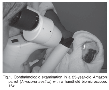
Ophthalmic examination was performed in a dark room and standard recording sheets were used to document the findings. The adnexa and the anterior ocular segments of both eyes were examined with a portable handheld biomicroscope7 (Fig.1). Direct8 and indirect ophthalmoscopy9 with a 30D indirect condensing lens10 was performed when pupillary diameter was considered sufficient. Both eyes were stained with fluorescein dye11 (Fig.2) and the excess dye flushed away with sterile saline12, so that any corneal abrasions could be detected. Topical anesthesia13 was instilled when necessary. Vision was assessed by menace and avoidance behavior test.
RESULTS
Amazon parrots were found the commonest admitted bird (29.53%, 57 in 193). Birds included male and female parrots, with ages varying from 8 months to 41 years old.
Cataracts was the most frequently reported disorder, present in 24 (21.05%) of the 114 examined eyes (Fig.3). In most cases, the cause of cataracts was not documented. Affected parrots were approximately 20 years old and mature cataract was its most observed development stage, observed in 12 eyes. Cataract surgery was successfully performed in two birds.
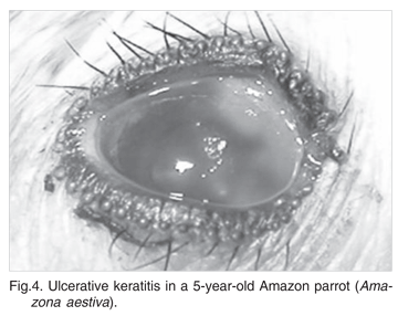
Uveitis, ulcerative keratitis (Fig.4) and keratoconjuncti-vitis were frequently diagnosed. Every reported disorder was listed in Table 1.
The cornea was the most affected ocular structure with most lesions diagnosed, with 25 eyes of 21 parrots with corneal involvement, presenting 28 reports (several parrots had multiple corneal disorders). Uveal disorders were commonly observed as well. Conjunctiva and eyelid disorders were diagnosed in lower frequency, as shown in Table 2.
DISCUSSION
The aim of this study was to report the occurrence of ocular disorders in Amazon parrots attended at the Ophthalmology Service of the Veterinary Teaching Hospital from 1997 to 2006. This bird was chosen because it is a common pet, being often brought to veterinary hospitals in Brazil. Authors believe that Amazona aestiva was the most frequent observed specie. Unfortunately, it could not be confirmed as birds registered as "Brazilian parrot" and "Amazona spp." may include Amazona ochrocephala or Amazona amazonica parrots.
Blepharospasm, ocular discharge, trauma, facial rubbing, eyelid swelling, periocular feather loss, fail to feed properly and blindness were frequently reported as cause of admission.
The most diagnosed disorder was cataracts in different stages of evolution, which also was observed in a similar survey realized in pet birds (Tsai et al. 1993) and in different bird species admitted at the same Veterinary Teaching Hospital where this study was conducted (Safatle et al. 2007). Causes of cataracts in birds described in the literature include: senescence, trauma, developmental abnor-malities, genetic disorders, nutritional deficiency, radiation, UV radiation and inflammatory factors (Gelatt 2007b). Cataracts was reported in a study conducted in birds, some of those may had been a developmental abnormality and some secondary to inflammation or trauma (Buyukmihci et al. 1988). Causes of most diagnosed cataracts were not defined. Four incipient, 6 immature, 12 mature and 2 hypermature cataracts were reported. Depending on the stage, it may cause an important visual deficit (Meyer 1986, Cubas 2007), what was commonly complaint during anamnesis in this survey. Lens extraction is the chosen treatment if evidence of other intraocular disorder is absent: synechiae, unresponsive mydriasis or extensive corneal opacity. If possible, electroretinography should also be performed to exclude any retinal dysfunction (Hendrix & Sims 2004). Lensectomy by needle discision and aspiration, and conventional extracapsular extraction or ultrasonic phacoemulsification can be successful in birds (Altman et al. 1997). Two lensectomies by needle discision and aspiration were successfully preformed in two parrots. When surgery is not performed, lens induced uveitis must be clinically monitored; such ophthalmopathy was reported in 8 of the 24 affected eyes (33.33%).
Most corneal problems in psittaciformes are due to epithelial defects secondary to trauma, keratitis secondary to abnormalities of the eyelid, infection, foreign bodies (such as sand, grass awns, seed husks and gravel), and dry eyes (Meyer 1986, Ritchie et al. 1994). In this survey, cornea was the most affected ocular structure (28 reports) and ulcerative keratitis secondary to trauma or infection some of the most reported corneal disorders.
Keratoconjunctivitis also was frequently observed. Keratoconjunctivitis (with blepharitis and periocular feather loss) occurs in psittacines in association with Chlamydia psittaci infection (Surman et al. 1974, Gelatt 1999a). Routine testing for clamydiosis, such as cytology and bacteriology is advised in any ocular discharge (Meyer 1986).
Keratoconjunctivitis sicca was suspected in 5 of the 7 reported eyes as ocular discharge and dry cornea were seen. Diagnose could not be confirmed as Schirmer tear test and phenol red thread tear test were not performed in any of the birds. Schirmer tear test is impractical for most but can be done in large birds; even then, normal values are not available and would need to be estimated from normal eyes of similar species (Altman et al. 1997). Phenol red thread tear test was already performed in Amazon parrots but its normal values were not established (Holt et al. 2006). Keratoconjunctivitis sicca in birds is often associated with hypovitaminosis A (Meyer 1986, Cubas 2007). Therefore, all affected parrots were successfully treated with oral vitamin A14 and topical cyclosporine A 0.2%15.
Corneal degeneration was observed in five eyes (Fig.5). In pet birds, crystal deposition in the corneal stroma was reported as the second most frequent ocular disorder in pet birds (Tsai et al. 1993). In all reported cases, neither cellular infiltration nor neovascularization was evoked and the cause was not defined, but poxvirus could be associated with the disorder (Tsai et al. 1993, Gelatt 2007b). Punctate and mycobacterial keratitis can occur in parrots but was not observed in this survey (Stanz et al. 1995, Gelatt 1999a).
Leucoma, keratitis, keratomalacia, descemetocele, keratouveitis, corneal abscesses and rupture of the globe were occasionally diagnosed.
Uveal tract was commonly affected. Trauma, penetrating injury, systemic disease, autoimmune condition and parasitic, mycotic, bacterial or viral disorders are causes of uveitis in birds (Meyer 1986). Most of diagnosed uveitis in this survey were due to unknown cause, lens induced, secondary to trauma or keratouveitis. Iris melanoma, confirmed after histopathologic examination, was reported in one eye (Guimarães et al. 2004).
Glaucoma and buphthalmia have rarely been reported in birds. Identification of glaucoma in most bird species is problematic because of the small size of their eyes, making even the use of applanation tonometers with small tips difficult or impossible (Altman et al. 1997). When glaucoma is found, it is usually due to trauma and resulting hyphaema, or iatrogenic associated with cataract surgery (Meyer 1986, Cubas 2007). Diagnosed buphthalmia in this survey was secondary to trauma and hyphaema.
Spontaneous lesions of the retina is rarely reported in birds, but is commonly observed in raptors after trauma. Retinal detachment and tearing, degeneration of the retina, pecten and choroid or optic nerve atrophy may occur (Altman et al. 1997). Subtle lesions involving the fundus may have been overlooked because of the absence of mydriasis in some of the birds. Posterior segment could be examined in a darkened room when there were no cataracts and the pupillary diameter was considered sufficient. This form of examination may not be ideal but is possible in some bird species, such as tawny owls (Bright 2000) and birds attended in typical veterinary teaching hospitals, where mydriatics are commonly found but are ineffective, due to the striated muscle in the iris, which allows voluntary control of their pupil size (Meyer 1986, Cubas 2007). Retinal detachment secondary to trauma was reported in 2 eyes and supposed amaurosis in 2 birds as any retinal degeneration was observed. Retinal degeneration of unknown cause has already been reported in a parakeet (Ritchie et al. 1994) but was not diagnosed in this survey.
Conjunctiva and eyelid disorders were relatively frequent observed. Blepharoconjunctivitis and conjunctivitis are common disorders in psittaciformes and has been associated with upper and lower respiratory tract infection, such as rhinitis and sinusitis or systemic and intra-ocular infections (Meyer 1986, Tully & Carter 1993, Altman et al. 1997, Gelatt 1999a). In this survey, 2 parrots with aero-sacculitis presented conjunctivitis and 1 with sinusitis presented an eyelid abscess. Other causes are: environ-mental factors (dust, drafty housing, irritating fumes), nutritional deficiencies (vitamin A is often cited, although no specific experimental evidence support this view) or trauma and can be associated to blepharitis or ulcerative keratitis (Shimakura et al. 1981, Meyer 1986, Altman et al. 1997, Gelatt 1999a, Rupley 1999, Cubas 2007).
Juvenile birds present blepharitis more often than adults, probably because the eyelids of juveniles are more exposed to trauma and other insults (Cousquer 2005). The reported parrot was 1 year old and presented, as a cause for the disorder, knemidocoptic mange (Knemidokoptes spp.).
Because of the presence of scleral ossicles, traumatic rupture of the eye does not result in collapse of the globe. In cases that cornea cannot be repaired, enucleation could be carried out, but only be performed as last option (Meyer 1986). Two traumatic globe ruptures were reported in this survey and enucleation was performed in one of the cases.
Orbital and intraocular neoplasms were rarely reported in this survey. Many case reports have described neoplasms in parrots (Paul-Murphy et al. 1985, Graham et al. 2003, Diaz-Figueroa et al. 2006, Simova-Curd et al. 2009). Diagnosed orbital neoplasm occurred along with lymphoma; fine needle cytology examination confirmed the diagnosis (Casagrande et al. 2005).
In cage birds, the majority of conditions reported have been associated with infection, what was frequently observed in this survey. Otherwise, in raptors, most ocular disorders are related to trauma and lesions are usually found in the posterior segment (Gelatt 1999a, Bright 2000).
Although microphthalmia, microphakia, cataracts, retinal dysplasia, malformation of the ciliary body, choroid, pecten and lentoid formation were already seen in wild-caught raptors (Buyukmihci et al. 1988), congenital developmental abnormalities are rare in birds. Just cataracts was observed in this survey, but eyes were not examined as the ones in the referred study.
Most examined parrots were first attended at the Avian Ambulatory Service of the same hospital for other reasons and when ophthalmological disorders were identified, parrots were refereed to the Ophthalmology Service. Ocular lesions may also be a particularly strong indication of systemic disorders, such as clamydiosis, micoplasmosis, poxvirosis or nutritional deficiency. A causative diagnosis of ocular lesions may be vital for the avian patient, not only as a basis for effective therapy of the primary disease but also for saving their vision, in which birds are primarily orientated (Korbel 1992, Gelatt 1999a).
Concluding, cataracts was the most observed disorder in Amazon parrots admitted at the Ophthalmology Service, Veterinary Teaching Hospital, School of Veterinary Medicine, University of São Paulo, Brazil, from 1997 to 2006. Uveitis, ulcerative keratitis and keratoconjunctivitis were frequently observed. Cornea, lens and uvea were the most affected ocular structures presenting different disorders. Results agree with other studies performed to record the occurrence of ophthalmologic disorders in pet birds (Tsai et al. 1993), free-living raptors (Murphy et al. 1982) and tawny owls (Cousquer 2005). Nonetheless, the data of this survey may serve as an indicator of occurrence and types of ophthalmological disorders in Amazon parrots, being helpful in future studies.
Acknowledgements.- To the staff of the Medical Records Department of the Veterinary Teaching Hospital, University of São Paulo, for their assistance on retrieving medical records used in this survey.
Received on May 19, 2009.
Accepted for publication on November 11, 2009.
- Altman R.B., Clubb S.L., Dorrestein G.M. & Quesenberry K. 1997. Avian Medicine and Surgery. W.B. Saunders, Pennsylvania, p.569-589.
- Bright P. 2000. Manual of Avian Medicine. Mosby, St Louis, p.264-312.
- Buyukmihci N.C., Murphy C.J. & Schulz T. 1988. Developmental ocular disease of raptors. J. Wildl. Dis. 24(2):207-213.
- Casagrande R.A., Nemer V.C., Torres L.N., Guimarães M.B., Sherlock T.M. & Matushima E.R. 2005. Linfoma em Amazona aestiva (papagaio-verdadeiro) domiciliado: relato de caso. Anais XII Encontro Nacional de Patologia Veterinária, Belo Horizonte, publicados em Arq. Bras. Med. Vet. Zootec. 57(Supl.1):79.
- Cousquer G. 2005. Ophthalmological findings in free-living tawny owls (Strix aluco) examined at a wildlife veterinary hospital. Vet. Res. 156(23):734-739.
- Cubas Z.S. 2007. Tratado de Animais Selvagens. Roca, São Paulo, p.1096-1099.
- Diaz-Figueroa O., Tully T.N., Williams J. & Evans D. 2006. Squamous cell carcinoma of the infraorbital sinus with fungal tracheitis and ingluvitis in an adult Solomon Eclectus Parrot (Eclectus roratus solomonensis). J. Avian Med. Surg. 20(2):113-119.
- Gancz A.Y., Malka S., Sandmeyer L., Cannon M., Smith D.A. & Taylor M. 2005. Horner's Syndrome in a Red-bellied Parrot (Poicephalus rufiventris). J. Avian Med. Surg. 19(1):30-34.
- Gelatt K.N. 1999a. Veterinary Ophthalmology. 3rd ed. Lippincott Williams and Wilkings, Pennsylvania, p.689-694.
- Gelatt K.N. 2007b. Veterinary Ophthalmology. Vol.2. 4th ed. Blackwell Publishing, Iowa, p.1370-1405.
- Graham J.E., Werner J.A., Lowenstine L.J., Wallack S.T. & Tell L.A. 2003. Periorbital Liposarcoma in an African Grey Parrot (Psittacus erithacus). J. Avian Med. Surg. 17(3):147-153.
- Guimarães M.B., Esteves C.A.L.G., Scholtz V.S., Contieri M.B., Mota E.F.F., Dagli M.L.Z. & Barros P.S.M. 2004. Melanoma ocular em papagaio verdadeiro (Amazona aestiva): relato de caso. VIII Congresso ABRAVAS e XIII Encontro da Associação Brasileira de Veterinários de Animais Selvagens, Jaboticabal. (Pôster)
- Hendrix D.V.H. & Sims M.H. 2004. Electroretinography in the Hispaniolan Amazon Parrot (Amazona ventralis). J. Avian Med. Surg. 18(2):89-94.
- Holt E., Rosenthal K. & Shofer F.S. 2006. The phenol red thread tear test in large Psittaciformes. Vet. Ophthalmol. 9(2):109-113.
- Hoppes S., Gurfield N., Flammer K., Colitz C. & Fisher P. 2000. Mycotic Keratitis in a Blue-fronted Amazon Parrot (Amazona aestiva). J. Avian Med. Surg. 14(3):185-189.
- Korbel R. 1992. Ocular manifestations of systemic diseases in birds. Tierärztl Praxis 20(4):385-394.
- Leber A.C. & Burge T. 1999. A dermoid of the eye in a blue-fronted Amazon parrot (Amazona aestiva). Vet. Ophthalmol. 2:133-135.
- Meyer D.B. 1986. Avian Physiology. 4th ed. Springer-Verlag, New York, p.38-48.
- Murphy C.J., Kern T.J., McKeever K., McKeever L. & MacCoy D. 1982. Ocular lesions in free-living raptors. J. Am. Vet. Med. Assoc. 181(11):1302-1304.
- Paul-Murphy J.R., Lowenstine L., Turrel J.M., Murphy C.J. & Fowler M.E. 1985. Malignant lymphoreticular neoplasm in an African gray parrot. J. Am. Vet. Med. Assoc. 187:1216-1217.
- Ritchie B.W., Harrison G.J. & Harrison L.R. 1994. Avian Medicine Principles and Applications. Winger Publishing, Lake Worth, p.673-677.
- Rupley A.E. 1999. Manual de Clínica Aviária. Roca, São Paulo, p.144-175.
- Safatle A.M.V., Guimarães M.B., Hvenegaard A.P. & Barros P.S.M. 2007. Prevalência de catarata em aves domésticas atendidas na FMVZ-USP entre os anos de 1997 e 2006. Anais 7ş Congresso Paulista de Clínicos Veterinários de Pequenos Animais, São Paulo, p.230-231.
- Shimakura S., Sawa H., Yamashita T. & Hirai K. 1981. An outbreak of ocular disease caused by staphylococcal infection in Amazon parrots (Amazona aestiva) imported into Japan. Jpn. J. Vet. Sci. 43:273-275.
- Sick H. 2001. Ornitologia Brasileira. Nova Fronteira, Rio de Janeiro, p.351.
- Stanz K.M., Miller P.E., Cooley J., Langenberg J.A. & Murphy C.J. 1995. Mycobacterial keratitis in a parrot. J. Am. Vet. Med. Assoc. 206(8):1177-1180.
- Simova-Curd S., Richter M., Hauser B. & Hatt J.M. 2009. Surgical Removal of a Retrobulbar Adenoma in an African Grey Parrot (Psittacus erithacus). J. Avian Med. and Surg. 23(1):24_28.
- Surman P.G., Schultz D.J. & Tham V.L. 1974. Keratoconjunctivitis and chlamydiosis in cage birds. Aust. Vet. J. 50:356-362.
- Tsai S.S., Park J.H., Hirai K. & Itakura C. 1993. Eye lesions in pet birds. Avian Pathol. 22: 95-112.
- Tully T.N. & Carter T.D. 1993. Bilateral supraorbital adscesses associated with sinusitis in an orange-winged Amazon parrot (Amazona amazonica). J. Avian Vet. Assoc. 7(3):157-158.
Publication Dates
-
Publication in this collection
20 Jan 2010 -
Date of issue
Dec 2009
History
-
Accepted
11 Nov 2009 -
Received
19 May 2009