Abstracts
A case-control study was carried out in litters of 1 to 7-day-old piglets to identify the main infectious agents involved with neonatal diarrhea in pigs. Fecal samples (n=276) from piglets were collected on pig farms in the State of Rio Grande do Sul, Brazil, from May to September 2007. Litters with diarrhea were considered cases (n=129) and normal litters (n=147) controls. The samples were examined by latex agglutination test, PAGE, conventional isolating techniques, ELISA, PCR, and microscopic methods in order to detect rotavirus, bacterial pathogens (Escherichia coli, Clostridium perfringens type A and C, and Clostridium difficile), and parasites (Coccidian and Cryptosporidium spp.). Outbreaks of diarrhea were not observed during sampling. At least one agent was detected in fecal samples on 25 out of 28 farms (89.3%) and in 16 farms (57.1%) more than one agent was found. The main agents diagnosed were Coccidia (42.86%) and rotavirus (39.29%). The main agents identified in litters with diarrhea were Clostridium difficile (10.6%), Clostridium perfringens type A (8.8%) and rotavirus (7.5%); in control litters, Clostridium difficile (16.6%) and Coccidian (8.5%). Beta hemolytic Escherichia coli and Clostridium perfringens type C were not detected. When compared with controls, no agent was significantly associated with diarrhea in case litters. These findings stress the need for caution in the interpretation of laboratorial diagnosis of mild diarrhea in neonatal pigs, as the sole detection of an agent does not necessarily indicate that it is the cause of the problem.
Swine; neonatal diarrhea; case-control; epidemiology; diagnosis
Um estudo de caso-controle em leitegadas de um a sete dias de idade foi realizado com o objetivo de identificar os principais agentes infecciosos envolvidos na diarreia neonatal de leitões. As amostras de fezes (n=276) foram coletadas em granjas de suínos no Estado do Rio Grande do Sul, Brasil, no período de maio a setembro de 2007. Leitegadas com diarreia foram consideradas casos (n=147) e leitegadas normais, controles (n=129). As amostras foram examinadas através do teste de aglutinação em látex, PAGE, cultivo, ELISA, PCR e métodos microscópicos para a excreção dos principais agentes de diarreia: virais (rotavirus), bacterianos (Escherichia coli, Clostridium perfringens tipos A e tipo C e Clostridium difficile) e parasitários (coccídeos e Cryptosporidium spp.). Durante o período do estudo não foram observados surtos e a diarréia, quando presente, apresentou-se leve. Pelo menos um agente foi identificado nas amostras fecais de 25 entre 28 granjas (89,3%) analisadas e em 16 granjas (57,1%) mais de um agente foi detectado. Os principais agentes encontrados nas granjas foram coccídeos (42,86%) e rotavírus (39,29%). Os principais agentes detectados nas leitegadas com diarreia foram Clostridium difficile (10,6%), Clostridium perfringens tipo A (8,8%) e rotavírus (7,5%). Por outro lado, nas leitegadas controle os agentes mais prevalentes foram Clostridium difficile (16,6%) e coccídeos (8,5%). E. coli Beta hemolítica e Clostridium perfringens tipo C não foram detectados. O presente estudo de caso-controle demonstrou que nenhum agente infeccioso esteve associado significativamente com diarreia (p>0.05). Esses achados reforçam a necessidade de que haja cuidado na interpretação de resultados de exames laboratoriais em materiais coletados de leitões com diarreia neonatal leve, pois a detecção isolada de um agente infeccioso não indica necessariamente que o mesmo seja a causa do problema.
Suínos; diarréia neonatal; caso-controle; epidemiologia; diagnóstico
LIVESTOCK DISEASES
Matched case-control study evaluating the frequency of the main agents associated with neonatal diarrhea in piglets1 1 . Part of the MSc Dissertation in Veterinary Science of the first author, defended at Unversidade Federal do Rio Grande do Sul (UFRGS), on February 27, 2008.
Estudo caso-controle pareado avaliando a frequência dos principais agentes causadores de diarréia neonatal em suínos no Rio Grande do Sul
Ricardo T. LippkeI; Sandra M. BorowskiII; Sandra M.T. MarquesIII; Suelen O. PaesiIV; Laura L. AlmeidaV; Andrea M. MorenoVI; Luís G. CorbelliniVII; David E.S.N. de BarcellosI* * Corresponding author: davidbarcellos@terra.com.br
ISetor de Suínos, Faculdade de Veterinária (Favet), UFRGS, Av. Bento Gonçalves 9090, Porto Alegre, RS 91540-000, Brazil
IIInstituto de Pesquisas Veterinárias Desidério Finamor (IPVDF), Estrada do Conde 6000, Eldorado do Sul, RS 92990-000, Brazil
IIILaboratório de Protozoologia, Favet-UFRGS, Porto Alegre, RS
IVLaboratório de Diagnóstico Molecular, Instituto de Biotecnologia, Universidade de Caxias do Sul, Rua Francisco Getúlio Vargas 1130, Caxias do Sul, RS 95070-560, Brazil
VSetor de Patologia Veterinária, Favet-UFRGS, Porto Alegre, RS
VILaboratório de Sanidade Suína, Faculdade de Medicina Veterinária e Zootecnia (FMVZ), Universidade de São Paulo (USP), Av. Prof. Dr. Orlando Marques de Paiva 87, São Paulo, SP 05508-270, Brazil
VIILaboratório de Epidemiologia (Epilab), Favet-UFRGS, Porto Alegre, RS
ABSTRACT
A case-control study was carried out in litters of 1 to 7-day-old piglets to identify the main infectious agents involved with neonatal diarrhea in pigs. Fecal samples (n=276) from piglets were collected on pig farms in the State of Rio Grande do Sul, Brazil, from May to September 2007. Litters with diarrhea were considered cases (n=129) and normal litters (n=147) controls. The samples were examined by latex agglutination test, PAGE, conventional isolating techniques, ELISA, PCR, and microscopic methods in order to detect rotavirus, bacterial pathogens (Escherichia coli, Clostridium perfringens type A and C, and Clostridium difficile), and parasites (Coccidian and Cryptosporidium spp.). Outbreaks of diarrhea were not observed during sampling. At least one agent was detected in fecal samples on 25 out of 28 farms (89.3%) and in 16 farms (57.1%) more than one agent was found. The main agents diagnosed were Coccidia (42.86%) and rotavirus (39.29%). The main agents identified in litters with diarrhea were Clostridium difficile (10.6%), Clostridium perfringens type A (8.8%) and rotavirus (7.5%); in control litters, Clostridium difficile (16.6%) and Coccidian (8.5%). Beta hemolytic Escherichia coli and Clostridium perfringens type C were not detected. When compared with controls, no agent was significantly associated with diarrhea in case litters. These findings stress the need for caution in the interpretation of laboratorial diagnosis of mild diarrhea in neonatal pigs, as the sole detection of an agent does not necessarily indicate that it is the cause of the problem.
Index terms: Swine, neonatal diarrhea, case-control, epidemiology, diagnosis
RESUMO
Um estudo de caso-controle em leitegadas de um a sete dias de idade foi realizado com o objetivo de identificar os principais agentes infecciosos envolvidos na diarreia neonatal de leitões. As amostras de fezes (n=276) foram coletadas em granjas de suínos no Estado do Rio Grande do Sul, Brasil, no período de maio a setembro de 2007. Leitegadas com diarreia foram consideradas casos (n=147) e leitegadas normais, controles (n=129). As amostras foram examinadas através do teste de aglutinação em látex, PAGE, cultivo, ELISA, PCR e métodos microscópicos para a excreção dos principais agentes de diarreia: virais (rotavirus), bacterianos (Escherichia coli, Clostridium perfringens tipos A e tipo C e Clostridium difficile) e parasitários (coccídeos e Cryptosporidium spp.). Durante o período do estudo não foram observados surtos e a diarréia, quando presente, apresentou-se leve. Pelo menos um agente foi identificado nas amostras fecais de 25 entre 28 granjas (89,3%) analisadas e em 16 granjas (57,1%) mais de um agente foi detectado. Os principais agentes encontrados nas granjas foram coccídeos (42,86%) e rotavírus (39,29%). Os principais agentes detectados nas leitegadas com diarreia foram Clostridium difficile (10,6%), Clostridium perfringens tipo A (8,8%) e rotavírus (7,5%). Por outro lado, nas leitegadas controle os agentes mais prevalentes foram Clostridium difficile (16,6%) e coccídeos (8,5%). E. coli Beta hemolítica e Clostridium perfringens tipo C não foram detectados. O presente estudo de caso-controle demonstrou que nenhum agente infeccioso esteve associado significativamente com diarreia (p>0.05). Esses achados reforçam a necessidade de que haja cuidado na interpretação de resultados de exames laboratoriais em materiais coletados de leitões com diarreia neonatal leve, pois a detecção isolada de um agente infeccioso não indica necessariamente que o mesmo seja a causa do problema.
Termos de indexação: Suínos, diarréia neonatal, caso-controle, epidemiologia, diagnóstico
INTRODUCTION
In the last three decades, a progressive change in pig production has occurred towards the increase of confinement and intensive rearing methods. In this new scenario, neonatal pig diarrhea gained importance, and depended on the combined effect of infectious agents and several risk factors (Barcellos 2002).
Neonatal diarrhea causes losses associated with mortality, decreased weight gain and excessive use of medication (Wittum et al. 1995). The main infectious agents involved may be viral: rotavirus and coronavirus; bacterial: Escherichia coli, Clostridium perfringens types A and C and Clostridium difficile; and parasitic: Isospora suis and Cryptosporidium spp. (Calderaro et al. 2001, Wieler et al. 2001, Yaeger et al. 2002).
Most studies on diagnosis of diarrhea in newborn piglets investigated few or a single infectious agent, and presented incomplete information about antibiotic, coccidiostat and/or probiotic use before sampling (Morin et al. 1983, Fitzgerald et al. 1988, Wieler et al. 2001). Consequently, an excessive bias can be set to a given infectious agent when, in fact, other factors could also be involved in the outcome of diarrhea, such as quality of the environment and hygiene. Observational studies such as case-control are used to identify risk factors and to estimate the quantitative effects of the various causes that may contribute to the occurrence of disease (Thrusfield 2007). With the use of this tool, the frequency of diarrhea associated with specific agents can be compared between affected and normal groups (Sobestiansky & Barcellos 2001).
The objective of the present study was to identify the main bacterial, parasitic and viral agents detected in litters with and without diarrhea, in pig farms located in the State of Rio Grande do Sul, Brazil, and to estimate the association between the presence of the agents and diarrhea.
MATERIALS AND METHODS
Characteristics of the farms, diseases and sampling
Feces and rectal swabs were collected from litters from 28 pig farms with 200 to 1000 sows, belonging to 6 integrated companies in the State of Rio Grande do Sul, Brazil, between May and September 2007. The farms presented high standards of housing and management with all-in/all-out practice and appropriate disinfection between batches of pigs. Farrowing crates had partially or fully slatted floors of plastic or iron bars. All farms vaccinated sows and gilts against Escherichia coli infection, using two doses of the vaccine in the last third of gestation. Sows and piglets in cases or control litters had not been previously treated with probiotics, antibiotics or coccidiostats. Data on parity order and age of occurrence of diarrhea were taken. Classification of fecal consistency was performed based on the chart proposed by Gheller et al. (2008). Piglets with ages between 1 and 7 days were sampled. Litters with two or more piglets with feces with creamy or liquid consistency (cases) were matched with litters presenting normal feces (controls). Case litters were matched to controls by age (same age or less than 1 day difference) with a case-control ratio of 1.1:1 (m:n matching that refers to varying numbers of cases and controls in the matched sets). The primary objective of matching is the elimination of biased comparison between cases and controls (Kuritz & Landis 1988). The following formula (Martin et al. 1987) and parameters for sampling size were applied:

Where, Zα is the value, which provides a/2 in each tail (for a 5% type I error Z is 1.96); Zb, which provides β in the lower tail (for a type II error of 0.2, Z value is 0.84); Pe is the estimate of response rate in the case group (10.9%) and Pc in the control group (2%) (Yaeger et al. 2007); P is the average response rate of case and control groups (i.e., 6%) and Q is the complement. To satisfy this assumption, 276 samples were necessary.
Samples consisted of pools of feces collected directly from the rectum or rectal swabs from two or more piglets of the same litter. Feces were stored in plastic tubes and swabs on Stuart Transport Medium, both kept at 2-8ºC for no longer than 24 hours. Swabs were collected for bacteriology and fecal samples divided in aliquots, two for parasitological examination and two frozen at -20ºC for rotavirus diagnosis and Clostridium difficile toxin assay.
Bacteriological examination
Rectal swabs were streaked into 5% sheep blood agar and MacConkey agar plates and incubated at 37ºC for 24 hours, in aerobic conditions. Diagnosis of neonatal colibacillosis was achieved when over 80% of colonies presented phenotypical characteristics compatible with E. coli, including the presence of profuse growth of lactose-fermenting colonies on Mac Conkey agar and hemolysis on blood agar, as suggested by Barcellos et al. (1980). For isolation of C. perfringens, samples were inoculated in selective SPS agar (Sulfite-polymyxin-sulfadiazine, HiMedia®, Mumbai, India) and incubated at 37ºC for 24 hours in an anaerobic jar, containing an atmosphere of 5% CO2, 5% H2 and 80% N2, in the presence of a palladium catalyst (Angelotti et al. 1962). Samples showing more than 20% of the colonies with black color (characteristic of C. perfringens in SPS agar) were frozen at -20ºC and submitted to a polymerase chain reaction (PCR) multiplex reaction. C. perfringens was diagnosed as type C in the presence of products for genes cpa and cpb and as type A if genes cpa and cpb2 were detected, following methods described by Moreno et al. (2003).
A subset of 132 fecal samples (66 from case litters and 66 controls) was examined for Clostridium difficile A and B toxins directly from the feces using a commercial enzyme linked immunoassay (ELISA) kit (Ridascreen Clostridium difficile toxin A/B®) following the recommendations of the producer (Biopharm, Darmstadt, Germany).
Parasitological examination
Oocysts of Cryptosporidium spp. were diagnosed from stained fecal smears using a modified Ziehl-Neelsen technique (Hen-riksen & Pohlens 1981), and observed in a optical micros-cope with 100x objective. Coccidia oocysts (Eimeria spp. and Isospora spp.) were diagnosed with a modified centrifugation and flotation technique in sucrose-saturated solution (Hoffman 1987) and observed in a fluorescence microscope using a wavelength of 340-365nm, with 20x objective (Daugschies et al. 2001).
Virological examination
Rotavirus diagnosis was performed with two tests: latex slide agglutination (ROTA Rich®) detecting type A rotavirus, according to manufacturer's instructions (Hemocor Indústria e Comércio, Brasil). Fecal samples (0.1g or 100mL) were diluted 1/6 in the kit's dilution buffer, homogenized and clarified (800 x g for 10 minutes). The test uses polystyrene latex particles sensitized with rabbit anti-rotavirus polyclonal serum and identifies type A rotavirus. As positive control inactivated rotavirus particles (strain SA-11) were used. Results were considered positive when visible agglutination occurred, and negative when the suspension remained milky after 2 minutes incubation.
For other types of rotavirus, polyacrilamide gel electro-phoresis (PAGE) was used according to Pereira et al. (1983) and the gels were stained with silver die (Herring et al. 1982). Fecal samples were first submitted to extraction by the phenol /chloroform method, starting with a 20% fecal suspension. In each round, a positive control was included (group A rotavirus from human origin, LDM 187). The migration pattern of the 11 bands of viral RNA was assessed after silver staining (Herring et al. 1982). Samples were considered as not type A rotavirus or atypical when the triplet typical of rotavirus type A was not detected. Positive results consisted of samples with positive results in latex agglutination tests and/or PAGE.
Statistical analysis
Conditional logistic regression for paired samples was used to assess if the differences found between case and control groups were due to chance or the presence of pathogenic agents. Conditional logistic regression is used to investigate the relationship between an outcome and a set of factors in matched case-control studies. The Proc PHREG procedure (SAS) for m:n matching was used, in which a stratum (i.e. age of litters) for each matched set was formed. The outcome was diarrhea (case) coded as 1 and no diarrhea (control) coded as 2 (variable status). A dummy survival time was created, so that all the cases in a matched set have the same event time in value, and the corresponding controls are censored at a later time (variable censor). Cases censor had a value of 1 and controls a value of 0. Additional explanatory variables tested were the parity order and size of litters.
Chi-square test for trend was used to test if the proportion of positive cases increases with the age of litters. Chi-square test was also used to compare proportions of positive cases according to stratum of age in days. Strata formed were: 1-2 (n=107); 3-4 (n=130) and >4 (n=39). These tests were performed using EpiInfo 6.0 (Center for Disease Control, Atlanta, USA).
RESULTS
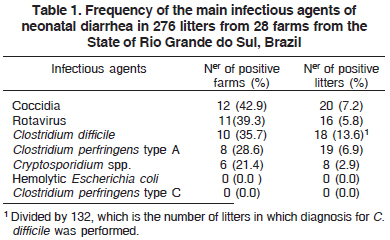
The diarrhea in the farms typically affected from 1 to 4 piglets in the litters and mortality was below 3%. All affected piglets presented mild diarrhea with creamy to pasty feces. The frequency of agents by farm and litters is presented in Table 1. At least one agent was diagnosed in fecal samples on 89.3% farms (25/28), in 57.1% (16/28) more than one agent was found. Considering farms, coccidiosis was the commonest diagnosis, positive in 42.9% farms (12/28, Table 1) followed by rotavirus, which was present in 39.3% farms (11/28).
In litters, the most prevalent agent detected was Clostridium difficile (13.6%, 95% CI: 7.8 -19.5%), followed by coccidian (7.2%, 95% CI: 4.2 -10.3%), Clostridium perfringens type A (6.9%, 95% CI: 3.9 -9.9%), rotavirus (5.8%, 95% CI: 3.0 -8.6%) and Cryptosporidium spp. (2.9%, 95% CI: 0.9 -4.9%, Table 1).
The main bacterial agent diagnosed was C. perfringens type A, found in 6.9% (n=19) samples. Its detection was higher in case (8.8%) than in control litters (4.6%), but the difference was not significant (p=0.47, Table 2).
C. difficile toxin was found in 7 case liters and 11 controls through ELISA, also without statistical significance (p=0.33). Hemolytic Escherichia coli and Clostridium perfringens type C were not found in samples from feces of control or case litters considering the criteria used to define its significance in the diagnosis.
Coccidia were present in 20 litters with a similar number in cases (n=9) and controls (n=11), and there was no statistical difference (Table 2). Cryptosporidium spp. was detected in 8 litters, without statistical difference between cases and controls (Table 2).
Rotavirus was diagnosed in 16 samples, 2 for type A using a Latex test and 14 with eletrophoretic pattern in PAGE other than Rotavirus A. However, the frequency in cases and controls was not statistically different (Table 2).
The overall frequency of positive samples for at least one infectious agent was 25.4% (95% CI: 20.2 -30.5%), which did not differ significantly (p>0.1) between cases 24.5% (36/147) and controls 26.3% (34/129). The frequency of infectious agents also did not differ between case and control litters (Table 2).
The distribution of agents related to age is presented in Table 3. Litter from 1 to 7 days old presented diarrhea (mode = 1; first quartile = 1-2 days, second quartile = 3-4 days, and third quartile >4 days). A significant trend in the frequency of positive toward older litters was found for protozoa infection (i.e. Coccidia and Cryptosporidium spp. together, p<0.01). Conversely, the proportion of positive litter for Clostridium perfringens type A was higher in young litters (p=0.003). Chi-Square to compare proportions demonstrated the higher frequency of rotavirus in the class >4 days when compared to younger age stratum as significant (p=0.008). No association was found between parity order and size of litter with diarrhea (p>0.1).
DISCUSSION
The main finding of the present work was the fact that no agent was significantly associated with diarrhea in case litters when compared to controls. This may imply that the non-diarrheic pigs were in the incubation phase of diarrhea development or that some pigs were able to resist infection without having diarrhea, as suggested by Pedersen et al. (2010). The fact that farms in which sampling took place experienced low-to-mild neonatal diarrhea episodes, affecting few animals, is also relevant. As neonatal diarrhea has a multifactorial etiology, other factors must be necessary for the outcome of clinical cases, including environmental, immunological and nutritional or management problems (Wittum et al. 1995). In fact, infection with the agents investigated here must be necessary but not sufficient to cause diarrhea in piglets, needing a range of component causes, as described in some causal models of disease (Thrusfield 2007). The etiological role of other agents not screened in the present work, such as enteric calicivirus (norovirus and sapovirus), cannot be excluded. However, norovirus is difficult to identify in pig feces and sapovirus is easily identified, but the infection is asymptomatic in swine (Barry et al. 2007, Halaihel et al. 2010). Another agent of swine diarrhea (coronavirus) has never been diagnosed in suckling pigs in Brazil (Brentano et al. 2002, Barthasson et al. 2009), and was not screened in the present work.
The occurrence of nutritional or non-infectious diarrhea, connected with changes in morphology and/ or physiology of the gastro-intestinal tract (Alexander 1981) cannot be ruled out either. Insufficient colostrum ingestion is considered a probable cause of non-infectious diarrhea in neonatal piglets (Jensen et al. 2001). In such cases, diarrhea occurs not only because of low colostrum ingestion, but also because of the lack of mucosal development and early intestinal maturation (Xavier et al. 2006). Excessive milk ingestion has been considered a significant cause of nutritional diarrhea in young piglets. Sows with elevated milk production and piglets that were very active when suckling were considered factors capable of influencing this type of diarrhea during lactation (Alexander 1981). However, new studies are needed to clarify the importance of nutritional causes in the outcome of diarrhea in piglets in the neonatal period.
Rotavirus was present in 39.3% of the farms. Previous studies in commercial pig farms in the State of São Paulo, Brazil, detected a higher frequency of rotavirus infection; 89.2% and 47.6% (San Juan et al. 1985, Calderaro et al. 2001). Similar studies in other Brazilian states (Rio Grande do Sul and Santa Catarina) detected rotavirus in most municipalities (72.7%), but not in all farms (Roehe et al. 1989). In comparison with our investigation, other Brazilian studies investigated outbreaks and estimated higher prevalences (22%, 18.6% and 10.9% respectively, San Juan et al. 1985, Roehe et al. 1989, Calderaro et al. 2001). Anther variable to be considered is the age of fecal collection. In the present survey we estimated a low prevalence (5.8%), analyzing piglets up to seven days old and, out of 16 rotavirus only 2 were classified as type A. This result could be related to the low age of the surveyed pigs. According to Morilla et al. (1991), rotavirus belonging to groups B, C, D and E generally infect younger piglets when compared to group A.
Coccidian was the commonest agent in production farms (42.9%). High levels of coccidiosis were also found in weanling piglets in other areas of Brazil, with positivity of 42.5% and 76.2% of the feces of weanling piglets in the States of Minas Gerais and São Paulo (Rostagno et al. 1999, Calderaro et al. 2001). In the present work, there was no difference in the presence of oocysts in feces of animals with or without diarrhea. The overall infection rate was low, but this would be understandable as most clinical cases of coccidiosis occur in piglets older than those examined in our study (Worliczek et al. 2009). According to the authors, age was the most important factor to determine the association between Isospora excretion and diarrhea, and infectious dose was not important to the outcome of the infection.
Cryptosporidium was identified in very few (1.36%) case litters and controls (4.65%), with no statistical difference. It has been previously demonstrated that there is no relationship between the occurrence of diarrhea and the detection of oocysts of Cryptosporidium in feces, especially in piglets with low levels of infection (Enemark et al. 2003). The possible explanation is that diarrhea is caused by pathogens other than Cryptosporidium sp. and this agent causes an opportunistic infection, inducing superficial lesions in few cells or in a limited epithelial area, resulting in lesions not severe enough to result in diarrhea (Vitovec et al. 1992).
The main bacterial agent diagnosed was Clostridium perfringens type A, found in 6.9% samples. These results are consistent with an increase in the association of this agent in the etiology of neonatal diarrhea (Vieira et al. 2008). However, its role as a cause of diarrhea in neonatal piglets is difficult to assess, as the agent belongs to the normal flora of the intestine and can already be found in the feces just a few hours after birth. The criterion for the classification of this agent in the present work was based on PCR identification of the genes cpa and cpb2 in C. perfringens isolated from fecal material. So far, it has been difficult to evaluate the patho-genicity of CpA based on the presence of toxin genes, as the toxin can be present even in C. perfringens isolates from healthy piglets (Bueschel et al. 2003).
All samples were negative for Clostridium perfringens type C. This agent was considered very important in preceding works on the etiology of neonatal diarrhea (Songer & Uzall 2005), but more recent surveys have given little importance to the association of this agent with neonatal diarrhea (Yaeger et al. 2002, Katsuda et al. 2006, Cruz-Junior 2010).
Clostridium difficile was detected in 18 fecal samples, but there was no statistical association between presence of toxins in feces from case and control pigs (p=0.33, Table 2). The infection caused by this agent is considered an emerging problem, and can be associated with a disruption of the normal intestinal flora by antibiotics (Yager et al. 2002). However, the presence of A and B toxins of C. difficilein feces is not consistently associated with diarrhea, because of this some authors have suggested the existence of sub clinical infections with the agents and the possible existence of an additional factor essential for the outcome of diarrhea (Yaeger et al. 2007). Apart from the present work, there is only one report about C. difficile infection in pigs in Brazil, in which 17% (10/60) of piglets with C. difficile toxin in the intestinal contents (Cruz-Junior 2010) were identified through ELISA. As with our study, there was no statistical difference between case and control piglets. These results suggest that C. difficile may be an emerging pathogen in pig herds in Brazil.
An unexpected finding was the absence of hemolytic Escherichia coli in significant levels in case and control litters, but the results must be interpreted considering the methodology used. In most blood agar plates, hemolytic colonies with characteristics of E. coli were present, but the amount of colonies did not fulfill the criterion chosen to define a positive result. Abundant growth of beta-hemolytic E. coli (Barcellos et al. 1980) is found especially in farms presenting clinically perceptible outbreaks of E. coli infection, with high morbidity and mortality. In the farms visited, sows were routinely vaccinated in the last third of gestation against Escherichia coli infection, and this may have interfered with the results, as suggested by Pedersen et al. (2010). Another study carried out recently in Brazil agreed with our results (Cruz-Junior 2010). In it, only 1 out of 30 piglets with neonatal diarrhea and 1 out 30 controls were positive both for fimbriae and toxins of E. coli using necropsy, bacterial isolation and PCR multiplex classification.
In Table 3, we present data regarding the relationship between age and the occurrence of diarrhea. As would be expected, most isolation of Clostridium perfringens type A was obtained from newborn piglets (1-2 days old). The higher frequency of infection at an early age may be associated with the low trypsin levels in the digestive tract of newborn piglets, influenced by the presence of trypsin inhibitors in the colostrum (Niilo 1988). Beta toxin of C. perfringens types A and C can benefit from this lack of inhibition during this period (Voth & Ballard 2005) and produce lesions associated with diarrhea. Clostridium difficile was also identified exclusively in very young piglets (1-4 days old). According to Songer & Uzall, 2005, a typical case of diarrhea caused by this agent will occur in a piglet 1-4 days of age, with diarrhea starting soon after birth. For rotavirus, the diagnosis of the agent was higher in piglets older than 4 days old. Cryptosporidium spp. was diagnosed in only one litter of very young piglets (1-2 days old), agreeing with previous results showing that the beginning of fecal oocysts excretion of this agent varies from 2 to 9 days after infection (Enemark et al. 2003).
In the present work, we did not attempt to carry out an extensive investigation of all causes of neonatal diarrhea in pigs; we concentrated on the most common agents detected in previous works. We found no difference of isolation of pathogens from piglets with diarrhea and normal littermates. These results stress the need for caution in the interpretation of the results of laboratory exams of diarrhea in neonatal pigs, as the sole detection of an agent does not necessarily indicate that it is causing the problem. The use of tests capable of quantifying organisms in stools or tissues and relating them to the causation of the lesion, such as quantitative PCR and immunohistochemistry, could allow a better discrimination between carrier and diseased animals.
Acknowledgements. To CAPES and CNPq for financial support and scholarships. To Priscila Zlotowski for help during the investigation.
Received on November 4, 2010.
Accepted for publication on February 22, 2011.
- Alexander T.J.L. 1981. Piglet diarrhea: A guide to diagnosis and control. Brit. Vet. J. 137:651-662.
- Angelotti R., Hall H.E., Foter M.J. & Lewis K.H. 1962. Quantitation of Clostridium perfringens in foods. Appl. Microbiol. 10:193-199.
- Barcellos D.E.S.N. 2002. Avaliação de medidas de controle para as principais formas de diarréia em suínos. Anais. I Simpósio Latino Americano de Suinocultura, Foz do Iguaçu, PR, p.130-142.
- Barcellos D.E.S.N., Guizzardi I.I. & Fallavena L.C.B. 1980. Frequência e causas de diarréias bacterianas em suínos nas zonas criatórias do Vale do Taquari e Missões, Rio Grande do Sul. Bolm IPVDF 80:27-37.
- Barry A.F., Alfieri A.F., Feronato C., Grieder W. & Alfieri A.A. 2007. Frequency of porcine enteric calicivirus infection in Brazilian pig herds. Anais. XIII Congresso da Abraves, Florianópolis, SC, p.343-344.
- Barthasson D.L., Brito W.M.E.D., Sobestiansky J., Caixeta J., Miranda T.M.T. & Silva L.A. 2009. Ocorrência de infecção por parvovírus suíno e gastroenterite transmissível em suínos, criados de forma extensiva, em Goiás. Arq. Bras. Med. Vet. Zootec. 61(5):1227-1229.
- Brentano L., Ciacci-Zanella J.R., Mores N. & Piffer I.A. 2002. Levantamento soroepidemiológico para coronavírus respiratório e da gastroenterite transmissível e dos vírus de influenza H3N2 e H1N1 em rebanhos suínos no Brasil. Comun. Téc. 306, Embrapa-CNPSA, Concórdia, SC. 6p.
- Bueschel D.M., Jost B.H., Billington S.J., Trinh H.T. & Songer J.G. 2003. Prevalence of cpb2, encoding beta2 toxin, in Clostridium perfringens field isolates: Correlation of genotype with phenotype. Vet. Microbiol. 94(2):121-129.
- Calderaro F.F., Baccaro M.R., Moreno A.M., Ferreira A.J.P., Jerez A.J. & Pena H.J.F. 2001. Frequência de agentes causadores de enterites em leitões lactentes provenientes de sistemas de produção de suínos do Estado de São Paulo. Arqs. Inst. Biológico, São Paulo, 68:29-34.
- Cruz-Junior E.C. 2010. Frequência de enteropatógenos em leitões de até sete dias de vida, na região do Triângulo Mineiro, Alto Paranaíba, Brasil. Dissertação de Mestrado, Escola de Veterinária, Universidade Federal de Minas Gerais, Belo Horizonte. 68p.
- Daugschies A., Bialek R., Joachim A. & Mundt H.C. 2001. Auto-fluorescence microscopy for the detection of nematode eggs and protozoa, in particular Isospora suis, in swine feces. Parasitol. Res. 87:409-412.
- Enemark H.L., Ahrens P., Bille-Hansen V., Heegaard P.M.H., Vigre H., Thamsborg S.M. & Lind P. 2003. Cryptosporidium parvum: Infectivity and pathogenicity of the porcine genotype. Parasitol. 126:407-416.
- Fitzgerald G.R., Barker T., Welter M.W. & Welter C.J. 1988. Diarrhea in young pigs: Comparing the incidence of the five most common infectious agents. Vet. Med. 83:80-86.
- Gheller N.B., Santi M., Lippke R.T., Gonçalves M.A.D., Ribeiro A.M.L. & Barcellos D.E.S.N. 2008. Visual assessment of fecal consistency in pigs. Proc. 20th IPVS Congress, Durban, South Africa, p.606.
- Halaihel N., Masia R.M., Fernandez-Jimenez M., Ribes J.M., Montava R., De Blas I., Gironés O., Alonso J.L. & Buesa J. 2010. Enteric calicivirus and rotavirus infections in domestic pigs. Epidemiol. Infect. 138:542-548.
- Henriksen S.A. & Pohlens J. 1981. Staining of Cryptosporidia by a modified Ziehl-Neelsen technique. Acta Vet. Scand. 22:594-596.
- Herring A.J., Inglis N.F., Ojeh C.K., Snodgrass D.R. & Menzies J.D. 1982. Rapid diagnosis of rotavirus infection by direct detection of viral nucleic acid in silver-stained polyacrylamide gels. J. Clin. Microbiol. 16:473-477.
- Hoffman R.P. 1987. Exame parasitológico de fezes, p.24-59. In: Hoffman R.P. (Ed.), Diagnóstico de Parasitismo Veterinário. Sulina, Porto Alegre. 156p.
- Jensen A.R., Elnif J., Burrin D.G. & Sangild P.T. 2001. Development of intestinal immunoglobulin absorption and enzyme activities in neonatal pigs is diet dependent. J. Nutr. 131:3259-3265.
- Katsuda K., Kohmoto M., Kawashima K. & Tsunemitsu H. 2006. Frequency of enteropathogen detection in suckling and weaned pigs with diarrhea in Japan. J. Vet. Diagn. Invest. 18:350-354.
- Kuritz S.J. & Landis J.R. 1988. Attributable risk estimation from matched case-control data. Biometrics 44:355-367.
- Martin S.W., Meek A.H. & Willeberg P. 1987. Veterinary Epidemiology, Principles and Methods. Iowa State University Press, Ames. 343p.
- Moreno A.M., Baccaro M.R., Ferreira A.J.P., Hirose F. & Campos D.S. 2003. Detection of b2 toxin gene from Clostridium perfringens isolated in diarrheic piglets. Arqs Inst. Biológico, São Paulo, 70:135-138.
- Morilla A., Arriaga C., Ruiz A., Martinez A.G., Cigarroa R. & Valazquez A. 1991. Association between diarrhoea and shedding of group A and atypical groups B to E rotaviruses in suckling pigs. Ann. Rech. Vet. 22:193-200.
- Morin M., Turgeon D., Jolette J., Ribinson Y., Phaneuf J.B., Sauvageau R., Beauregard M., Teusher E., Higgins R. & Lariviére S. 1983. Neonatal diarrhea of pigs in Quebec: Infectious causes of significant outbreaks. Can. J. Comp. Med. 47:11-17.
- Niilo L. 1988. Clostridium perfringens type C enterotoxemia. Can. Vet. J. 29:658-664.
- Pedersen K.S., Angen O., Jorsal S.E., Stege H. & Nielsen J.P. 2010. In intestinal pathogens in acute diarrhea in weaners: A case control study in 20 Danish herds. Proc. 21st IPVS Congress, Vancouver, Canada, p.756.
- Pereira H.G., Azeredo R.S., Leite J.P.G., Candeias J.A.N., Racz M.L., Linhares A.C., Gabbay Y.B. & Trabulsi J.R. 1983. Electrophoretic study of the genome of humam rotaviruses from Rio de Janeiro, São Paulo and Pará, Brazil. Hyg. 90:117-126.
- Roehe P.M., Cunha A.C., Salvo E.O., Martins R.M., Rosa J.C., Oliveira L. & Hoffmann V.L. 1989. Rotavírus em suínos na Região Sul do Brasil. Pesq. Vet. Bras. 9:45-49.
- Rostagno M.H., Bicalho K.A., Lage A.P., Martins N.E., Leite R.C. & Leite R.C. 1999. Prevalência de Isospora suis em leitões de granjas comerciais de ciclo completo. Anais IX Congresso da Abraves, Belo Horizonte, MG, p.195.
- San Juan C.S., Waack R.S., Bellinzoni R. & Scodeller E.A. 1985. Ocorrência de rotavirose em leitões em rebanhos da região leste do Estado de São Paulo. Anais II Congresso de Abraves, Rio de Janeiro, RJ, p.135.
- Sobestiansky J. & Barcellos D.E.S.N. 2001. Clínica veterinária em sistemas intensivos de produção de suínos e relatos de casos clínicos. Art 3. Impressos Especiais, Goiânia. 153p.
- Songer J.G. & Uzall F.A. 2005. Clostridial enteric infections in pigs. J. Vet. Diagn. Invest. 17:528-536.
- Thrusfield M., 2007. Veterinary Epidemiology. 3rd ed. Blackwell Science Publication, Oxford. 610p.
- Vieira A.A.S., Guedes R.M.C., Salvarani F.M., Silva R.O.S., Assis R.A. & Lobato F.C.F. 2008. Genotipagem de Clostridium perfringens isolados de leitões diarréicos. Arqs. Inst. Biológico, São Paulo, 75:513-516.
- Vitovec J. & Koudela B. 1992. Pathogenesis of intestinal cryptosporidiosis in conventional and gnotobiotic piglets. Vet. Parasitol. 43:25-36.
- Voth D.E. & Ballard J.D. 2005. Clostridium difficile toxins: Mechanism of action and role in disease. Clin. Microb. Rev. 18:247-263.
- Wieler L.H., Ilieff A., Herbst W., Bauer C., Vieler E., Bauerfeind R., Failing K., Klös H., Wengert D., Baljer G. & Zahner H. 2001. Prevalence of enteropathogens in suckling and weaned piglets with diarrhoea in southern Germany. J. Vet. Med. B 48:151-159.
- Wittum T.E., Dewey C.E., Hurd H.S., Dargatz D.A. & Hill G.W. 1995. Herd and litter-level factors associated with the incidence of diarrhea, morbidity and mortality in piglets 1 to 3 days of age. J. Swine Hlth Prod. 3:99-104.
- Worliczek H.L., Mundt H.C., Ruttkowski B. & Joachim A. 2009. Age, not infection dose, determines the outcome of Isospora suis infections in suckling piglets. Parasitol. Res. 105:157-162.
- Xavier E.G., Rutz F. & Roll V.F.B. 2006. Imunonutrientes na produção de suínos. Anais I Simpósio UFRGS de Produção Reprodução e Sanidade Suína, Porto Alegre, RS, p.174-195.
- Yaeger M.J., Funk N. & Hoffman L. A. 2002. A survey of agents associated with neonatal diarrhea in Iowa swine including Clostridium difficile and porcine reproductive and respiratory syndrome virus. J. Vet. Diagn. Invest. 14:281-287.
- Yaeger M.J., Kinyon J.M. & Songer J.G. 2007. A prospective, case control study evaluating the association between Clostridium difficile toxins in the colon of neonatal swine and gross and microscopic lesions. J. Vet. Diagn. Invest. 19:52-59.
Publication Dates
-
Publication in this collection
11 July 2011 -
Date of issue
June 2011
History
-
Accepted
22 Feb 2011 -
Received
04 Nov 2010