Abstracts
Differences in the microscopic morphology of the hoof in forelimbs and hindlimbs of horses have been scarcely reported in the literature, especially concerning the distribution of primary and secondary epidermal laminae in the different regions. This study aimed to determine the density of primary and secondary epidermal laminae in the hoof of horses. For this, it was used fore and hindlimbs of 16 adult mixed breed horses. With a cross section 0.5 cm above the sole, it was quantified the primary epidermal laminae in the regions of the toe, and of lateral and medial quarters. Fragments with about 1cm ³ were taken from the proximal, middle and distal thirds of the hooves, in the different regions, subjected to conventional histological techniques and examined with an optical microscope. Data were statistically analyzed in relation to the fore and hindlimbs and between their various regions. The density of primary epidermal laminae varied around the hoof circumference, with greater values in the hoof toe, which gradually decreased towards the bulb of the hoof, without difference between thoracic and pelvic limbs. The average density of the secondary epidermal laminae per primary epidermal lamina does not change around the circumference of the hoof. Our findings indicated that the density of epidermal laminae is not different between fore and hindlimbs. The variation in the density of primary epidermal laminae around the hoof seems to be part of an adaptive response to different stresses in each region. A better understanding of the structural morphology contributes to a better understanding of the diagnosis, pathophysiology, and treatment of disorders that affect the hoof.
Epidermal laminae; Equus caballus; hoof; morphology
Diferenças na morfologia microscópica dos cascos dos membros pélvicos e torácicos dos equinos têm sido pouco relatadas na literatura, principalmente no tocante a distribuição de lâminas epidérmicas primárias e secundárias nas diversas regiões. O propósito deste estudo foi quantificar a densidade de lâminas epidérmicas primárias e secundárias no casco de equinos. Foram utilizados membros torácicos e pélvicos de oito equinos adultos e sem raça definida. Em uma secção transversal de aproximadamente 0,5cm de altura da sola dos cascos foi quantificada a densidade das lâminas epidérmicas primárias tanto na região da pinça quanto dos quartos lateral e medial. Fragmentos com aproximadamente 1cm³ foram retirados dos terços proximal, médio e distal do casco, nas diferentes regiões e submetidos a técnica histológica convencional, a densidade de lâminas epidérmicas secundárias foi quantificada com auxilio de microscópio óptico. Os dados foram analisados estatisticamente em relação aos membros torácicos e pélvicos e entre suas diversas regiões. A densidade de lâminas epidérmicas primárias varia ao redor da circunferência do casco, sendo maior na região da pinça do casco e diminui gradualmente em direção ao bulbo do casco, não existindo diferença entre membros pélvicos e torácicos. A densidade média de lâminas epidérmicas secundárias por lâmina epidérmica primária não varia em torno da circunferência dos cascos, assim como, quando comparada entre os membros torácicos e pélvicos. A variação da densidade das lâminas epidérmicas primárias em torno do casco parece fazer parte de uma resposta adaptativa às diferentes tensões existentes em cada região. O melhor entendimento da morfologia das estruturas do casco contribui na melhor compreensão do diagnóstico, fisiopatologia e tratamento das afecções que as acometem.
Lâminas epidérmicas; Equus caballus; casco; morfologia
ANIMAL MORPHOPHYSIOLOGY
Density of primary and secondary epidermal laminae of equine hoof
Densidade das lâminas epidérmicas primárias e secundárias nos cascos de equinos
André R.C. Barreto-Vianna; Luana S. Oliveira; André S. Leonardo; Marcelo I. Santana; Roberta F. Godoy; Eduardo M.M. de Lima* * Corresponding author: limaemm@unb.br
Departamento de Anatomia Veterinária, Faculdade de Agronomia e Medicina Veterinária, Universidade de Brasília (UnB), ICC Ala Sul, Campus Darcy Ribeiro, Cx. Postal 4508, Brasília, DF 70760-701, Brazil
ABSTRACT
Differences in the microscopic morphology of the hoof in forelimbs and hindlimbs of horses have been scarcely reported in the literature, especially concerning the distribution of primary and secondary epidermal laminae in the different regions. This study aimed to determine the density of primary and secondary epidermal laminae in the hoof of horses. For this, it was used fore and hindlimbs of 16 adult mixed breed horses. With a cross section 0.5 cm above the sole, it was quantified the primary epidermal laminae in the regions of the toe, and of lateral and medial quarters. Fragments with about 1cm ³ were taken from the proximal, middle and distal thirds of the hooves, in the different regions, subjected to conventional histological techniques and examined with an optical microscope. Data were statistically analyzed in relation to the fore and hindlimbs and between their various regions. The density of primary epidermal laminae varied around the hoof circumference, with greater values in the hoof toe, which gradually decreased towards the bulb of the hoof, without difference between thoracic and pelvic limbs. The average density of the secondary epidermal laminae per primary epidermal lamina does not change around the circumference of the hoof. Our findings indicated that the density of epidermal laminae is not different between fore and hindlimbs. The variation in the density of primary epidermal laminae around the hoof seems to be part of an adaptive response to different stresses in each region. A better understanding of the structural morphology contributes to a better understanding of the diagnosis, pathophysiology, and treatment of disorders that affect the hoof.
Index terms: Epidermal laminae, Equus caballus, hoof, morphology.
RESUMO
Diferenças na morfologia microscópica dos cascos dos membros pélvicos e torácicos dos equinos têm sido pouco relatadas na literatura, principalmente no tocante a distribuição de lâminas epidérmicas primárias e secundárias nas diversas regiões. O propósito deste estudo foi quantificar a densidade de lâminas epidérmicas primárias e secundárias no casco de equinos. Foram utilizados membros torácicos e pélvicos de oito equinos adultos e sem raça definida. Em uma secção transversal de aproximadamente 0,5cm de altura da sola dos cascos foi quantificada a densidade das lâminas epidérmicas primárias tanto na região da pinça quanto dos quartos lateral e medial. Fragmentos com aproximadamente 1cm³ foram retirados dos terços proximal, médio e distal do casco, nas diferentes regiões e submetidos a técnica histológica convencional, a densidade de lâminas epidérmicas secundárias foi quantificada com auxilio de microscópio óptico. Os dados foram analisados estatisticamente em relação aos membros torácicos e pélvicos e entre suas diversas regiões. A densidade de lâminas epidérmicas primárias varia ao redor da circunferência do casco, sendo maior na região da pinça do casco e diminui gradualmente em direção ao bulbo do casco, não existindo diferença entre membros pélvicos e torácicos. A densidade média de lâminas epidérmicas secundárias por lâmina epidérmica primária não varia em torno da circunferência dos cascos, assim como, quando comparada entre os membros torácicos e pélvicos. A variação da densidade das lâminas epidérmicas primárias em torno do casco parece fazer parte de uma resposta adaptativa às diferentes tensões existentes em cada região. O melhor entendimento da morfologia das estruturas do casco contribui na melhor compreensão do diagnóstico, fisiopatologia e tratamento das afecções que as acometem.
Termos de indexação: Lâminas epidérmicas, Equus caballus, casco, morfologia.
INTRODUCTION
Horses are still commonly used as a force of traction for wagons in urban areas. These animals are subjected to an exhausting and repetitive work routine, without food and rest compatible to the physical activity developed every day (Velho 2007, Maciel et al. 2008). Many of these animals are affected by locomotor disorders, mostly related to their hoofs. In order to understand the physiopathology, diagnosis, and treatment of these disorders it is necessary to know the hoof morphology.
Inside the wall of the hoof there are primary epidermal laminae, from which arise secondary epidermal laminae. Likewise, the dermis has primary and secondary epidermal laminae (Pollitt 2001). Dermal and epidermal laminae interdigitize and attach firmly to each other, and in turn the dermis is attached to the periosteum of the third phalanx through connective tissue. This laminar arrangement has the purpose to increase the contact surface, increasing thus the attachment between the wall and adjacent tissues (Pollitt 2001, Thomason et al. 2005). The architecture of the hoof laminae is the histopathologic basis of the laminitis process (Pollitt 1996).
The laminar junction in horses plays a key role in transferring the forces of weight bearing between the epidermis of the hoof wall and the third phalanx, but the way this is done is poorly understood (Douglas et al. 1998). This junction encompasses a complex mixture of tissues with many other different mechanical properties (Pollitt 2004).
There is evidence that stress of laminar junction influences the modeling of primary epidermal laminae, determining a higher density laminar (Thomason et al., 2005). Changes in laminar morphology found in healthy horses cannot be considered normal or pathological, since it is not known how these findings represent the normal range or subclinical disease manifestations (Lancaster et al. 2007). In and trotters and Thoroughbred horses (Thomason et al., 2008), there is a remodeling, induced by stress, in the primary epidermal laminae, but without a complete characterization and understanding of the relationship between stress, strain and remodeling process. Redden (2003) mentioned that distortion of a component of the capsule of hoof changes all other components and adjacent tissues. Thus, the large surface of the connections between the dermal and epidermal laminaes creates good mechanical strength, however, the cellular level, these connections are quite fragile and vulnerable to ischemia and enzymatic destruction (Redden 2003).
Aiming to add knowledge about the hoof morphology, the goal of the present study was to quantify the density of primary and secondary epidermal laminae in the various regions of the forelimb and hindlimb hooves of horses.
MATERIALS AND METHODS
For this study, it was used forelimbs and hindlimbs of 16 adult mixed breed horses, eight males and eight females, with 334 kg in average weight.
To collect the hooves, the distal ends of the limbs were disarticulated at the metacarpophalangeal and metatarsophalangeal joints, for the forelimbs and hindlimbs, respectively.
All the animals died in the Veterinary Hospital of the University of Brasília due to problems not related to the locomotor system. The experiments were conducted in accordance with the guidelines of the Ethics Committee of Animal Use in the Institute of Biological Sciences of the University of Brasília, Brazil - Protocol number 51203/2010.
Data processing. By using a band saw, eight hooves were transversally sectioned at about 0.5cm above the sole, and from the other eight hooves, it was taken fragments with around 1cm³ of the proximal, middle, and distal thirds, in the regions of the toe, and of the lateral and medial quarters (Fig.1), according to the protocol described by Pollitt (1996).
Density of primary epidermal laminae. The density of primary epidermal laminae was measured according to Lancaster et al. (2007), using a cross segment, fixed in 10% formaldehyde solution, taken from the hoof sole which was analyzed with the aid of a magnifying glass (Olympus SZ40). The primary epidermal laminae were counted on the cutting surface along the entire perimeter of the hoof. Initially, a needle marked the midpoint of the toe, between the lateral and medial face, and then another two needles were fixed, one 25 primary epidermal laminae away in the medial direction, and the other in the lateral direction. The region between these two needles had 50 primary epidermal laminae, and was called zone I. From these needles, others were fixed, 50 primary epidermal laminae away, both medially and laterally, and the intervals between two consecutive needles delimited the pair zones (II, IV, VI and VIII), always medial, and the odd zones (III, V, VII and IX), always lateral (Fig. 2A). Afterwards, with an electronic digital caliper (Starret®), it was measured the length of the zones previously set. The density of the primary epidermal laminae (number of laminae per centimeter) was achieved by dividing the number of laminae existing in the zone concerned (50 laminae) by its length.
Density of secondary epidermal laminae. The fragments taken from the different regions of the hoof were fixed in 10% formaldehyde solution. The area occupied by the laminae was separated from the hoof wall and from the distal phalanx, histologically processed by dehydration in ethanol, diafanization in xylene, embedded in paraffin, and cut in 4µm-thickness sections with a hand microtome (Zeiss®, Hyrax M15). The slides were mounted and stained with hematoxylin-eosin, analyzed under optical microscope (Labomed® ALX400) coupled to a video camera (Labomed® iVu3000) and image analysis software (ProgRes® Capture Pro 2.5).
In order to measure the density, the amount of secondary epidermal laminae originated by primary epidermal laminae was counted, and the total number was divided by the total number of primary epidermal laminae measured, obtaining thus the density of secondary epidermal laminae per primary epidermal laminae.
Statistical analysis. Density data of primary and secondary epidermal laminae of the forelimbs and hindlimbs were tabulated and analyzed with the software GraphPad Prism®5 using descriptive statistics, Pearson correlation, analysis of variance (ANOVA) followed by Tukey's test, being set the significance level at P<0.05.
RESULTS
Density of primary epidermal laminae
After dividing the internal stratum, it was possible to form from seven to nine zones (depending on the length of hoof circumference) with 50 primary epidermal laminae each (Table 1, Fig.2A). No significant difference (p<0.05) was found in density of primary epidermal laminae between forelimb and hindlimb (p=0.24), nor between zones corresponding to lateral and medial quarters.
The density of primary epidermal laminae in the toe area was significantly different from the other regions of the hoof, with a decrease in density from the hoof toe towards the bulbs of the hoof in the forelimbs and hindlimbs (Fig.3A,B). A high positive correlation was found between the density of primary epidermal laminae of the toe area and of the quarter lateral, for either forelimbs (r=0.86) or hindlimbs (r=0.98).
Density of secondary epidermal laminae
The secondary epidermal laminae were observed along the entire length of the primary epidermal laminae. For both forelimbs (Fig.3C) and hindlimbs (Fig.3D) no significant difference was verified in density of these laminae around the hoof circumference.
Comparing the limbs (Table 2), no significant difference was detected in the density of secondary epidermal laminae between the quarters medial (P=0.79) and between the hoof toes (P=0.09), but a statistical difference was found between the quarters lateral (P<0.001). In relation to the thirds, it was verified a poor correlation without significant difference between the proximal thirds (r=-0.04; P=0.34) and between the middle thirds (r=0.21; P=0.44), however a significant difference was registered between the distal thirds (r=0.18; P<0.001).
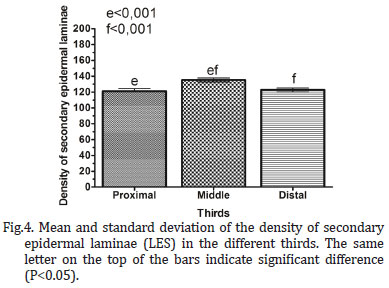
The density of secondary epidermal laminae on the proximal third was 121.2+35.34, on the middle third, was 135.3+26.98, and on the distal third, 122.8+26.83. It was possible to observe significant difference between proximal and middle thirds (P<0.001), as well as between the middle and distal thirds (P<0.001), which was not found between the proximal and distal thirds (P=0.69) (Fig.4).
By analyzing the density of secondary epidermal laminae, the Pearson correlation performed between the hoof toe, medial and lateral quarters with the corresponding thirds, pointed out weak correlations, both positive and negative, and a only a single strong negative correlation, between the proximal and distal thirds of the medial quarter (Table 3).
DISCUSSION
This study was conducted to further elucidate functional morphologic features and quantify regional primary and secondary laminar density in the feet of adult mixed breed horses. Results yielded new information regarding distribuition of this laminaes in different regions of the hoof in both fore and hindlimbs
Stashak & Hood (2005) verified homogeneity in morphology and distribution of primary epidermal laminae around the hoof perimeter. Contrastingly, our results showed that both in forelimbs (Fig.2A) and hindlimbs (Fig.2B), the region of the hoof toe had a greater concentration of primary epidermal laminae, corroborating the studies of Bowker (2003), Bidwell & Bolker (2006) Lancaster et al. (2007) and Thomason et al. (2008). Douglas & Thomason (2000) mentioned that the hoof toe is the region with the highest stress between the hoof wall and the distal phalanx, the gradual reduction in density of primary epidermal laminae towards the hoof bulb, as observed in the present study, probably is an adaptive response to different stresses in the several regions, once under physiological situations the force imposed in the quarters region would be lower than required in the hoof toe, as well as the stress related to the hoof bulbs is lower than to the quarters.
The precise mechanisms of this adaptive response have not been completely described, but Bowker (2003) suggested that the changes, both morphological and quantitative, occur through bifurcation of the primary and secondary epidermal laminae. In this way, it was a response to mechanical stress, with a direct relationship between the laminae arrangement and hoof functionality.
According to Lancaster et al. (2007), given a disturbance in the balance among the laminae distribution, such as a great amount of laminae agglomerated in a region or a low amount in another one, may generate weakened or injured areas in the hoof, justifying thus more detailed studies on the mechanisms of modifications in the hoof morphology, focusing to improve the understanding and treatment of pathologies.
Much of the literature consulted (Douglas & Thomason 2000, Thomason et al. 2005, Lancaster et al. 2007, Thomason et al. 2008, Kawasako et al. 2009) have analyzed only the forelimbs. Comparing forelimbs and hindlimbs, it was found no significant difference regarding the density of primary epidermal laminae, also observed by Bidwell and Bowker (2006). From this result, it is suggested that despite the center of mass of the horses had been diverted to closer to the forelimbs, producing a higher stress on them, this fact did not interfere with the density of these laminae.
Different from Lancaster et al. (2007) and Douglas & Thomason (2000), which found a higher density of primary epidermal laminae at the lateral quarter compared to the medial quarter, in the present study, no significant difference was observed between the quarters.
In agreement with Pollitt (2004) and Stashak & Hood (2005), each primary epidermal laminae give rise to 20-150 secondary epidermal laminae. In the present study, in the forelimbs, the number of secondary epidermal laminae ranged from 31 to 108, and in the hindlimbs, from 58 to 195. There is little literature about the distribution of secondary epidermal laminae, with only data on the total number of laminae, like in Pollitt (2004) and Stashak & Hood (2005), without respect to the location.
The lack of significant difference in the density of secondary epidermal laminae between the regions that comprise the hoof toe and the medial and lateral quarters, showed the absence of variation in the amount of laminae originated by primary laminae, i.e., each primary lamina, regardless if located at the hoof clamp or in the quarters, remained giving rise to the same amount of secondary laminae, but the quantity of these laminae in the several regions had varied owing the heterogeneous number of primary epidermal laminae around the hoof. Regarding the density of secondary laminae in the proximal, middle, and distal thirds of the hoof, it was possible to verify variations, but further studies are necessary to elucidate their implications on the hoof morphology. Although the fore and hindlimbs not have difference in the density of primary and secondary laminaes, Bidwell and Bowker (2006) found that in the forelimbs these laminaes have higher length. Demonstrating, the fact that the center of mass in horses be moved toward the front limbs does not cause change in density of these laminaes but couse in length. There are no studies regarding the growth and density of laminaes, but we believe that these should be related in accordance with the degree of tension that is exerted on the hoof. More studies are important to understand this possible relationship.
CONCLUSIONS
Forelimbs and hindlimbs presented the same distribution pattern for primary and secondary epidermal laminae around the hoof.
The density of primary epidermal laminae varied according to the region, with a higher concentration at the hoof toe, and a gradual reduction towards the bulbs, both for forelimbs and hindlimbs. This variation in the distribution around the hood circumference was not observed for secondary epidermal laminae, which have varied only in the proximal-distal direction. The observed changes in laminar architecture both in primary and secondary laminaes mainly along the hoof circumference for primary laminaes , indicate the potential of the laminae to change and remodel in response to variation in regional stresses. Probably the variation in the density of primary laminae around the hoof had been a consequence of the adaptive response to different stresses between the hoof wall and the distal phalanx in the several regions.
Received on July 20, 2012.
Accepted for publication on February 25, 2013.
- Bidwell L.A. & Bowker R.M. 2006. Evaluation of changes in architecture of the stratum internum of the hoof wall from fetal, newborn, and yearling horses. Am. J. Vet. Res. 67:1947-1955.
- Bowker R.M. 2003. The growth and adaptive capabilities of the hoof wall and sole: Functional changes in response to stress. Proc. Am. Assoc. Equine Pract. 49:146-168.
- Douglas J.E. & Thomason J.J. 2000. Shape, orientation and spacing of the primary epidermal laminae in the hooves of neonatal and adult horses (Equus caballus). Cells Tissues Organs 166:304-318.
- Douglas J.E., Biddick T.L., Thomason J.J. & Jofriet J.C. 1998. Stress/strain behaviour of the equine laminar junction. J. Exp. Biology 201:2287-2297.
- Kawasako K., Higashi T., Nakaji Y., Komine M., Hirayama K. & Matsuda K. 2009. Histologic evaluation of the diversity of epidermal laminae in hooves of horses without clinical signs of laminitis. Am. J. Vet. Res. 70:186-193.
- Lancaster L.S., Bowker R.M. & Mauer W.A. 2007. Density and morphologic features of primary epidermal laminae in the feet of three-year-old racing Quarter Horses. Am. J. Vet. Res. 68:11-19.
- Maciel R.M., Lopes S.T.A., Martins D.B., Merini L.P., Franciscato C. & Veiga A.P.M. 2008. Perfil hematológico e parasitológico de equinos utilizados na tração de carroças no município de Santa Maria-RS. 38ş Combravet, Gramado, RS. (Resumo)
- Pollitt C.C. 1996. Basement membrane pathology: A feature of acute equine laminitis. Equine Vet. J. 28:38-46.
- Pollitt C.C. 2001. Equine Laminitis. Art John McDougall, Queensland. 129p.
- Pollitt C.C. 2004. Anatomy and physiology of the inner hoof wall. Clin. Tech. Equine Practice 3:3-21.
- Redden R.F. 2003. Hoof Capsule Distortion: Understanding the mechanisms as a basis for rational management. Vet. Clin. North Am. Equine Pract. 19(2):443-462.
- Stashak T.S. & Hood D.M. 2005. Evaluation of architectural changes along the proximal to distal regions of the dorsal laminar interface in the equine hoof. Am. J. Vet. Res. 66:277-283.
- Thomason J.J., McClinchey H.L., Faramarzi B. & Jofriet J.C. 2005. Mechanical behavior and quantitative morphology of the equine laminar junction. Anat. Rec. A, Discov. Mol. Cell Evol. Biol. 283:366-379.
- Thomason J.J., Faramarzi B., Revill A. & Sears W. 2008. Quantitative morphology of the equine laminar junction in relation to capsule shape in the forehoof of Standardbreds and Thoroughbreds. Equine Vet. J. 40:473-480.
- Velho J. 2007. Inserção do Médico Veterinário nas Comunidades Carentes de Pelotas/RS. 2ş Salão de Extensão e Cultura, Pelotas/RS.
Publication Dates
-
Publication in this collection
22 May 2013 -
Date of issue
Apr 2013
History
-
Received
20 July 2012 -
Accepted
25 Feb 2013