Abstracts
The purpose of this paper was to study the etiology of mastitis, determine the antimicrobial susceptibility profile of Staphylococcus spp. and to identify the risk factors associated with infection in dairy cows in the states of Bahia and Pernambuco, Brazil. From the 2,064 milk samples analyzed, 2.6% were associated with cases of clinical mastitis and 28.2% with subclinical mastitis. In the microbiological culture, Staphylococcus spp. (49.1%) and Corynebacterium spp. (35.3%) were the main agents found, followed by Prototheca spp. (4.6%) and Gram negative bacilli (3.6%). In the antimicrobial susceptibility testing, all 218 Staphylococcus spp. were susceptible to rifampicin and the least effective drug was amoxicillin (32.6%). Multidrug resistance to three or more drugs was observed in 65.6% of Staphylococcus spp. The risk factors identified for mastitis were the extensive production system, not providing feed supplements, teat drying process, not disinfecting the teats before and after milking, and inadequate hygiene habits of the milking workers. The presence of multiresistant isolates in bovine milk demonstrates the importance of the choice and appropriate use of antimicrobial agents. Prophylactic and control measures, including teat antisepsis and best practices for achieving hygienic milking should be established in order to prevent new cases of the disease in herds.
Staphylococcus spp.; multidrug resistance; mammary gland; risk factors
Objetivou-se estudar a etiologia da mastite, determinar o perfil de sensibilidade dos Staphylococcus spp. aos antimicrobianos e identificar os fatores de risco associados à infecção em vacas leiteiras nos estados da Bahia e Pernambuco. Das 2.064 amostras de leite analisadas, 2,6% estavam associadas a casos de mastite clínica e 28,2% à mastite subclínica. No exame microbiológico, Staphylococcus spp. (49,1%) e Corynebacterium spp. (35,3%) foram os principais agentes isolados, seguidos de Prototheca spp. (4,6%) e bacilos Gram negativos (3,6%). No teste de sensibilidade aos antimicrobianos, todos os 218 Staphylococcus spp. apresentaram-se sensíveis à rifampicina e a droga menos eficaz foi a amoxicilina (32,6%). A resistência simultânea a três ou mais drogas foi observada em 65,6% dos Staphylococcus spp. Os fatores de risco identificados para a mastite foram o sistema de criação extensivo, não realização de suplementação alimentar, processo de secagem dos tetos, não realização de desinfecção dos tetos antes e após a ordenha e hábitos higiênicos inadequados dos ordenhadores. A presença de isolados multirresistentes no leite bovino demonstra a importância da escolha e da utilização adequada de antimicrobianos. Medidas de controle e profilaxia, incluindo a antissepsia dos tetos e boas práticas para a obtenção de ordenha higiênica devem ser estabelecidas, com o intuito de prevenir novos casos da doença nos rebanhos.
Staphylococcus spp.; multirresistência; glândula mamária; fatores de risco
LIVESTOCK DISEASES
Etiology, antimicrobial susceptibility profile of Staphylococcus spp. and risk factors associated with bovine mastitis in the states of Bahia and Pernambuco
Etiologia, perfil de sensibilidade dos Staphylococcus spp. aos antimicrobianos e fatores de risco associados à mastite bovina nos estados da Bahia e Pernambuco
Carina C. KrewerI; Izabela P. de S. LacerdaII; Evandro S. AmansoII; Noelly B. CavalcanteII; Rodolfo de M. PeixotoIII; José W. Pinheiro JúniorIV; Mateus M. da CostaII; Rinaldo A. MotaI,* * Corresponding author: rinaldo.mota@hotmail.com
IDepartamento de Medicina Veterinária, Universidade Federal Rural de Pernambuco (UFRPE), Rua Dom Manoel de Medeiros s/n, Dois Irmãos, Recife, PE 51171-900, Brazil
IILaboratório de Microbiologia e Imunologia Animal, Universidade Federal do Vale do São Francisco (Univasf), Rodov. BR 407 Km 12, Lote 543, Projeto de Irrigação Nilo Coelho s/n C1, Petrolina, PE 56300-000, Brazil
IIIInstituto Federal de Educação, Ciência e Tecnologia do Sertão Pernambucano, Campus Floresta, Rua Projetada s/n, Caetano II, Floresta, PE 56400-000, Brazil
IVUnidade Acadêmica de Garanhuns, UFRPE, Av. Bom Pastor s/n, Boa Vista, Garanhuns, PE 55296-901, Brazil
ABSTRACT
The purpose of this paper was to study the etiology of mastitis, determine the antimicrobial susceptibility profile of Staphylococcus spp. and to identify the risk factors associated with infection in dairy cows in the states of Bahia and Pernambuco, Brazil. From the 2,064 milk samples analyzed, 2.6% were associated with cases of clinical mastitis and 28.2% with subclinical mastitis. In the microbiological culture, Staphylococcus spp. (49.1%) and Corynebacterium spp. (35.3%) were the main agents found, followed by Prototheca spp. (4.6%) and Gram negative bacilli (3.6%). In the antimicrobial susceptibility testing, all 218 Staphylococcus spp. were susceptible to rifampicin and the least effective drug was amoxicillin (32.6%). Multidrug resistance to three or more drugs was observed in 65.6% of Staphylococcus spp. The risk factors identified for mastitis were the extensive production system, not providing feed supplements, teat drying process, not disinfecting the teats before and after milking, and inadequate hygiene habits of the milking workers. The presence of multiresistant isolates in bovine milk demonstrates the importance of the choice and appropriate use of antimicrobial agents. Prophylactic and control measures, including teat antisepsis and best practices for achieving hygienic milking should be established in order to prevent new cases of the disease in herds.
Index terms:Staphylococcus spp., multidrug resistance, mammary gland, risk factors.
RESUMO
Objetivou-se estudar a etiologia da mastite, determinar o perfil de sensibilidade dos Staphylococcus spp. aos antimicrobianos e identificar os fatores de risco associados à infecção em vacas leiteiras nos estados da Bahia e Pernambuco. Das 2.064 amostras de leite analisadas, 2,6% estavam associadas a casos de mastite clínica e 28,2% à mastite subclínica. No exame microbiológico, Staphylococcus spp. (49,1%) e Corynebacterium spp. (35,3%) foram os principais agentes isolados, seguidos de Prototheca spp. (4,6%) e bacilos Gram negativos (3,6%). No teste de sensibilidade aos antimicrobianos, todos os 218 Staphylococcus spp. apresentaram-se sensíveis à rifampicina e a droga menos eficaz foi a amoxicilina (32,6%). A resistência simultânea a três ou mais drogas foi observada em 65,6% dos Staphylococcus spp. Os fatores de risco identificados para a mastite foram o sistema de criação extensivo, não realização de suplementação alimentar, processo de secagem dos tetos, não realização de desinfecção dos tetos antes e após a ordenha e hábitos higiênicos inadequados dos ordenhadores. A presença de isolados multirresistentes no leite bovino demonstra a importância da escolha e da utilização adequada de antimicrobianos. Medidas de controle e profilaxia, incluindo a antissepsia dos tetos e boas práticas para a obtenção de ordenha higiênica devem ser estabelecidas, com o intuito de prevenir novos casos da doença nos rebanhos.
Termos de indexação:Staphylococcus spp., multirresistência, glândula mamária, fatores de risco.
INTRODUCTION
Brazil occupies sixth position in milk production worldwide. The Northeast region is responsible for approximately 10% of all bovine milk produced in the country, with production especially in the states of Bahia, Pernambuco and Ceara (IBGE 2011). In these locations, a large part of dairy activity is directed to the subsistence of small rural properties, playing an important economic and social role (Vilela et al. 2002). From the technological point of view, the quality of the raw material is one of the greatest obstacles to the development and consolidation of the dairy industry in Brazil (Guimarães & Langoni 2009).
Bovine mastitis is associated with reduction in milk production and causes changes in milk composition, and it is recognized as one of the main illnesses that affect the profitability of dairy farms (Bradley 2002). In addition, the disease presents a public health risk through the possibility of transmission of pathogenic microorganisms, toxins or antimicrobial residues through the milk (Fagundes & Oliveira 2004). Among the agents of contagious origin, Staphylococcus spp. are the bacteria most frequently isolated from clinical and subclinical cases (Taponen & Pyörälä 2009, Mota et al. 2012). Other microorganisms including Corynebacterium bovis, Streptococcus spp., Escherichia coli, Klebsiella pneumoniae, algae and yeasts are also reported in the etiology of intramammary infections (Corbellini et al. 2001, Langoni et al. 2011).
In Brazil there are few studies on the risk factors associated with mastitis (Souza et al. 2005, Oliveira et al. 2012), which are important for knowledge of effective measures in a prevention and control program for the disease (Coentrão et al. 2008). In addition to the adoption of hygiene-health management practices, antibiotic therapy may be an effective strategy in reduction of mastitis rates in a herd (Barlow 2011).
Considering the high prevalence of the illness in herds and the losses to the milk production chain, the purpose of this paper was to study the etiology of mastitis, determine the antimicrobial susceptibility profile of Staphylococcus spp. and identify the risk factors associated with infection in dairy cows in the states of Bahia and Pernambuco, Brazil.
MATERIALS AND METHODS
This study was based on analysis of 2064 bovine milk samples originating from 525 lactating cows from eight properties (coded from A to H) in the states of Bahia (n=426) and Pernambuco (n=1638), with seven located in the Lower Middle São Francisco Valley region and one in the Agreste of Pernambuco. At the time of visit to the properties, a questionnaire was applied consisting of objective questions to herd managers to obtain data regarding general characteristics of the property, animal management, hygiene management practices during milking and the milking workers profile. Which antimicrobial drugs were used in treatment of mastitis and other diseases in the herds under study were also checked.
Initially, physical examination of the mammary gland and milk from the animals was made and then the California Mastitis Test (CMT) was performed (Schalm & Noorlander 1957). Samples for microbiological examination were collected after washing the teats with soap and water, drying with paper towel and undertaking antisepsis of the ostium of the teats with alcohol at 70%. Regardless of the reaction of the milk samples in the CMT, on average, 5 mL of milk from each mammary quarter for each animal was collected in labeled sterile containers.
Ten microliters aliquots of cows' milk were streaked in 5% sheep blood agar and then the plates were incubated at 37°C for 48 hours. Microorganisms were identified by means of morphological (coloring, size, presence or absence of hemolysis of the colonies), tinctorial (Gram staining) and biochemical characteristics, according to Quinn et al. (1994). The samples that showed isolation of three or more different microorganisms were considered contaminated (National Mastitis Council 1999).
The in vitro susceptibility profile was determined in 218 isolates of Staphylococcus spp. by the disk diffusion method (Bauer et al. 1966). Disks with the following antimicrobial agents: amoxicillin (10µg), ampicillin (10µg), cephalexin (30µg), ciprofloxacin (5µg), doxycycline (30µg), enrofloxacin (5µg), erythromycin (15µg), streptomycin (10µg), gentamicin (10µg), lincomycin (2µg), oxacillin (1µg), penicillin (10µg), rifampicin (5µg), trimethoprim-sulfamethoxazole (25µg) and tetracycline (30µg). Results were interpreted through reading of the inhibition zones observed (CLSI 2008). For quality control of the technique and of the disks used, Staphylococcus aureus ATCC 25923 was used. Isolates that showed resistance to three or more drugs tested were considered multiresistant.
To identify the risk factors associated with infection, univariate analysis of the variables of interest was performed by the Pearson chi-square test or Fisher's exact test, when necessary. After that, logistic regression was performed considering the microbiological examination (positive or negative) as the dependent variable. The independent or explanatory variables considered in the model were those that showed statistical significance (<0.2). This probability was stipulated so that possible risk factors of the event would not be excluded from analysis (Hosmer & Lemeshow 1989). Statistical calculations were carried out through use of the program Epi Info, version 3.5.1, Centers for Disease Control and Prevention (CDC).
RESULTS
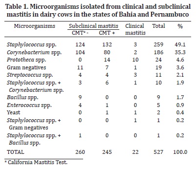
From the milk samples analyzed, 53 (2.6%) were associated with clinical mastitis and 584 (28.2%) showed reactions of one, two or three scores on the CMT. In the microbiological exam, 527 (25.5%) samples were positive and 662 (32.1%) showed contamination. In 875 (42.4%), any agent was identified. The microorganisms isolated from clinical and subclinical cases of mastitis are shown in Table 1.
The lowest percentage of susceptibility of Staphylococcus spp. to the antimicrobial drugs was for amoxicillin (32.6%), followed by ampicillin (33%), penicillin (34%), tetracycline (82.6%), streptomycin (88.1%), doxycycline (88.6%), trimethoprim-sulfamethoxazole (97.8%), erythromycin (98.2%), lincomycin (98.2%), oxacillin (98.2%), ciprofloxacin (99.1%), cephalexin (99.5%), enrofloxacin (99.5%) and gentamicin (99.5%). All the isolates were susceptible to rifampicin and 61 (28%) showed susceptibility to all the drugs tested. The multidrug resistance profile of the Staphylococcus spp. according to the properties studied may be observed in Table 2.
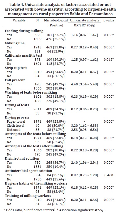
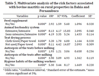
In Tables 3 and 4 the results are made available of univariate analysis of the factors of interest associated with the microbiological examination. In logistic regression, the following were identified as risk factors: animal husbandry system (OR=8.13; p=0.000), feed supplementation (OR=2.92; p=0.000), teat drying process (OR=8.11; p=0.000), antisepsis of the teats before (OR=1.93; p=0.005) and after (OR=3.09; p=0.000) milking, and hygiene habits of the milking workers (OR=5.3; p=0.000) (Table 5).
DISCUSSION
The frequencies of clinical and subclinical mastitis are highly esteemed parameters in evaluation of the health of the bovine mammary gland (Fonseca & Santos 2001). In this study, the rate (2.6%) of mammary quarters that showed signs of inflammation or alterations in the milk were greater than that found by Freitas et al. (2005) in dairy cows in the Agreste of Pernambuco (1%) and less than that observed for cows in Mato Grosso (5.8%) (Martins et al. 2010). The greater prevalence of subclinical infection in relation to clinical infection was also verified in other herds of different states (Bueno et al. 2002, Oliveira et al. 2009).
Of the mammary quarters with clinical mastitis and among the reagents in the CMT, 41.5% and 42% were positive in the microbiological culture respectively. According to the literature, this examination may be negative in approximately 15 to 40% of the samples with clinical alterations and is associated with factors such as low concentration or low elimination of pathogens in the milk, intracellular localization of certain agents, spontaneous elimination of the infection and, in some cases, non-infectious mastitis (Olde Reikerink et al. 2008). Furthermore, 260 (12.6%) samples analyzed were negative in the CMT but positive in the culture. This result reinforces the fact that even when triage tests are used on the dairy farms, there are animals that may harbor mastitis causing agents (Kapronezai et al. 2005).
The microorganisms identified in this study were similar to those reported by other researchers in different regions of Brazil (Barbalho & Mota 2001, Ferreira et al. 2007, Langoni et al. 2011). Staphylococcus spp. and Corynebacterium spp. were the main agents diagnosed on all the properties visited, where management faults were verified, such as inadequate hygiene practices of the hands of the milking workers (farms D, E, F) and of the milking equipment (B, G, H), as well as lack of performing antisepsis of the teats after milking (B, D, E, F). These procedures are closely associated with transmission of contagious mastitis during milking (Fonseca & Santos 2001) and may explain the significant number of these pathogens in the samples studied.
In spite of reports regarding the isolation of Prototheca spp. in cases of mastitis in Brazil (Mota et al. 1999, Amorim et al. 2010), it is not a very common finding. In this study, this alga appeared as the main agent recovered from clinical infections, corresponding to 45.4% of the cases in which there was microbial growth. All the isolates of Prototheca spp. came from the H property, where a large number of cows were milked mechanically and the grazing areas were excessively dirty with organic matter. As the collection of samples was performed in a rainy period, this suggests that the infections occurred due to broad dissemination of algae in the environment of the animals (Bueno et al. 2006). Furthermore, the presence of Gram negative bacteria in the samples evaluated indicates the opportunistic behavior of these microorganisms in the establishment of mastitis, among which E. coli and K. pneumoniae have been most observed in clinical and subclinical cases of the disease (Langoni et al. 2011).
Low rates of in vitro susceptibility to amoxicillin, ampicillin and penicillin were also found in Staphylococcus spp. from bovine mastitis in Pernambuco (Freitas et al. 2005) and São Paulo (Nader Filho et al. 2007). In most of the properties studied, the beta-lactams were the drugs of choice for therapy of intramammary infections, such that frequent and often inadequate use of these medications has probably contributed to selection of resistant bacteria in the herds. On the other hand, the microorganisms showed susceptibility percentages above 80% for the rest of the drugs evaluated, especially for cephalexin, gentamicin and enrofloxacin. Other authors (Nader Filho et al. 2007, Medeiros et al. 2009) described similar findings, suggesting that these antimicrobial agents could provide good in vivo effectiveness in the treatment of staphylococcus mastitis. Furthermore, susceptibility to all the active ingredients tested was seen in 28% of the isolates of this study; this situation was described in 37.8% of the Staphylococcus spp. of mastitis milk analyzed by Medeiros et al. (2009).
Resistance to three or more drugs was observed in 65.6% of the Staphylococcus spp., differing from Nader Filho et al. (2007) and Ribeiro et al. (2009), who reported percentages of 48.6% and 39.6% for the bacteria analyzed, respectively. In spite of the small number of isolates coming from properties A (n=5) and D (n=9), all of them showed multidrug resistance; this was observed in 90.5% and 71.7% in farms G and B, respectively. In all the herds, most (80.4%) of the bacteria were resistant to three or four antimicrobial agents simultaneously, principally from the beta-lactam class. Multidrug resistance to five, six or seven drugs was found in 28 (19.6%) Staphylococcus spp., 67.9% of which came from farm B. Such isolates also showed a characteristic resistance profile for the tetracyclines and streptomycin, which was little pronounced on the other properties and is associated with the use of these medications for treatment of infectious diseases in the animals of the herd in question.
Some production characteristics were identified as risk factors for the occurrence of mastitis on the properties. A significantly greater frequency of positive samples was seen on the microbiological examination for animals maintained in an extensive production system (OR=8.13; p<0.05) or semi-intensive production system (OR=4.19; p<0.05). Some authors affirmed that cows raised intensively are more susceptible to the development of intramammary infections through the greater concentration of animals and exposure to organic matter and to pathogenic microorganisms (Kalmus et al. 2006). In spite of that, we believe that the results of this study are associated with deficiencies in nutritional and hygiene-health management of the animals and of the facilities, with little adoption of measures for control and prevention of mastitis in the herds analyzed. In addition, the lack of feed supplements (OR=2.92; p<0.05) was also indicated as a risk factor for the disease, considering that insufficient ingestion of certain vitamins and minerals may negatively affected the immunological resistance of the cows through causing alterations in the mechanisms related to the leukocyte function and to the integrity of the mammary tissue (Heinrichs et al. 2009).
Among the variables associated with milking management, the teat drying process, the lack of performing antisepsis of the teats and inadequate hygiene habits of the milking workers constituted risk factors for mastitis (Table 5). The use of cloth towels for drying the teats after washing is not recommended due to the possibility of transmission of microorganisms to the udder; such microorganisms may be disseminated among the animals, especially if they are used for multiple cows. On the other hand, the practices of disinfection of the teats before and after milking proved to be effective in elimination of surface agents of the mammary gland, contributing to reduction in the incidence of mastitis in the herds. Moreover, adjustment of the hygiene habits of the milking workers, directing them to wash their hands with soap and water before and during milking, is an essential measure for prevention of intramammary infections (Fonseca & Santos 2001).
In spite of not being confirmed as risk factors, other factors that showed significant association (p<0.2) are worthy of note. The percentage of positive microbiological examinations was greater for the samples in which milking was performed with the presence of the calf. According to Brito et al. (2000), sucking by the calf promotes an increase in the colonization of microorganisms from the oral cavity in the skin of the teats; nevertheless, Oliveira et al. (2011) affirmed that this procedure may reduce the rates of mastitis due to the removal of residual milk from the mammary gland and the antimicrobial action of the saliva. In addition, these authors observed that the feeding of animals during milking contributed to the increase in the occurrence of infection in cows from Minas Gerais (Souza et al. 2005) and Pernambuco (Oliveira et al. 2012) since soon after the milking, the teat sphincter remains open, favoring the entrance of environmental pathogens.
CONCLUSIONS
On the rural properties studied, there is a predominance of subclinical and clinical infections caused by Staphylococcus spp. and Prototheca spp. respectively.
The presence of multiresistant isolates in bovine milk shows the importance of adequate choice and use of antimicrobial agents, with a view towards success in the treatment of mastitis.
The risk factors identified are mainly associated with deficiencies in management during milking.
Control and prevention measures, including antisepsis of the teats and good practices for achieving hygienic milking should be established for the purpose of preventing new cases of the disease in the herds.
Received on March 19, 2013.
Accepted for publication on April 10, 2013.
- Amorim R.N.L., Souza A.O.G., Lima P.M., Bezerra F.S.B., Alves N.D. & Feijó F.M.C. 2010. Mastite clínica em bovino causada por Prototheca zopfii no estado do Ceará. Acta Vet. Bras. 4:307-311.
- Barbalho T.C.F. & Mota R.A. 2001. Isolamento de agentes bacterianos envolvidos em mastite subclínica bovina no Estado de Pernambuco. Revta Bras. Saúde Prod. Anim. 2:31-36.
- Barlow J. 2011. Mastitis therapy and antimicrobial susceptibility: a multispecies review with a focus on antibiotic treatment of mastitis in dairy cattle. J. Mammary Gland. Biol. Neoplasia. 16:383-407.
- Bauer A.W., Kirby W.M., Sherris J.C. & Turck M. 1966. Antibiotic susceptibility testing by a standardized single disc method. Am. J. Clin. Pathol. 45:493-496.
- Bradley A. 2002. Bovine mastitis: an envolving disease. Vet. J. 164:116-128.
- Brito J.R.F., Paiva e Brito M.A.V. & Verneque R.S. 2000. Contagem bacteriana da superfície de tetas de vacas submetidas a diferentes processos de higienização, incluindo a ordenha manual com participação do bezerro para estimular a descida do leite. Ciência Rural 30:847-850.
- Bueno V.F.F., Nicolau E.S., Mesquita A.J., Ribeiro A.R., Silva J.A.B., Costa E.O., Coelho K.O. & Neves R.B. 2002. Mastite bovina clínica e subclínica na região de Pirassununga, SP: frequências e redução na produção. Ciênc. Anim. Bras. 3:47-52.
- Bueno V.F.F., Mesquita A.J. & Dias Filho F.C. 2006. Prototheca zopfii: importante patógeno na etiologia da mastite bovina no Brasil. Ciênc. Anim. Bras. 7:273-283.
- CLSI 2008. Performance Standards for Antimicrobial Disk and Dilution Susceptibility Tests for Bacteria Isolated from Animals: approved standard. 3rd ed. CLSI, Wayne.
- Coentrão C.M., Souza G.N., Brito J.R.F., Paiva e Brito M.A.V. & Lilenbaum W. 2008. Fatores de risco para mastite subclínica em vacas leiteiras. Arq. Bras. Med. Vet. Zootec. 60:283-288.
- Corbellini L.G., Orlemeier D., Cruz C., Dias M.M. & Ferreiro L. 2001. Bovine mastitis due to Prototheca zopfii: clinical, epidemiological and pathological aspects in a Brazilian dairy herd. Trop. Anim. Health Prod. 6:463-473.
- Fagundes H. & Oliveira C.A.F. 2004. Infecções intramamárias causadas por Staphylococcus aureus e suas implicações em saúde pública. Ciência Rural 34:1315-1320.
- Ferreira J.L., Lins J.L.F.H.A., Cavalcant T.V., Macedo N.A. & Borjas A.R. 2007. Prevalência e etiologia da mastite bovina no município de Teresina, Piauí. Ciênc. Anim. Bras. 8:261-266.
- Freitas M.F.L., Pinheiro Júnior J.W., Stamford T.L.M., Rabelo S.S.A., Silva D.R., Silveira Filho V.M., Santos F.G.B., Sena M.J. & Mota R.A. 2005. Perfil de sensibilidade antimicrobiana in vitro de Staphylococcus coagulase positivos isolados do leite de vacas com mastite no Agreste do estado de Pernambuco. Arqs Inst. Biológico, São Paulo, 72:171-177.
- Fonseca L.F. & Santos M.V. 2001. Qualidade do Leite e Controle de Mastite. Lemos Editorial, São Paulo. 175p.
- Guimarães F.F. & Langoni H. 2009. Leite: alimento imprescindível, mas com riscos para a saúde pública. Vet. Zootec. 16:38-51.
- Heinrichs A.J., Costello S.S. & Jones C.M. 2009. Control of heifer mastitis by nutrition. Vet. Microbiol. 134:172-176.
- Hosmer D. & Lemeshow S. 1989. Applied Logistic Regression. John Wiley and Sons, New York. 322p.
- IBGE 2011. Banco de dados agregados - Sistema IBGE de recuperação automática. Available at <ftp://ftp.ibge.gov.br/Producao_Pecuaria/Producao_da_Pecuaria_Municipal/2011/> Accessed on Dec. 20, 2012.
- Langoni H., Penachio D.S., Citadella J.C.C., Laurino F., Faccioli-Martins P.Y., Lucheis S.B., Menozzi B.D. & Silva A.V. 2011. Aspectos microbiológicos e de qualidade do leite bovino. Pesq. Vet. Bras. 31:1059-1065.
- Kalmus P., Viltrop A., Aasmãe B. & Kask K. 2006. Occurrence of clinical mastitis in primiparous Estonian dairy cows in different housing conditions. Acta Vet. Scand. 48:21.
- Kapronezai J., Melville P. & Benites N.R. 2005. Análise microbiológica, teste de tamis e California Mastitis Test realizados em amostras de leite de fêmeas bubalinas pertencentes a rebanhos do estado de São Paulo. Arqs Inst. Biológico, São Paulo, 72:183-187.
- Martins R.P., Silva J.A.G., Nakazato L., Dutra V. & Almeida Filho E.S. 2010. Prevalência e etiologia da mastite bovina na microrregião de Cuiabá, MT. Ciênc. Anim. Bras. 11:181-187.
- Medeiros E.S., Mota R.A., Santos M.V., Freitas M.F.L., Pinheiro Júnior J.W. & Teles J.A.A. 2009. Perfil de sensibilidade microbiana in vitro de linhagens de Staphylococcus spp. isoladas de vacas com mastite subclínica. Pesq. Vet. Bras. 29:569-574.
- Mota R.A., Sá M.E.P., Oliveira A.A.F., Silva L.B.G. & Souza M.I. 1999. Mastite bovina por Prototheca zopfii no Estado de Pernambuco. Anais do Encontro de Pesquisadores em Mastites, Botucatu, SP, p.162. (Resumo)
- Mota R.A., Medeiros E.S., Santos M.V., Pinheiro Júnior J.W., Moura A.P.B.L. & Coutinho L.C.A. 2012. Participação dos Staphylococcus spp. na etiologia das mastites em bovinos leiteiros no estado de Pernambuco (Brasil). Ciênc. Anim. Bras. 13:124-130.
- Nader Filho A., Ferreira L.M., Amaral L.A., Rossi Junior O.D. & Oliveira R.P. 2007. Sensibilidade antimicrobiana dos Staphylococcus aureus isolados no leite de vacas com mastite. Arqs Inst. Biológico, São Paulo, 74:1-4.
- National Mastitis Council 1999. Laboratory Handbook and Bovine Mastitis. The National Mastitis Council, Arlington. 222p.
- Olde Reikerink R.G., Barkema H., Kelton D. & Scholl D. 2008. Incidence rate of clinical mastitis on Canadian dairy farms. J. Dairy Sci. 91:1366-1377.
- Oliveira A.A., Melo C.B. & Azevedo H.C. 2009. Diagnóstico e determinação microbiológica da mastite em rebanhos bovinos leiteiros nos tabuleiros costeiros de Sergipe. Ciênc. Anim. Bras. 10:226-230.
- Oliveira C.M.C., Sousa M.G.S., Silva N.S., Mendonça C.L., Silveira J.A.S., Oaigen R.P., Andrade S.J. & Barbosa J.D. 2011. Prevalência e etiologia da mastite bovina na bacia leiteira de Rondon do Pará, estado do Pará. Pesq. Vet. Bras. 31:104-110.
- Oliveira J.M.B., Vanderlei D.R., Moraes W.S., Brandespim D.F., Mota R.A., Oliveira A.A.F., Medeiros E.S. & Pinheiro Júnior J.W. 2012. Fatores de risco associados à mastite bovina na microrregião Garanhuns, Pernambuco. Pesq. Vet. Bras. 32:391-395.
- Quinn P.J., Carter M.E., Markey B. & Carter G.R. 1994. Clinical Veterinary Microbiology. Wolfe, London. 648p.
- Ribeiro M.G., Geraldo J.S., Langoni H., Lara G.H.B., Siqueira A.K., Salerno T. & Fernandes M.C. 2009. Microrganismos patogênicos, celularidade e resíduos de antimicrobianos no leite bovino produzido no sistema orgânico. Pesq. Vet. Bras. 29:52-58.
- Schalm O.W. & Noorlander D.O. 1957. Experiments and observations leading to development of the California mastitis test. J. Am. Vet. Med. Assoc. 130:199-204.
- Souza G.N., Brito J.R.F., Moreira E.C., Brito M.A.V.P. & Bastos R.R. 2005. Fatores de risco associados à alta contagem de células somáticas do leite do tanque em rebanhos leiteiros da Zona da Mata de Minas Gerais. Arq. Bras. Med. Vet. Zootec. 57:251-260.
- Taponen S. & Pyörälä S. 2009. Coagulase-negative staphylococci as cause of bovine mastitis - not so different from Staphylococcus aureus? Vet. Microbiol. 134:29-36.
- Vilela D., Bressan M., Gomes A.T., Leite J.L.B., Martins M.C. & Nogueira Netto V. 2002. O Agronegócio do Leite e Políticas Públicas para o seu Desenvolvimento Sustentável. Embrapa Gado de Leite, Juiz de Fora, MG, 546p.
Publication Dates
-
Publication in this collection
02 July 2013 -
Date of issue
May 2013
History
-
Received
19 Mar 2013 -
Accepted
10 Apr 2013