Abstracts
The objective of this study was to evaluate the culture of equine bone marrow mononuclear fraction and adipose tissue - derived stromal vascular fraction cells in two different cell culture media. Five adult horses were submitted to bone marrow aspiration from the sternum, and then from the adipose tissue of the gluteal region near the base of the tail. Mononuclear fraction and stromal vascular fraction were isolated from the samples and cultivated in DMEM medium supplemented with 10% fetal bovine serum or in AIM-V medium. The cultures were observed once a week with an inverted microscope, to perform a qualitative analysis of the morphology of the cells as well as the general appearance of the cell culture. Colony-forming units (CFU) were counted on days 5, 15 and 25 of cell culture. During the first week of culture, differences were observed between the samples from the same source maintained in different culture media. The number of colonies was significantly higher in samples of bone marrow in relation to samples of adipose tissue.
Horses; bone marrow; adipose tissue; culture medium
O objetivo deste estudo foi avaliar o cultivo de células da fração mononuclear da medula óssea e da fração vascular estromal do tecido adiposo de equinos em dois diferentes meios. Cinco cavalos foram submetidos à aspiração da medula óssea do esterno e à coleta de tecido adiposo da região glútea, próxima à inserção da cauda. A fração mononuclear e a fração vascular estromal foram obtidas das amostras e cultivadas em meio DMEM suplementado com soro fetal bovino a 10% ou em meio AIM-V. As culturas foram observadas uma vez por semana com um microscópio de luz invertida, com o intuito de se realizar uma análise qualitativa das características morfológicas das células, bem como do aspecto geral do cultivo celular. As unidades formadoras de colônia (CFU) foram contadas nos dias 5, 15 e 25 do cultivo celular. Durante a primeira semana, foram observadas diferenças entre amostras obtidas de mesma origem mantidas em diferentes meios. O número de colônias foi significativamente maior nas amostras de medula óssea em relação às amostras de tecido adiposo.
Cavalos; medula óssea; tecido adiposo; meios de cultura
ANIMAL MORPHPHYSIOLOGY
Culture of equine bone marrow mononuclear fraction and adipose tissue-derived stromal vascular fraction cells in different media
Cultivo de células da fração mononuclear da medulla óssea e da fração vascular stromal do tecido adipose de equinos em diferentes meios
Gesiane RibeiroI, * * Corresponding author: gesiane@usp.br ; Cristina O. MassocoII; José Corrêa de Lacerda NetoIII
IFaculdades Metropolitanas Unidas (FMU), Campus Ponte Estaiada, Rua Ministro Nelson Hungria 541, Real Parque-Morumbi, São Paulo, SP 05690-050, Brazil. E-mail: gesiane@usp.br
IIDepartamento de Patologia, Faculdade de Medicina Veterinária e Zootecnia da Universidade de São Paulo (USP). Av. Prof. Dr. Orlando Marques de Paiva 87, Cidade Universitária, São Paulo, SP 05508-270, Brasil. E-mail: cmassoco@usp.br
IIIDepartamento de Clínica e Cirurgia Veterinária, Faculdade de Ciências Agrárias e Veterinárias, Universidade Estadual Paulista (Unesp), Via de Acesso Prof. Paulo Donato Castellane s/n, Jaboticabal, SP 14884-900, Brazil. E-mail: jlacerda@fcav.unesp.br
ABSTRACT
The objective of this study was to evaluate the culture of equine bone marrow mononuclear fraction and adipose tissue - derived stromal vascular fraction cells in two different cell culture media. Five adult horses were submitted to bone marrow aspiration from the sternum, and then from the adipose tissue of the gluteal region near the base of the tail. Mononuclear fraction and stromal vascular fraction were isolated from the samples and cultivated in DMEM medium supplemented with 10% fetal bovine serum or in AIM-V medium. The cultures were observed once a week with an inverted microscope, to perform a qualitative analysis of the morphology of the cells as well as the general appearance of the cell culture. Colony-forming units (CFU) were counted on days 5, 15 and 25 of cell culture. During the first week of culture, differences were observed between the samples from the same source maintained in different culture media. The number of colonies was significantly higher in samples of bone marrow in relation to samples of adipose tissue.
Index terms: Horses, bone marrow, adipose tissue, culture medium.
RESUMO
O objetivo deste estudo foi avaliar o cultivo de células da fração mononuclear da medula óssea e da fração vascular estromal do tecido adiposo de equinos em dois diferentes meios. Cinco cavalos foram submetidos à aspiração da medula óssea do esterno e à coleta de tecido adiposo da região glútea, próxima à inserção da cauda. A fração mononuclear e a fração vascular estromal foram obtidas das amostras e cultivadas em meio DMEM suplementado com soro fetal bovino a 10% ou em meio AIM-V. As culturas foram observadas uma vez por semana com um microscópio de luz invertida, com o intuito de se realizar uma análise qualitativa das características morfológicas das células, bem como do aspecto geral do cultivo celular. As unidades formadoras de colônia (CFU) foram contadas nos dias 5, 15 e 25 do cultivo celular. Durante a primeira semana, foram observadas diferenças entre amostras obtidas de mesma origem mantidas em diferentes meios. O número de colônias foi significativamente maior nas amostras de medula óssea em relação às amostras de tecido adiposo.
Termos de indexação: Cavalos, medula óssea, tecido adiposo, meios de cultura.
INTRODUCTION
The bone marrow aspirate concentrate (BMAC) has been used to induce bone formation in humans with osseous defects, nonunion, or osteonecrosis. In horses BMAC has been used to improve cartilage, bone, and tendon repair, with good results (Gutierrez-Nibeyro 2011). Oliveira et al. (2011) utilized bone marrow mononuclear fraction cells in the treatment of induced tendinitis in horses and observed improvement in tissue organization, with greater alignment of fibers and higher percentage of non-lesioned area.
Also adipose tissue - derived stromal vascular fraction cells have been used to improve tendon and ligament repair, with apparently good results (Gutierrez-Nibeyro 2011). In a Nixon et al. (2008) study that compared intralesional adipose tissue - derived stromal vascular fraction cells and saline solution in a collagenase induced model of superficial digital flexor tendonitis, treated tendons had significantly better histologic organization and reduced inflammatory cell infiltrate than controls at 6 weeks.
In veterinary medicine, knowledge of these cells is still very limited, and they are utilized based on the hypothesis that their essential characteristics, such as time of multiplication and potential for differentiation, are the same for all species (Vidal et al. 2007). However, according to Fraser et al. (2008), stem cells from adipose tissue and from bone marrow of the same individual are similar but not identical. The advent of stem cells and stem cell-based therapies for specific diseases requires particular knowledge of laboratory procedures, which not only guarantee the continuous production of cells, but also provide them an identity and integrity as close as possible to their origin (Renzi et al. 2012).
The first works with tissue cultures were carried out with organic fluids of animals, such as lymph. When Eagle (1955) formulated a basic culture medium containing amino acids, carbohydrates, vitamins and minerals, he noted that the supplementation of the medium with organic fluids was still necessary, since such fluids contained factors that were undefined but essential for cell growth. The supplementation of basic culture medium with 20% animal serum was widely utilized. Due to the presence of growth factors and a small quantity of gammaglobulin, fetal bovine serum (FBS) is one of the most utilized. FBS is used at a concentration of 10%, but it can be increased or decreased depending on the culture (Gonçalves 2002).
The main advantages of utilizing FBS are the following: presence of the majority of factors necessary for the proliferation and maintenance of cells, its efficacy in different types of cell cultures and its buffering action, preventing adverse effects such as change in pH. On the other hand, the addition of FBS to the medium can have some disadvantages such as: cost, availability, origin and quality, besides the risk of contamination by bacteria, fungi and viruses (Gonçalves 2002).
Many other cell culture media have been developed from Eagle's basic medium, for example, Dulbecco's modified medium Eagle's (DMEM), with increased amounts of vitamins and amino acids. DMEM was utilized for the first time for cultivating rat embryonic cell, and it is currently used in the culture of various mammalian cell strains, mainly those that grow as adherent monolayers. However, cell growth in DMEM still needs supplementation with FBS (Morgan & Darling 1995).
The possibility of interference from unknown factors present in FBS has led to the increasing use of some culture media without the addition of serum. AIM-V from GIBCO is one of these new media. However, often the use of medium without added serum requires the addition of specific supplements that have not been fully determined, and while some types of cells can be grown in medium without serum, some characteristics can be negatively affected (Morgan & Darling 1995).
Thus, the objective of this work was to evaluate the characteristics of equine bone marrow mononuclear fraction and adipose tissue - derived stromal vascular fraction cells cultivated in the presence or absence of FBS.
MATERIALS AND METHODS
This study was approved by the Committee of Animal Ethics and Well-being (CEBEA) of the School of Agricultural and Veterinary Sciences of São Paulo State University (Unesp), Jaboticabal Campus/SP, Brazil, under Protocol No. 013041-5.
Animals. Five male mixed-breed horses with a mean age of 11±3.71 years and weight varying from 400 to 500 kg were used. They were from the Serum Production Farm of Butantan Institute, located in the city of São Roque, SP, Brazil. These horses were healthy animals that were selected after a general clinical examination and that were not being utilized in immunization protocols at the Institute.
Each animal was submitted to two types of tissue collection. First, bone marrow was aspirated and then subcutaneous adipose tissue was obtained. Tissue collection and processing were done on the same day, and samples were transported to the laboratory refrigerated.
Bone marrow. The bone marrow samples were collected from the sternum of each horse by needle aspiration biopsy. The procedure was done with the animal standing, restrained with a halter.
The biopsy site was shaved and prepared for sterile collection. The skin and tissues adjacent to the sternebrae were anesthetized by infiltration of 5mL of 2% lidocaine hydrochloride with vasoconstrictor (Xilestesin®, Cristália, Itapira, SP, Brazil). The puncture was performed with the introduction of a 14G catheter needle (Nipro Medical Ltda, Sorocaba, SP, Brazil) into the fifth sternebra, and the aspiration was carried out with a 20-mL syringe containing 0.2mL sodium heparin (Liquemine®, Roche Brasil, Rio de Janeiro, RJ, Brazil).
Adipose tissue. The samples of adipose tissue were collected from the fat located under the dorsal surface of the gluteus muscle, close to the insertion of the tail. The procedure was carried out with the animal standing and kept in upper torso restraints.
The region was shaved, anesthetized with local infiltration of 2% lidocaine hydrochloride with vasoconstrictor (Xilestesin®, Cristália, Itapira, SP, Brazil), and prepared for sterile tissue collection. An incision was made in the skin of approximately 5.0cm, slightly parting the skin to expose the subcutaneous tissue and to obtain a sample of adipose tissue. Next, the skin was closed with 0 nylon suture (Brasuture, São Sebastião da Grama, SP, Brazil) in separate simple stitches. The material collected was placed in tubes containing RPMI-1640 culture medium (Cultilab, Campinas, SP, Brazil) and kept refrigerated in a thermal container while transported to the laboratory.
Isolation of cells from the bone marrow mononuclear fraction and adipose tissue-derived stromal vascular fraction. The samples of bone marrow aspirate were diluted in HBSS (Hank's Balanced Salt Solution; Cultilab, Campinas, SP, Brazil) in a proportion of 1:1 and then processed for the isolation of mononuclear cells. Ficoll-Paque (1070g/L) (Amersham, São Paulo, SP, Brazil) was slowly added in a proportion of 1:1 and the tubes centrifuged at 350g for 30 minutes. After centrifugation, a white layer formed on top of the Ficoll layer containing the mononuclear cells, which were aspirated and washed twice with HBSS.
To obtain the stromal vascular fraction, the samples of adipose tissue of each horse were fragmented in a sterile Petri dish (Corning, Midland, MI, USA), using scalpels (Embramac, Itapira, SP, Brazil) and placed in RPMI-1640 medium (Cultilab, Campinas, SP, Brazil) containing 0.3mg/mL type I collagenase (Sigma-Aldrich, St. Louis, MO, USA) to digest the connective tissue at 37oC for approximately two hours with mixing. Next, the cell suspension was filtered using a nylon mesh filter with 100-micron pores (Millipore, USA) and centrifuged at 300 g for ten minutes at 4°C. The pellet recovered after centrifugation was resuspended in 8 mL of HBSS.
The number of viable cells in the bone marrow mononuclear fraction and stromal vascular fraction of adipose tissue was determined on the basis of exclusion of the vital stain trypan blue (0.2%; Gibco Invitrogen, São Paulo, SP, Brazil).
Primary culture and sub-culture. The cells derived from adipose tissue and bone marrow were designated AD and BM, respectively.
The cells were incubated in 25-cm2 cell culture flasks at an initial concentration of 5x10⁵ cells/mL at a final volume of 3mL of RPMI-1640, in a 5% CO2 incubator at 37°C.
After 72 hours of incubation, that is, of primary culture, as non-adherent cells were removed by washing the flasks with Ca2+- and Mg2+-free Hanks (Hanks' balanced solution, Cultilab, Campinas, SP, Brazil). The adherent cells were dislodged with 0.25% trypsin (Sigma-Aldrich, Sto Louis, MO, USA) - 0.01% EDTA (Sigma Chemicals) for 5 minutes or until the visible detachment of all cells from the bottom of the flasks. Trypsin action was stopped by resuspending the cells RPMI-1640 medium with 10% FBS (Cultilab), and cells were sub-cultured 24-well plates, with the addition of 1x10⁴ cells/mL per well in a total volume of 1.0 mL DMEM medium (Cultilab) supplemented with 10% FBS or AIM-V medium (GIBCO Invitrogen) without FBS. Cell passages were always performed when cells reached 90% confluence utilizing trypsin/EDTA solution as described above, and after the fourth passage, the cells were transferred from wells to 25-cm2 flasks.
Morphological analysis and colony-forming units (CFU). Cultures of cells were examined once a week using an inverted light microscope, to perform a qualitative analysis. This analysis consisted of the description of the morphological characteristics of the cells and general appearance of the cell culture.
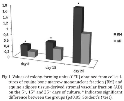
The number of CFU was obtained by counting the cell groupings, where five or more cells were considered a colony. CFU were counted on days five, 15 and 25 of sub-culture, where five random fields were examined per culture flask of each animal.
Statistical analysis. The CFU obtained were analyzed by Student's t test, using the GraphPad InStat 3.0 program. The level of significance was set at 5% (P<0.05).
RESULTS
CFU counts
In relation to the number of CFU observed in cultures of cells obtained from adipose tissue (AD) and of cells obtained from bone marrow (BM), there were no differences between the samples of the same origin cultivated in different media, indicating that the culture medium had no influence on this characteristic. However, significant differences were found between the samples of AD and BM, where bone marrow samples showed a higher number of CFU than did samples from adipose tissue (Fig.1), suggesting that bone marrow cells had a greater colony-forming capability.
Morphological analysis
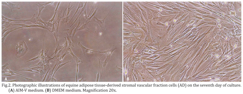
At the beginning of morphological evaluations (day zero), AD and MO cultures showed similar characteristics, with rounded cells of different sizes and very heterogeneous shapes. However, on the third day of culture, after removal of the non-adherent cells, AD cultures showed a population of fusiform and firmly adhered cells, while the MO cells were round and fewer in number.
During the first week of culture, it was possible to detect differences between the cultures maintained in DMEM (containing 10% FBS) and those grown in AIM-V. AD cells maintained a predominantly fusiform shape and showed greater cellularity in DMEM compared to cells cultivated in AIM-V (Fig.2). Meanwhile, in cultures of MO, there was a population of adhered cells with greater numbers and more refringence in AIM-V.
As of the 14th day of culture, differences between the two culture media were no longer found. AD cells maintained a fusiform shape and showed disorganized proliferation. The samples of MO displayed the presence of rounded cells and cells with a fusiform appearance, and they formed colonies or cell aggregates.
At 21 days of culture, the AD samples contained strongly adhered cells with a fusiform appearance, which showed organized proliferation. In MO cultures, there was a greater refringence and presence of rounded cells and fusiform cells.
The last morphological evaluation was done at 32 days of culture, at which time AD cultures showed the same characteristics as previously seen and those of MO had a predominance of adhered fusiform cells.
DISCUSSION
The methods used to collect equine bone marrow and adipose tissue, as well as the techniques used for the isolation of cells from the bone marrow mononuclear fraction and adipose tissue-derived stromal vascular fraction were considered simple and easy, and had already been described in detail (Ribeiro et al. 2012).
The cell population with the fusiform appearance described here in samples from both adipose tissue and bone marrow were morphologically similar to mesenchymal stem cells from human (Zuk et al. 2001, You et al. 2009), sheep (Fadel et al. 2011), horse (Fortier et al. 1998, Smith et al. 2003, Hegewald et al. 2004, Vidal et al 2006, 2007, Maia et. al 2012, De Vita et al. 2013), and other animals reported in many studies, in which these cells were described as star-shaped or fibroblast-like.
In relation to cell expansion, it was observed in the course of culture that AD samples always reached 90% confluence before MO samples, and thus, AD cells were passaged about every three days, whereas MO cells every five days. These observations corroborated the findings of Colleoni et al. (2009), indicating that, in horses, cells from adipose tissue show a greater capacity for proliferation in culture compared to cells obtained from bone marrow. Similar results were obtained by Cowan et al. (2004) who studied murine mesenchymal stem cells and reported that the rate of expansion of cells derived from adipose tissue was five times greater in the first seven days compared to bone marrow cells.
Besides, it was possible to observe that the cells from adipose tissue had more homogeneous cell populations while the cells from bone marrow showed a more heterogeneous culture for a longer time. Fortier et al. (1998) were able to complete the removal of blood cells and non-adherent hematopoietic stem cells of the mesenchymal stem cell culture by the ninth day of culture, when the population of predominant cells showed a similar morphology as the cells found in the present work. Maia et al. (2012) had adherent cells from bone marrow mononuclear fraction between 24 and 48 hours of culture, and fibroblast-like cells morphology by the fourth day of culture. Therefore, in this study, it appears that the non-adherent cells were successfully removed from the adipose tissue cultures in the first exchange of medium, while the cell cultures from bone marrow still showed non-adherent cells, although in decreasing amounts, up to the last evaluation, thus explaining the heterogeneous appearance of the culture.
De Vita et al. (2013), studying mesenchymal stem cells from equine amniotic fluid, reported an initial difficulty in cell isolation, and achieved better cell adhesion on the culture plate by decreasing the amount of medium in the primary culture to leave the cells closest to the plate, and by increasing the time to the first exchange of medium from 2 to 7 days, to prevent the removal of cells that were capable of adhesion and were still in suspension.
Vidal et al. (2007) also observed that cell populations obtained from adipose tissue did not form as many colonies as cells from bone marrow, and that they were distributed more uniformly and adhered more firmly to the culture plate, as observed in this study.
Mesenchymal stem cell cultures with DMEM medium supplemented with fetal bovine serum (FBS) are often used today (De Vita et al. 2013, Maia et. al 2012). However, to avoid the potential risk of exposing human patients to cells cultivated in the presence of FBS, some investigators have searched for a substitute supplement (Bieback et al. 2009) because, according to the results of Pal et al. (2009), the presence of serum is essential for maintaining and expanding mesenchymal stem cell cultures. In the present work, despite samples coming from adipose tissue, they showed greater cellularity in DMEM supplemented with FBS in the first two weeks, but afterwards, there is no longer any difference in relation to grown in AIM-V medium without FBS, suggesting that this can be an alternative for the culture of cells destined for transplantation.
In a current study about the potential of neural differentiation of mesenchymal stem cells from equine bone marrow, Maia et al. (2012) surprisingly found that control cells culture that have not undergone any treatment, also expressed neural markers. These researchers suggested that FBS present in the culture medium may have influenced the differentiation in the control group, because it is known that FBS is rich in steroids, such as glucocorticoids and estrogen, which may have interacted with other growth factors, also present in the serum, driving a cascade of events to cell differentiation. Therefore, if FBS presence in mesenchymal stem cells culture could induce cell differentiation, it is very important more studies about it are carried out and other alternatives of culture media able to support the mesenchymal stem cells culture without FBS are analyzed.
CONCLUSIONS
The AIM-V culture medium is an alternative for the culture of equine bone marrow mononuclear fraction and adipose tissue - derived stromal vascular fraction cells in the absence of FBS.
Equine adipose tissue - derived stromal vascular fraction cells have a greater capacity for proliferation in culture compared to bone marrow mononuclear fraction cells.
Equine bone marrow mononuclear fraction cells have a greater capacity for agglutination and formation of colonies compared to adipose tissue - derived stromal vascular fraction cells.
Received on October 11, 2013
Accepted for publication on November 19, 2013
- Bieback K., Hecke A., Kocaömer A., Lanner H., Schallmose K., Strun D. & Klüte H. 2009. Human alternatives to fetal bovine serum for the expansion of mesenchymal stromal cells from bone marrow. Stem Cells 27:2331-2341.
- Colleoni S., Bottani E., Tessaro I., Mari G., Merlo B., Romagnoli N., Spadari A., Galli C. & Lazzari G. 2009. Isolation, growth and differentiation of equine mesenchymal stem cells: effect of donor, source, amount of tissue and supplementation with basic fibroblast growth factor. Vet. Res. Commun. 33:811-821.
- Cowan C.M., Shi Y.Y. & Aalami O.O. 2004. Adipose-derived adult stromal cells heal critical-size mouse calvarial defects. Nat. Biotechnol. 22:560-567.
- De Vita B., Campos L.L., Listoni A.J., Maia L., Sudano M.J., Curcio B.R., Landim-Alvarenga F.C. & Prestes N.C. 2013. Isolamento, caracterização e diferenciação de células-tronco mesemquimais do líquido amniótico equino obtido em diferentes idades gestacionais. Pesq. Vet. Bras. 33(4):535-542.
- Eagle H. 1955. Nutrition needs of mammalian cells in tissue culture. Science 122:501-514.
- Fadel L., Viana B.R., Feitosa M.L., Ercolin A.C., Roballo K.C., Casals J.B., Pieri N.C., Meirelles F.V., Martins D.S., Miglino M.A. & Ambrósio C.E. 2011. Protocols for obtainment and isolation of two mesenchymal stem cell sources in sheep. Acta Cir. Bras. 26(4):267-273.
- Fortier L.A., Nixon A.J., Williams J. & Christina S.C. 1998. Isolation and chondrocytic differentiation of equine bone marrow-derived mesenchymal stem cells. Am. J. Vet. Res. 59:1182-1187.
- Fraser J.K., Zhu M., Wulur I. & Alfonso Z. 2008. Adipose-derived stem cells. Meth. Mol. Biol. 449:59-67.
- Gonçalves R.F. 2002. Cultivo de células epiteliais da tuba uterina de bovinos na presença e ausência de soro fetal bovino: análise bioquímica, morfológica e morfométrica. Tese de Doutorado, Faculdade de Medicina Veterinária e Zootecnia, Universidade de São Paulo, São Paulo.
- Gutierrez-Nibeyro S.D. 2011. Commercial cellbased therapies for musculoskeletal injuries in horses. Vet. Clin. Equine 27:363-371.
- Hegewald A.A., Ringe J., Bartel J., Krüger I., Notter M., Barnewitz D., Kaps C. & Sittinger M. 2004. Hyaluronic acid and autologous synovial fluid induce chondrogenic differentiation of equine mesenchymal stem cells: a preliminary study. Tissue and Cell 36:431-438.
- Maia L., Landim-Alvarenga F.C., Golim M.A., Sudano M.J., Taffarel M.O., De Vita B., Freitas N.P.P. & Amorim R.M. 2012. Potencial de transdiferenciação neural das células-tronco mesenquimais da medula óssea de equino. Pesq. Vet. Bras. 32(5):444-452.
- Morgan S.J. & Darling D.C. 1995. Cultivo de Celulas Animales. Acribia, Zaragoza. 159p.
- Nixon A.J., Dalgren L.A., Haupt J.L. Yeager A.E. & Ward D.L. 2008. Effect of adipose-derived nucleated cell fractions on tendon repair in horses with collagenase-induced tendinitis. Am. J. Vet. Res. 69:928-937.
- Oliveira P.G.G., Alves A.L.G., Carvalho A.M., Hussni C.A., Watanabe R.L., Rodrigues M.M.P. & Mota L.S. 2011. Uso de células mononucleares da medula óssea no tratamento de tendinites induzidas experimentalmente em equinos. Arq. Bras. Med. Vet. Zootec. 63:1391-1398.
- Pal R., Hanwate M., Jan M. & Totey S. 2009. Phenotypic and functional comparison of optimum culture conditions for upscaling of bone marrow-derived mesenchymal stem cells. J. Tissue Eng. Reg. Med. 3:163-174.
- Ribeiro G., Massoco C.O. & Lacerda Neto J.C. 2012. Viabilidade celular da fração mononuclear da medula óssea e fração vascular estromal do tecido adiposo de equinos após o processo de congelamento e descongelamento. Pesq. Vet. Bras. 32(Supl.1):118-124.
- Renzi S., Lombardo T., Dotti S., Dessi S.S., De Blasio P. & Ferrari M. 2012. Mesenchymal stromal cell cryopreservation. Biopreserv. Biobanking 10:276-281.
- Smith R.K.W., Korda M., Blunn G.W. & Goodship A.E. 2003. Isolation and implantation of autologous equine mesenchymal stem cells from bone marrow into the superficial digital flexor tendon as a potential novel treatment. Equine Vet. J. 35:99-102.
- Vidal M.A., Kilroy G.E., Johnson J.R., Lopez M.J., Moore R.M. & Gimble J.M. 2006. Cell growth characteristics and differentiation frequency of adherent equine bone marrow-derived mesenchymal stromal cells: adipogenic and osteogenic capacity. Vet. Surg. 35:601-610.
- Vidal M.A., Kilroy G.E., Lopez M.J., Johnson J.R., Moore R.M. & Gimble J.M. 2007. Characterization of equine adipose tissue-derived stromal cells: adipogenic and osteogenic capacity and comparison with bone marrow-derived mesenchymal stromal cells. Vet. Surg. 36:613-622.
- You Q., Tong X., Guan Y., Zhang D., Huang M., Zhang Y. & Zheng J. 2009. The biological characteristics of human third semester amniotic fluid stem cells. J. Int. Med. Res. 37:105-112.
- Zuk P.A., Zhu M., Mizuno H., Huang J., Futrell J.W., Katz A.J., Benhaim P., Lorenz H.P. & Hedrick M.H. 2001. Multilineage cells from human adipose tissue: implication for cell-based therapies. Tissue Eng. 7:211-228.
Publication Dates
-
Publication in this collection
07 Apr 2014 -
Date of issue
Dec 2013
History
-
Received
11 Oct 2013 -
Accepted
19 Nov 2013