Abstracts
In order to determine auscultatory and electrocardiographic characteristics of Crioulo horses, one hundred animals ranging between one and twenty-six years of age (21 stallions, nine geldings, 27 pregnant mares e 43 not pregnant mares) were evaluated. The cardiac auscultation was performed during the clinical examination of the cardiovascular system, evaluating frequency, rate, normal and abnormal heart sounds (heart murmurs). The electrocardiographic examination followed the bipolar base-apex derivative system with animals at rest, by using an ECG-PC TEB equipment. The cardiac frequency, heart rate, morphology, duration, wave and complex amplitudes and interval durations were determined. The results were submitted to ANOVA and Tukey tests with an error probability of 5%. The cardiac auscultation revealed presence of functional systolic and diastolic murmur (10.00%) and systolic murmur compatible with tricuspid regurgitation besides normal heart sounds S1 (100.0%), S2 (100.0%), S3 (19.0%) and S4 (34.0%). The cardiac frequency obtained the average of 43.64 bpm, observing significative differences in relation to sexual and age factors and training level. The sinus rhythm was the most frequent (57.00%), followed by sinus tachycardia (38.00%) and sinus arrhythmia (5.00%), being observed rhythm disturbances in 16% of tracings. The P and T waves were observed more frequently in their forms P bifida positive (95.00%) and biphasic T (91.00%), being variable at tracing. There were also observed Q waves in 12.00% of the tracings. Thus, it was concluded that the auscultatory characteristics of Crioulo horses are according to the described in the literature for the species and the sexual factor, category, age factor and training level can influence some electrocardiographic parameters.
Electrocardiogram; ECG; cardiology; horses; cardiovascular
Com o objetivo de determinar as características auscultatórias e eletrocardiográficas de equinos da raça Crioulo foram avaliados 100 animais (21 garanhões, nove machos castrados, 27 fêmeas prenhes e 43 fêmeas vazias) com idades entre um e 26 anos. A auscultação cardíaca foi realizada junto ao exame clínico do sistema cardiovascular, avaliando-se frequência, ritmo e sons cardíacos normais e anormais (sopros). O exame eletrocardiográfico seguiu o sistema de derivação bipolar base-ápice, com os animais em repouso, utilizando-se um aparelho ECG-PC TEB. Foram determinadas a frequência (FC) e ritmo cardíaco, morfologia, duração e amplitude de ondas e complexos e duração de intervalos. Os resultados foram submetidos ao teste ANOVA e teste Tukey com probabilidade de erro de 5%. A auscultação cardíaca evidenciou além dos sons cardíacos normais S1 (100,0%), S2 (100,0%), S3 (19,0%) e S4 (34,0%), presença de sopro funcional sistólico e diastólico (10,00%) e sopro sistólico compatível com regurgitação tricúspide. A FC obteve valores médios de 43,64 bpm, observando-se diferenças significativas em relação ao fator sexual, etário e nível de treinamento. O ritmo sinusal foi o mais frequente (57,00%), seguido de taquicardia sinusal (38,00%) e arritmia sinusal (5,00%), sendo as alterações do ritmo observadas em 16% dos traçados. A análise de ondas, complexos e intervalos foram observadas diferenças significativas quanto a fatores sexual, etário e nível de treinamento. As ondas P e T foram observadas com maior frequência em suas formas P bifida positiva (95,00%) e T bifásica (91,00%), sendo variável no traçado. Também foram observadas ondas Q em 12,00% dos traçados. Conclui-se que as características auscultatórias de equinos da raça Crioula estão de acordo com o descrito na literatura para a espécie e que o fator sexual, a categoria, o fator etário e o nível de treinamento podem influenciar alguns parâmetros eletrocardiográficos.
Eletrocardiograma; ECG; cardiologia; equinos; cardiovascular
MORFOFISIOLOGIA/ANIMAL MORPHOPHYSIOLOGY
Auscultatory and electrocardiographic characteristics of Crioulo horses
Características auscultatórias e eletrocardiográficas de equinos da raça Crioula
Jackson SchadeI; Michele F.S. SchadeII; Joandes H. FontequeIII* * Corresponding author: fonteque@hotmail.com
IGraduando em Medicina Veterinária, Bolsista de Iniciação Científica (PROBIC/UDESC), Departamento de Medicina Veterinária (DMV), Centro de Ciências Agroveterinárias (CAV), Universidade do Estado de Santa Catarina (UDESC), Av. Luiz de Camões 2090, Bairro Conta Dinheiro, Lages, SC 88520-000, Brazil
IIGraduando em Medicina Veterinária, DMV/CAV-UDESC, Lages, SC
IIIDocente do Departamento de Medicina Veterinária, DMV/CAV-UDESC, Av. Luiz de Camões 2090, Bairro Conta Dinheiro, Lages, SC 88520-000
ABSTRACT
In order to determine auscultatory and electrocardiographic characteristics of Crioulo horses, one hundred animals ranging between one and twenty-six years of age (21 stallions, nine geldings, 27 pregnant mares e 43 not pregnant mares) were evaluated. The cardiac auscultation was performed during the clinical examination of the cardiovascular system, evaluating frequency, rate, normal and abnormal heart sounds (heart murmurs). The electrocardiographic examination followed the bipolar base-apex derivative system with animals at rest, by using an ECG-PC TEB equipment. The cardiac frequency, heart rate, morphology, duration, wave and complex amplitudes and interval durations were determined. The results were submitted to ANOVA and Tukey tests with an error probability of 5%. The cardiac auscultation revealed presence of functional systolic and diastolic murmur (10.00%) and systolic murmur compatible with tricuspid regurgitation besides normal heart sounds S1 (100.0%), S2 (100.0%), S3 (19.0%) and S4 (34.0%). The cardiac frequency obtained the average of 43.64 bpm, observing significative differences in relation to sexual and age factors and training level. The sinus rhythm was the most frequent (57.00%), followed by sinus tachycardia (38.00%) and sinus arrhythmia (5.00%), being observed rhythm disturbances in 16% of tracings. The P and T waves were observed more frequently in their forms P bifida positive (95.00%) and biphasic T (91.00%), being variable at tracing. There were also observed Q waves in 12.00% of the tracings. Thus, it was concluded that the auscultatory characteristics of Crioulo horses are according to the described in the literature for the species and the sexual factor, category, age factor and training level can influence some electrocardiographic parameters.
Index terms: Electrocardiogram, ECG, cardiology, horses, cardiovascular.
RESUMO
Com o objetivo de determinar as características auscultatórias e eletrocardiográficas de equinos da raça Crioulo foram avaliados 100 animais (21 garanhões, nove machos castrados, 27 fêmeas prenhes e 43 fêmeas vazias) com idades entre um e 26 anos. A auscultação cardíaca foi realizada junto ao exame clínico do sistema cardiovascular, avaliando-se frequência, ritmo e sons cardíacos normais e anormais (sopros). O exame eletrocardiográfico seguiu o sistema de derivação bipolar base-ápice, com os animais em repouso, utilizando-se um aparelho ECG-PC TEB. Foram determinadas a frequência (FC) e ritmo cardíaco, morfologia, duração e amplitude de ondas e complexos e duração de intervalos. Os resultados foram submetidos ao teste ANOVA e teste Tukey com probabilidade de erro de 5%. A auscultação cardíaca evidenciou além dos sons cardíacos normais S1 (100,0%), S2 (100,0%), S3 (19,0%) e S4 (34,0%), presença de sopro funcional sistólico e diastólico (10,00%) e sopro sistólico compatível com regurgitação tricúspide. A FC obteve valores médios de 43,64 bpm, observando-se diferenças significativas em relação ao fator sexual, etário e nível de treinamento. O ritmo sinusal foi o mais frequente (57,00%), seguido de taquicardia sinusal (38,00%) e arritmia sinusal (5,00%), sendo as alterações do ritmo observadas em 16% dos traçados. A análise de ondas, complexos e intervalos foram observadas diferenças significativas quanto a fatores sexual, etário e nível de treinamento. As ondas P e T foram observadas com maior frequência em suas formas P bifida positiva (95,00%) e T bifásica (91,00%), sendo variável no traçado. Também foram observadas ondas Q em 12,00% dos traçados. Conclui-se que as características auscultatórias de equinos da raça Crioula estão de acordo com o descrito na literatura para a espécie e que o fator sexual, a categoria, o fator etário e o nível de treinamento podem influenciar alguns parâmetros eletrocardiográficos.
Termos de indexação: Eletrocardiograma, ECG, cardiologia, equinos, cardiovascular.
INTRODUCTION
The horse has its origin in equine population from the Iberian Peninsula, which had been brought to America by Spanish and Portuguese people. From the seventeenth century many of these horses have been lost or abandoned, starting to create wild herds distributed throughout America. Since then, the breed was formed by natural selection during four centuries, being shaped in a hostile environment, where only the strongest survived and managed to pass on their genes to future generations. Thus, the horse acquired its own characteristics, achieving worldwide notoriety from the twentieth century on, when several associations were created (ABCCC 2011).
According to Beck (1989), the horse can tolerate hard tests, with eminently practical and economical quality. It presents qualities as great fertility, genotypic prepotency, adaptability, ability to guide cattle and docility, concluding that the main reason for its breeding lies in its endurance, sobriety and rusticity. Cardiology has assumed great importance in the Veterinary Medicine, not only for the detection of cardiac abnormalities and appropriate treatments, but also for establishing prognoses both athletic life as survival, rationalizing the athletic campaign or the activity that is intended (Diniz 2006).
In horses, cardiac abnormalities are referred to as the third major cause of performance degradation in athletes, after musculoskeletal and respiratory alterations (Martin, Reef & Parente 2000). Cardiac auscultation is an extremely important component of cardiovascular clinical examination (Speirs 1999), because it allows the diagnosis of several cardiac disorders or the detection changes such as arrhythmias, murmurs, pericardial friction rub, splitting of heart sounds and other changes, either pathological or physiological (Mendes Neto 2008). The murmurs are audible as vibrations produced by turbulent blood flow during a cardiac cycle normally silent (Speirs 1999). According to Marr (2005), heart murmurs are very common in horses, occurring in 65-80% of horse breeds athletes. Although most cardiac murmurs are physiological, others provide evidence of heart disease and may require further investigation (Bonagura & Reef 2000). Functional heart murmurs (physiological) may be present without evident cardiac disease, being associated with left ventricular ejection, hypoproteinemia and anemia (Marr 2005). Pathological heart murmurs include valve regurgitation, septal defects, vascular lesions and less commonly in the horse, valve stenosis (Bonagura & Reef 2000). To assist in their identification, murmurs can be classified to auscultation, being used as a reference for subsequent examinations (Patteson 1996).
Cardiac arrhythmias are very common in horses, but their presence does not imply directly into heart disease (Marr 2005). According to Robertson (1992) healthy horses incidence of arrhythmias are about 25% to 30%, where these are attributed to the high variation of vagal tone at rest, being considered benign or physiological. But they must be differentiated from pathological conditions such as atrial fibrillation, third degree atrioventricular and premature atrial and ventricular blocks (Hilwig 1977, Miller 1989). The electrocardiogram (ECG) is an important diagnostic method in recognizing the formation and conduction disturbances of the cardiac impulse, being able to identify many arrhythmias and should always be interpreted in conjunction with accurate clinical examination of the cardiovascular system (White & Rhode 1974). However, orientation, amplitude and duration of the ECG waves depend on several factors, including the animal's age, breed, sex, derivation examined and fitness (Reef 1992). In the equine species the ECG is valid for determining the frequency, rhythm and conduction times. The determination of heart size by evaluating the complex is limited, since the genesis of the ECG is different from that which occurs in humans and small animals (Reef 1985) and horse shows some particularities in cardiac depolarization (Bonagura & Reef 2000). Therefore, the determination of arrhythmias and conduction disturbances is still the main indication for the realization of ECG in horses (Reef 1985). The bipolar base-apex derivative system is one of the systems used in horses that has great practicality for the location of electrode placement, besides presenting regular tracings with complex of large amplitude, facilitating thus the reading (Ayala et al. 1994, Patteson 1996).
Due to the scarce literature about cardiovascular evaluation in Crioulo horses, the purpose of this study is to determine the auscultatory and electrocardiographic characteristics of horses from this breed in relation to age, reproductive period and level of training.
MATERIALS AND METHODS
One hundred equines aged between 1 and 26 years were used, represented by 21 stallions, nine geldings, 27 pregnant mares (middle third of pregnancy) and 43 not pregnant mares. The animals were obtained from five ranches located in Cabanha OCA, Mafra-SC; Cabanha Impulso, São Bento do Sul-SC; Estância Rio da Pedra, Rio Negrinho-SC; Cabanha JM, Rio Negrinho-SC; Cabanha Rio Bonito, Ponta Grossa-PR. The horses were distributed in four groups according to age: group 1, consisted of animals younger than two years old (n=15); group 2, animals from three to five years (n=28); group 3, animals from six to ten years (n=25) and group 4, animals over 11 years (n=32). To evaluate the influence of the level of training, horses were grouped into: animals in training (n=31); untrained animals (n=15); animals handled (n=9) and animals unhandled (n=12).
First, the horses were evaluated by clinical examination of the cardiovascular system according to Speirs (1999), with the aid of a stethophon for auscultation and evaluation of frequency, rhythm, normal and abnormal heart sounds (murmurs). In the presence of murmurs, these were classified as time, duration, location, intensity and quality according to Speirs (1999). The presence of any health disorder was used as a criterion for exclusion of animals. For the electrocardiographic examination was used the ECG-PC TEB equipment, with AC 177 cable connected to a voltage stabilizer. The equipment was adjusted for the speed of 25mm/s and sensitivity of 1cm=1mV, being registered tracings during a five-minute period. The examinations were performed without any chemical tranquilization, with the animal contained in standing position on a rubber floor in order to minimize interference. The arrangement of the electrodes followed the bipolar base-apex derivative system described by Patteson (1996), connected to the animal's skin through metallic conductors humidified with alcohol 70% to produce more conductivity between the electrodes and the skin of the animal. The tracings were analyzed in derivation I (DI) to determine the frequency (bpm) and heart rate, morphology, duration (sec) and amplitude (mV.) of the waves and complex, and leveling of ST segment (mV). Statistical analysis of data was performed by means of ANOVA and Tukey test to compare the averages, assuming a probability of error of 5% according to Curi (1998). The project was filed and approved by the Ethics Committee on Animal Experiments (CETEA) from Universidade de Santa Catarina (UDESC) under number 1.18.11.
RESULTS AND DISCUSSION
The results concerning the relative frequency (%) of normal heart sounds S1, S2, S3 and S4 (heart sounds) listened individually and combined (Table 1), agreed with the statements of Patteson (1996), Speirs (1999), Bonagura & Reef (2000), Mendes Neto (2008) and Marr & Bowen (2010), where three (S1, S2, S3 or S1, S2, S4) or four (S1, S2, S3, S4) heart sounds may be evidenced on auscultation in normal horses. However, in other species, such as dogs and cats, the presence of S3 and S4 sounds are considered abnormal. Thus, the presence of three or four heart sounds constitutes the evidence of cardiac diseases in these species (Tilley & Goodwin 2002). The frequency of S3 and S4 in auscultation shows that these sounds may be auscultated in some, but not in all horses, according to Patteson (1996). According to Marr & Bowen (2010) S3 and S4 heart sounds are commonly listened in pure blood racing horses and the S4 sound less frequent in ponies. Patteson (1996) states that the S3 sound is most often auscultated in equine athletes and less common in older horses and ponies. The current study reveals lower frequency of S3 sound in senile horses (15.79% -3/19), corroborating the statements of Patteson (1996).
Diastolic murmurs with functional features focus on mitral valve were auscultated in three equines (Table 1) (Patteson 1996, Bonagura & Reef 2000, Marr 2005). Functional diastolic murmurs occur in early in the third or middle part of diastole and are heard on the base of the heart or left atrioventricular area, corresponding to the mitral valve (Marr 2005), being generally mild (Bonagura & Reef 2000). According to Patteson (1996), Bonagura & Reef (2000), Marr (2005) and Marr & Bowen (2010) these murmurs can often be strident or musical, a fact that was not observed in this current study.
Systolic murmur over the focus of the aortic valve was identified in one horse, being classified as systolic ejection murmur, which is considered the most common functional murmur in horses (Patteson 1996, Bonagura & Reef 2000). According to Patteson (1996) and Marr & Bowen (2010), systolic ejection murmurs may be auscultaded in 60% and 66% of normal horses respectively, and according to Patteson (1996) they are more evident in athletic horses and foals. Patteson & Cripps (1993) found a high prevalence of systolic ejection murmurs (53%) in racing horses. According to the obtained in this current study, horses showed low frequency of this type of murmur. Physiological or functional murmurs are common in horses without heart disease (Patteson 1996, Bonagura & Reef 2000, Marr 2005), and may be related to the large size and the huge volumes of inflow and flow of the heart in this species (Bonagura & Reef 2000). According to Patteson (1996), Speirs (1999), Bonagura & Reef (2000) and Marr (2005) physiological murmurs may also be associated with high speed of blood flow related to decreased of blood viscosity in cases of anemia, hypoproteinemia, high sympathetic activity (colic, exercise), fever or peripheral vasodilation. Due to the fact that the evidence of murmur may be related to congenital heart disease or valve insufficiency, it is important that physiologic murmurs are properly distinguished from pathological murmurs (Marr 2005).
Systolic murmurs over tricuspid valve area have been evidenced in the cardiac auscultation of seven animals (Table 1). The characteristics of these murmurs indicate that these are related to the tricuspid valve regurgitation. According to Marr & Reef (1995), Marr (2005) and Marr & Bowen (2010) valve regurgitation may occur in the absence of valvular pathology, being described as physiological regurgitation, showing no clinical significance. This type of regurgitation is associated to small regurgitant jets which produce low-intensity soft murmurs (Marr & Reef 1995). The factors that influence the physiological regurgitation are not well elucidated yet, and may involve not only neuroendocrine factors, but also changes in the geometry of the ventricles as part of the adaptation of the heart to exercise and training (Marr & Bowen 2010).
A grade II holosystolic murmur in tricuspid area growing, compatible with tricuspid regurgitation murmur associated with this valve disease, which, according Bonagura & Reef (2000) and Marr & Bowen (2010) may be the pathologic murmur detected more frequently in horses has been observed in one of the seven animals cited. According to Patteson (1996) and Bonagura & Reef (2000) auscultation of a horse with this change reveals a holosystolic murmur of grade II to V/VI with maximal intensity over the area of the tricuspid valve, consistent with the one observed in this study. For Marr & Bowen (2010) the murmur may present growing character, however, Patteson (1996) and Bonagura & Reef (2000) cite that the murmur has character of plateau and decrescendo, respectively. It is important to remember that the horse in question was clinically healthy, with no signs of declining performance. According to Patteson (1996), Bonagura & Reef (2000) and Marr & Bowen (2010) tricuspid regurgitation is rarely associated to the impairment of performance in athletic horses. For Bonagura & Reef (2000) the systolic murmur of tricuspid regurgitation may be an incidental finding detected during routine examination, however it becomes more problematic when identified in a horse with low performance. Therefore we can suppose that the horse in question may present a mild valve alteration, which produced no apparent hemodynamic consequences.
In relation to cardiac rhythm identified on auscultation, the most common was the sinus, followed by sinus tachycardia (Table 1). Pauses during the auscultation were identified in three horses (Table 1). In two of these animals, the pauses were characterized by the periodic absence of S1 and S2, with currently absolute silence, where isolates S4 were not identified. These findings characterize sinus arrest (Patteson 1996, Speirs 1999). Periodic pauses characterized by the absence of S1 and S2 and presence of isolate S4 sounds were identified in one of the three mentioned horses, characterizing second-degree atrioventricular block (AVB 2nd degree) according to Patteson (1996) and Speirs (1999).
Presence of dysrhythmia has been evidenced on auscultation in three horses (Table 1). Of these, two were characterized by cyclic irregularity in heart rate, which were not associated with breathing, indicating sinus arrhythmia (Patteson 1996). According to Patteson (1996) sinus arrhythmia can, but is not necessarily associated, to breathing. Atrial premature complex (APC) has been identified on auscultation of an equine, characterized by regular sinus rhythm interrupted by a premature beat (S1 followed by S2), followed by a diastolic longer pause (Patteson 1996, Bonagura & Reef 2000). All cardiac rhythm disturbances detected by auscultation were later confirmed by ECG, which emphasizes the importance of an accurate cardiac auscultation.
In the analysis of the electrocardiographic tracing, heart rate or cardiac rhythm most frequently observed was the sinus (Table 2), similar to observations of Fernandes et al. (2004), Diniz et al. (2008), Dumont et al. (2010) and Diniz et al. (2011), where this rhythm was the most common in horses. This result is in accordance with Patteson (1996), who considered it as a physiological rhythm in this species. Sinus tachycardia was the second most frequent rhythm followed by sinus arrhythmia (Table 2). Diniz et al. (2011) also found sinus tachycardia as the second most frequent rhythm (23.0%), followed by sinus arrhythmia (20.0%) in jumping horses. However Diniz et al. (2008) found sinus arrhythmia (28.2%), followed by sinus tachycardia (6.7%) in equines of Mangalarga Marchador breed.
The sinus tachycardia is considered a common rhythm in equines, which may be physiological in the absence of other clinical signs being associated with pain, fear, excitation or exercise (Fregin 1992, Patteson 1996). The frequent presence of this rhythm in this study may be associated with the excitation of the animals at the moment of the physical and electrocardiographic examination. Diniz et al. (2008) related the excitation resulting from the placement of the electrodes and the consequent delay in the return of heart rate (HR) to baseline values to the frequent presence of tachycardia. In this current study in Crioulo horses sinus tachycardia has been identified more often on auscultation (Table 1) in relation to the electrocardiographic tracing (Table 2). This result may have been influenced by the moment of the auscultation, in the early period of the examination. At this time the animals could be frightened and not accustomed to the environment where the test was conducted. It should also be taken into consideration that the recording of the electrocardiogram was obtained during five minutes. At this period we can observe gradual reduction in HR of the animals, reaching values closer to the baseline.
Sinus arrhythmia can be normally found in equines at rest (Fregin 1992), being associated to changes in vagal tone (Patteson 1996). Ayala et al. (1995) found sinus arrhythmia as the most common rhythm in equines of the Andalusian breed. However Patteson (1996) states that this rhythm has a low frequency in equines at rest being more common during the recovery phase after exercise, especially after light exercise. Only two of the five horses with sinus arrhythmia (Table 2) were identified by auscultation (Table 1). This fact may have occurred due to changes in the autonomic nervous system, which in the period of auscultation could not be manifesting. Raekallio (1992) observed periods of sinus arrhythmia in most of the evaluated horses using Holter monitoring, apparently caused by variation in the autonomic nervous system. Alterations in heart rate have been observed in the evaluation of the electrocardiographic tracing in 16.0% of the animals (Table 2).
Sinus arrest, AVB 2nd degree and wandering pacemaker are considered physiological arrhythmias to equine species, being influenced by the high vagal tone, therefore they should be abolished with exercise or excitation (Hilwig 1977, Reimer 1992, Mitten 1996, Patteson 1996, Marr & Bowen 2010). According to Patteson (1996) blocking sinus is rarely pathological in equines. Patteson (1996) and Marr & Bowen (2010) argue that this arrhythmia is not uncommon in this species, but is less frequent than AVB, a fact that was not observed in animals of this current study. The AVB 2nd degree is considered the most common arrhythmia in equines, which can be observed in up to 30% of horses at rest (Patteson 1996) and according to Marr & Bowen (2010) is detected in more than 40% of equines at rest using Holter monitoring. The current study revealed low frequency of this arrhythmia in equines from the Crioulo breed (Table 2). Diniz et al. (2008) also found a low frequency of AVB 2nd degree in equines from Mangalarga Marchador breed, attributing to the fact that the horses were not being physically conditioned. It must be considered that in this study 31% of the evaluated animals were being physically conditioned, and observation of AVB 2nd degree occurred in an unconditioned animal. Thus, there may be breed influences regarding the occurrence of this arrhythmia. The AVB 2nd degree observed in the current study in Crioulo horses is of the type Mobitz type 1, which according to Patteson (1996) is the most common in horses.
Wandering pacemaker is commonly observed in horses, and according to Hilwig (1977) and Raekallio (1992) may be present in 30% of healthy horses and at rest. In a study conducted with jumping horses, Diniz et al. (2011) observed wandering pacemaker in 25% of animals. The current study shows a low frequency of this arrhythmia in Crioulo horses (Table 2), consonant with Diniz et al. (2008) who found 6.7% of this arrhythmia in equines from Mangalarga Marchador breed.
Non physiological variations in heart rate were found which were characterized by atrial premature complexes (APC) and premature ventricular complexes (PVC) (Table 2). According to Patteson (1996) APC is an supraventricular arrhythmia that is relatively common in horses and may be seen in clinically healthy animals. According to Mitten (1996), the APC may occur with equines at rest, with no association to exercise intolerance, and can decrease or disappear during exercise. Patteson (1996) cites that the APC may be more frequent during exercise than at rest, or can be detected during exercise in animals that had no APC at rest. However, they may show signs of myocardial disease, electrolyte imbalance, toxemia, sepsis, hypoxia or chronic valvular disease, being a significant finding in animals with low athletic performance (Patteson 1996). The PVCs are relatively uncommon in equines in comparison to other species (Patteson 1996) being an indicative of myocardial disease (Mitten 1996). However Patteson (1996) asserts that in the absence of other abnormalities, the presence of an isolated PVC on an evaluation by Holter may be of no significance, however it must alert to the possibility of myocardial disease. Mitten (1996) asserts that the PVC is an indicative of myocardial disease even in the absence of other abnormalities and it must be considered meaningful if the rate increases during exercise or presents multiform. It is noteworthy that the animals in which APC and PVC were observed clinically healthy and without any complaint of low athletic performance, therefore, they can be considered clinical findings for the sample. Only one Crioulo horse with APC of the nine horses that had APC and PVC has been identified on auscultation, a fact that can be attributed to the low frequency of these changes during the electrocardiographic tracing. Thus, during the auscultation the APC and PVC may not have manifested.
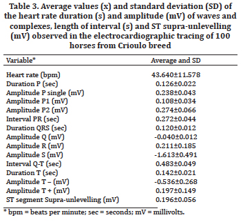
The average HR (Table 3) found itself within the range for adult horses at rest described by other authors as Hilwig (1977) and Fregin (1992), in which the values cited are 22 to 50bpm and 26 to 50bpm respectively. According to Diniz et al. (2011) HR is a parameter that can vary according to the sport, gender and breed of the animal. Significant differences were observed (P<0.05) in relation to gender and pregnant mare category for HR (Table 4). This result differs from those found by Vincenzi (1995), Diniz et al. (2008) and Diniz et al. (2011), which found no sexual influences on HR in other breeds. This difference may be due to the difference between male and female body, since such characteristics are higher in males. Significant differences have been observed in relation to HR of pregnant mares in relation to other categories such as not pregnant mares, stallions and geldings (Table 4). Fernandes et al. (2004) found no difference between pregnant mare and other categories such as foals and yearling animals. According to Liberatori Filho, Lopes & Lopes (1998), adaptive changes occur in the maternal organism during pregnancy which according to Campos Filho (1992) are particularly meaningful in the circulatory system. These physiological adjustments include an increase on blood volume, heart rate, cardiac output, besides reduction of systemic vascular resistance, resulting in the development of a hemodynamic state of high output. Thus, the current study in Crioulo horses, demonstrates that this physiological behavior can also be observed in the equine species.
There were significant differences (P<0.05) for HR in relation to age group (Table 5). This result corroborates with the results observed by Fernandes et al. (2004) where there was a decrease in HR when compared to one-year old foals and yearling animals. Thus, we can affirm that the body development observed until the age of two has influence on HR at rest. Animals older than 11 years showed differences (P<0.05) when compared to animals between 3-5 years and 6-10 years. This result may be due to the large number of pregnant mares present in this group, which contributed to increase the average. The level of training presented no significant difference (P>0.05) between animals in training and untrained animals. However, differences were observed (P<0.05) among unhandled animals in relation to animals in training and untrained animals (Table 6). These results may be associated to physiological adaptations of the myocardium during exercise, characterized by ventricular hypertrophy and consequent decrease in HR (Young 2005). Therefore, we might suppose that these physiological adaptations are of minor magnitude or absent. However, it must be taken into account that unhandled animals may be more frightened and clinical and electrocardiographic examination can lead to more excitation, increasing HR.
The average values concerning the duration of waves, intervals and complex (Table 3) were found according to the standards described by Patteson (1996) for equine species. These results found themselves in similar parts to those found by Ayala et al. (1994) and Diniz et al. (2008) in horses from Andalusian and Mangalarga Marchador breeds, respectively. Thus it is probable that the breed has not influence on the variation of these parameters, agreeing with Diniz et al. (2008). Regarding the amplitude of the waves (Table 3), Ayala et al. (1994) found different values in relation to these parameters. However Diniz et al. (2008) found similar values for R wave amplitude, however other variables showed great difference especially on T positive and negative waves amplitude. Thus, we can suggest that the breed is a factor of variation on electrocardiographic parameters. However, statistical inferences would be needed for attesting this influence in Crioulo horses, requiring future studies on the issue.
There were significant differences (P<0.05) concerning gender and category in relation to the amplitude of P2, PR interval, QRS duration, R-wave amplitude, S wave amplitude , QT interval, T positive and negative waves amplitude and ST supra-unlevelling (Table 4). Diniz et al. (2008) found sexual influences regarding R and S waves amplitude in equines from Mangalarga Marchador breed using the base-apex derivation system. The authors attributed the result to body difference between males and females. Thus, there may be sexual influences on electrocardiographic variables. Regarding the category pregnant mare, the differences may have occurred due to the interference of pregnancy on the positioning of the heart, in agreement with observations made by Fernandes et al. (2004). As regards the age factor, there was statistic difference (P<0.05) compared to the P2 amplitude, PR interval, QRS duration, QT interval, T positive and negative waves duration (Table 5). Diniz et al. (2008) did not obtain significant differences in electrocardiographic variables in relation to age factor in equines from Mangalarga Marchador breed. Diniz et al. (2011) observed the influence of age on electrocardiographic parameters, however the authors used the classical system of derivation of members.
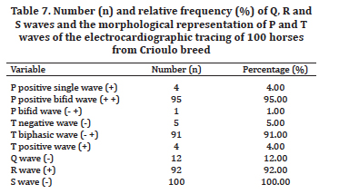
Significant differences (P<0.05) have also been observed in relation to the level of training being obtained for P2 amplitude, QRS complex duration, QT interval and T wave duration (Table 6). These results demonstrate that performance level may influence the electrocardiographic variables in horses. Generally between groups of gender, age and performance, it was observed that the behavior of QT interval occurred in a manner inversely proportional to HR, confirming affirmations made by Vincenzi et al. (2000), Fernandes et al. (2004) Diniz et al. (2011). Thus, the differences (P<0.05) observed for this variable may have been influenced by variations in HR in each group. Agreeing with Diniz et al. (2008) the P and T waves have been observed in 100% of the traces and they have a morphological pattern consistent with Patteson (1996) described. The frequency of morphological representations of P and T waves observed in the electrocardiographic tracing (Table 7) was different from that found by Diniz et al. (2008), wich revealed P wave bifid (68.3%), single (25.0%) and biphasic (6.7%), and T positive wave (11.0%), negative (20%) and biphasic (61.7%). Thus, we can observe a large variation in the distribution of morphological forms of these waves between Crioulo and Mangalarga Marchador breeds. It was observed in the analysis of the electrocardiographic tracing great variation concerning the morphology of the T wave with spontaneous changes during the trace, agreeing with affirmations made by Patteson (1996) and Sheard (1998). This fact has not been described in equines from Mangalarga Marchador breed (Diniz et al. 2008). According to Patteson (1996) T wave changes can also be seen in animals with heart disease, electrolyte disturbances and systemic disease. However these changes are highly variable and nonspecific, and thus they are of minor diagnostic utility.
Variations have also been observed in relation to the morphology of the P wave in the tracing which have been influenced by the HR. With the increase of the HR, the P wave changed their bifid positive form to positive only due to the fusion of the P1 and P2 components. A similar phenomenon has been observed by Senta, Smetzer & Smith (1970) in trotting equines during exercise. The results obtained in this study showed that P wave morphology can be influenced by alterations in HR of Crioulo horses at rest. The frequency of observation of waves Q, R and S in the electrocardiographic tracing obtained in this study (Table 7) differed from those found by Diniz et al. (2008), for Q (3.3%) and R waves (73.3%). Other authors did not observe the presence of Q wave in trotting equines from Andalusian breed, at the same bipolar base-apex derivation (Ayala et al. 1994). Diniz et al. (2008) suggested that the presence of Q wave on the electrocardiographic tracing could be a racial particularity.
CONCLUSIONS
According to the content exposed in the current study, we can conclude that the auscultatory characteristics of Crioulo horses are according to the described in the literature for the species.
The functional and pathological heart murmurs were found in low frequency, and part of the arrhythmias were detected during auscultation. The analysis of the electrocardiographic tracing showed low frequency of arrhythmias being sinus arrhythmia the most common. The sexual factor, category, age factor and level of training may influence some electrocardiographic parameters.
Considering that the evaluated horses presented some peculiarities in comparison to other breeds, it was possible to obtain an auscultatory and electrocardiographic pattern for the horses of Crioulo breed.
Acknowledgements
To the owners of Cabanhas OCA, Mafra/SC; Cabanha Impulso, São Bento do Sul/SC; Estância Rio da Pedra, Rio Negrinho/SC; Cabanha JM, Rio Negrinho/SC e Cabanha Rio Bonito, Ponta Grossa/PR.
REFERÊNCIAS
ABCCC 2011. História do cavalo Crioulo, <http//:www.abccc.com.br> Acesso em 01 mar. 2011.
Ayala I., Montes A., Bernal L.J. & Sandoval J.A. 1995. Electrocardiographic values in Spanish-bred horses in different ages. Aust. Vet. J. 72(6):225-226.
Ayala I., Montes A., Fernandez del Palacio M.J. & Gutiérrez Panizo C. 1994. Aportaciones al estúdio electrocardiográfico del caballo. An. Vet. (Murcia) 9(10):25-35.
Beck S.L. 1989 Eqüinos: raças, manejo, equitação. 2ª ed. Editora dos Criadores, São Paulo. 703p.
Bonagura J.D. & Reef V.B. 2000. Doenças cardiovasculares, p.250-319. In: Reed S.M. & Bayly W.M. (Eds), Medicina Interna Equina. Guanabara Koogan, Rio de Janeiro.
Campos Filho O. 1992. Estudo Doppler-ecocardiográfico do comportamento fisiológico dos fluxos valvares na gravidez e puerpério. Tese de Doutorado em Medicina, Escola Paulista de Medicina, São Paulo, SP. 152p.
Curi P.R. 1998. Metodologia e Análise da Pesquisa em Ciências Biológicas. 2ª ed. Gráfica e Editora Tipomic, Botucatu. 263p.
Diniz M.P. 2006. Perfil eletrocardiográfico de eqüinos de salto criados em São Paulo. Dissertação de Mestrado em Medicina Veterinária, Faculdade de Medicina Veterinária e Zootecnia, Universidade de São Paulo, São Paulo, SP. 134p.
Diniz M.P., Muzzi R.A.L., Muzzi L.A.L. & Alves G.E.S. 2008. Estudo eletrocardiográfico de equinos da raça Mangalarga Marchador. Arq. Bras. Med. Vet. Zootec. 60(3):536-542.
Diniz M.P., Michima L.E.S. & Fernandes W.R. 2011. Estudo eletrocardiográfico de equinos de salto sadios. Pesq. Vet. Bras. 31(4):355-361.
Dumont C.B.S., Leite C.R., Moraes J.M., Alves R.O., Godoy R.F. & Lima E.M.M. 2010. Parâmetros eletrocardiográficos de equinos Puro Sangue Árabe submetidos a exercício prolongado de enduro. Ciência Rural 40:1966-1973.
Fernandes W.R., Larsson M.H.M.A., Alves A.L.G., Fantoni D.T. & Belli C.B. 2004. Características eletrocardiográficas em eqüinos clinicamente normais da raça Puro Sangue Inglês. Arq. Bras. Med. Vet. Zootec. 56(2):143-149.
Fregin G.F. 1992. Medical evaluation of the cardiovascular system. Vet. Clin. North Am., Equine Pract. 8:329-346.
Hilwig R.W. 1977. Cardiac arrhythmias in the horse. J. Am. Vet. Med. Assoc. 170:153-163.
Liberatori Filho A.W., Lopes M.D. & Lopes A.C. 1998. Alterações funcionais do sistema circulatório na gravidez. Revta Bras. Clin. Terap. 27:123-127.
Marr C.M. 2005. Cardiac Murmurs: why worry? 9th Congress on Equine Medicine and Surgery, Geneva. In: Chuit P. & Montavon S. (Eds), Intern. Vet. Inf. Serv. (www.ivis.org), Ithaca, New York.
Marr C.M. & Bowen I.M. 2010. Cardiology of the Horse. 2nd ed. W.B. Saunders, London. 294p.
Marr C.M. & Reef V.B. 1995. Physiological valvular regurgitation in clinically normal young racehorses: prevalence and two-dimensional charac-
teristics. Equine Vet. J. 19:56-62.
Martin B.B., Reef V.B. & Parente E.J. 2000. Causes of poor performance of horses during training, racing, or showing: 348 cases (1992-1996). J. Am. Vet. Med. Assoc. 216(4):554-558.
Mendes Neto D. 2008. Sistema circulatório: semiologia do sistema circulatório de equinos e ruminantes, p.201-245. In: Feitosa F.L.F. (Ed.), Semiologia Veterinária, a arte do diagnóstico: cães, gatos, equinos, ruminantes e silvestres. 2ª ed. Roca, São Paulo.
Miller M.S. 1989. The equine electrocardiogram: usage in equine practice. Proc. Am. Assoc. Equine Pract. 34:577-586.
Mitten L.A. 1996. Cardiovascular causes of exercise intolerance. Vet. Clin. North Am., Equine Pract. 12(3):473-494.
Patteson M.W. & Cripps P.J. 1993. A survey of auscultation findings in horses.
Equine Vet. J. 25(5):409-415.
Patteson M.W. 1996. Equine Cardiology. Blackwell Science, Oxford. 254p.
Raekallio M. 1992. Long term ECG recording with holter monitoring in clinically healthy horses. Acta Vet. Scand. 33(1):71-75.
Reef V.B. 1985. Evaluation of the equine cardiovascular system. Vet. Clin. North Am., Equine Pract. 1:275-288.
Reef V.B. 1992. Cardiovascular problems associated with poor performance, p.381-410. In: Robinson N.E. (Ed.), Current Therapy in Equine Medicine. 3rd ed. W.B. Saunders, London.
Reimer J.M. 1992. Cardiac arrhythmias, p.382-393. In: Robinson N.E. (Ed.), Current Therapy in Equine Medicine. 3rd ed. W.B. Saunders, London.
Robertson S.A. 1992. Electrocardiography for the equine practioner. Vet. Annual 32:192-200.
Senta T., Smetzer D.L. & Smith C.R. 1970. Effects of exercise on certain eletrocardiographic parameters and cardiac arrhythmias in the horse: a radiotelemetric study. Cornell Vet. 60:552-569.
Sheard P.W.T. 1998. Cardiovascular system, p.295-438. In: Colahan P.T. (Ed.), Equine Medicine and Surgery. 5th ed. Mosby, St Louis.
Speirs V.C. 1999. Exame Clínico de Eqüinos. Artmed, Porto Alegre. 366p.
Tilley L.P. & Goodwin J.K. 2002. Manual de Cardiologia para Cães e Gatos. 3rd ed. Roca, São Paulo. 489p.
Vincenzi R.C. 1995. Determinação dos parâmetros eletrocardiográficos de equinos da raça Mangalarga, criados no Estado de São Paulo. Dissertação de Mestrado em Medicina Veterinária, Faculdade de Medicina Veterinária e Zootecnia, Universidade de São Paulo, São Paulo, SP. 73p.
Vincenzi R.C., Larsson M.H.M.A. & Fernandes W.R. 2000. Parâmetros eletrocardiográficos em equinos clinicamente normais da raça Mangalarga. III. Amplitude e duração dos complexos e intervalos. Revta Bras. Med. Vet. 22(5):194-198.
White N.A. & Rhode E.A. 1974. Correlation of electrocardiographic findings to clinical disease in the horse. J. Am. Vet. Med. Assoc. 164:46-56.
Young L.E. 2005. The effect of athletic training on the equine heart. 9th Congress on Equine Medicine and Surgery, Geneva. In: Chuit P. & Montavon S. (Eds), Intern. Vet. Inf. Serv. (www.ivis.org), Ithaca, New York.
Received on October 22, 2013.
Accepted for publication on February 23, 2014.
- ABCCC 2011. História do cavalo Crioulo, <http//:www.abccc.com.br> Acesso em 01 mar. 2011.
- Ayala
- Ayala
- Beck
- Bonagura
- Campos
- Curi
- Diniz
- Diniz
- Diniz
- Dumont
- Fernandes
- Fregin G.F. 1992.
- Hilwig
- Liberatori
- Marr C.M. 2005.
- Marr
- teristics. Equine Vet. J. 19:56-62.
- Martin B.B., Reef
- Mendes Neto D. 2008. Sistema circulatório: semiologia do sistema circulatório de equinos e ruminantes, p.201-245. In: Feitosa F.L.F. (Ed.), Semiologia Veterinária, a arte do diagnóstico: cães, gatos, equinos, ruminantes e silvestres. 2ª ed. Roca, São Paulo.
- Miller M.S. 1989. The
- Mitten L.A. 1996.
- Patteson
- Raekallio
- Reef
- Reef
- Reimer
- Robertson
- Senta
- Sheard
- Speirs
- Tilley L.P. &
- Vincenzi R.C. 1995. Determinação dos parâmetros eletrocardiográficos
- Vincenzi R.C., Larsson M.H.M.A. & Fernandes W.R. 2000. Parâmetros
- White
- Young L.E. 2005. The
Publication Dates
-
Publication in this collection
16 May 2014 -
Date of issue
Mar 2014
History
-
Accepted
23 Feb 2014 -
Received
22 Oct 2013