Abstracts
A survey was undertaken aiming to obtain an overview of ocular and periocular lesions diagnosed in domestic mammals over a period of 50 years in a veterinary pathology diagnostic laboratory in the Central Region of the State of Rio Grande do Sul, Brazil. In this lab, 33,075 histophatological exams had been performed over the period surveyed, of which 540 (1.6%) concerned ocular and periocular lesions. For various reasons ninety specimens were excluded from the study and the remaining 450 consisted of samples from dogs (53.5%), cattle (28.2%), cats (11.1%), horses (5.1%) sheep (1.3%), rabbits (0.4%), and pig (0.2%). The eyelids were the most prevalent (248/450) site of lesions in each of the species studied, followed by third eyelid (73/450), and conjunctiva (27/450). In dogs (241 samples) lesions in sebaceous glands (including Meibomian glands) were the most common findings (75/241), followed by melanocytic tumors (52/241) and nonspecific conjunctivitis (13/241). Squamous cell neoplasms, both benign and malignant, were relatively common. In cattle, anatomical sites affected by ocular and periocular lesions, in decreasing order of frequency, were eyelid, cornea and third eyelid. Squamous cell carcinoma (SCC) alone accounted for 80.3% of all diagnoses, while all neoplastic lesions made up for 85.0% of the lesions diagnosed in cattle. Neoplasia accounted for most of the lesions diagnosed in cats (39/50 cases); all of these were malignant, and SCC, hemangiosarcoma and fibrosarcoma were the most common types diagnosed. In horses, 19 out of 23 submissions were neoplasms and most were sarcoid (8/23) and SCC (8/23). There were six submissions from sheep with unpigmented skin, all of which represented SCC of the eyelids (5) and third eyelid (1).
Ophthalmology; palpebra; neoplasm
Foi realizada uma investigação para obter-se uma visão geral das lesões oculares e perioculares de mamíferos domésticos diagnosticadas ao longo de um período de 50 anos num laboratório de diagnóstico de patologia veterinária da Região Central do Rio Grande do Sul. Nesse laboratório, durante o período pesquisado foram realizados 33.075 exames histopatológicos, 540 dos quais (1,6%) eram de lesões oculares e perioculares. Por várias razões, 90 espécimes foram excluídos do estudo. As 450 amostras restantes consistiam espécimes de cães (53,5%), bovinos (28,2%), gatos (11,1%), cavalos (5,1%), ovelhas (1,3%), coelhos (0,4%), e porco (0,2%). As pálpebras foram o local mais prevalente (248/450) de ocorrência das lesões, seguidas da terceira pálpebra (73/450) e conjuntiva (27/450). Em cães (241 diagnósticos) as lesões nas glândulas sebáceas (incluindo as glândulas meibomianas) consistiram nos achados mais comuns (75/241), seguidos dos tumores melanocíticos (52/241) e de conjuntivites inespecíficas (13/241). Neoplasmas de células escamosas, tanto benignos como malignos, foram achados relativamente comuns. Em bovinos, os locais anatômicos afetados por lesões perioculares e oculares, em ordem decrescente de frequência, foram pálpebra, córnea e terceira pálpebra. Somente o carcinoma de células escamosas (CCE) perfez 80,3% de todos os diagnósticos, enquanto todas as lesões neoplásicas juntas perfizeram 85,0% das lesões diagnosticadas em bovinos. Em gatos, a maioria (39/50 casos) das lesões diagnosticadas era de neoplasia maligna e CCE, hemangiossarcoma e fibrosarcoma foram os diagnósticos mais frequentes. Em equinos 19 de 23 submissões eram neoplasmas e os mais comuns foram sarcoide (8/23) e CCE (8/23). Em ovinos foram encontradas seis submissões, todas casos de CCE de pálpebra (5/6) ou terceira pálpebra (1/6) de ovinos de pele despigmentada.
Oftalmologia; pálpebra; neoplasma
PEQUENOS ANIMAIS / SMALL ANIMAL DISEASES
Fifty years in the blink of an eye: a retrospective study of ocular and periocular lesions in domestic animals
Cinquenta anos num piscar de olhos: um estudo retrospectivo sobre lesões oculares e perioculares em animais domésticos..
Tessie Beck Martins; Claudio S.L. Barros* * Corresponding author: claudioslbarros@uol.com.br
Programa de Pós-Graduação em Medicina Veterinária, área de concentração em Patologia Veterinária, Centro de Ciências Rurais (CCR), Universidade Federal de Santa Maria (UFSM), Avenida Roraima 1000, Camobi, Santa Maria, RS 97105-900, Brazil
ABSTRACT
Martins T.B. & Barros C.S.L. 2014. Pesquisa Veterinária Brasileira 34(12):1215-1222. Programa de Pós-Graduação em Medicina Veterinária, Universidade Federal de Santa Maria, Camobi, Av. Roraima 1000, Santa Maria, RS 97105-900, Brazil. E-mail: claudioslbarros@uol.com.br
A survey was undertaken aiming to obtain an overview of ocular and periocular lesions diagnosed in domestic mammals over a period of 50 years in a veterinary pathology diagnostic laboratory in the Central Region of the State of Rio Grande do Sul, Brazil. In this lab, 33,075 histophatological exams had been performed over the period surveyed, of which 540 (1.6%) concerned ocular and periocular lesions. For various reasons ninety specimens were excluded from the study and the remaining 450 consisted of samples from dogs (53.5%), cattle (28.2%), cats (11.1%), horses (5.1%) sheep (1.3%), rabbits (0.4%), and pig (0.2%). The eyelids were the most prevalent (248/450) site of lesions in each of the species studied, followed by third eyelid (73/450), and conjunctiva (27/450). In dogs (241 samples) lesions in sebaceous glands (including Meibomian glands) were the most common findings (75/241), followed by melanocytic tumors (52/241) and nonspecific conjunctivitis (13/241). Squamous cell neoplasms, both benign and malignant, were relatively common. In cattle, anatomical sites affected by ocular and periocular lesions, in decreasing order of frequency, were eyelid, cornea and third eyelid. Squamous cell carcinoma (SCC) alone accounted for 80.3% of all diagnoses, while all neoplastic lesions made up for 85.0% of the lesions diagnosed in cattle. Neoplasia accounted for most of the lesions diagnosed in cats (39/50 cases); all of these were malignant, and SCC, hemangiosarcoma and fibrosarcoma were the most common types diagnosed. In horses, 19 out of 23 submissions were neoplasms and most were sarcoid (8/23) and SCC (8/23). There were six submissions from sheep with unpigmented skin, all of which represented SCC of the eyelids (5) and third eyelid (1).
Index terms: Ophthalmology, palpebra, neoplasm.
RESUMO
Foi realizada uma investigação para obter-se uma visão geral das lesões oculares e perioculares de mamíferos domésticos diagnosticadas ao longo de um período de 50 anos num laboratório de diagnóstico de patologia veterinária da Região Central do Rio Grande do Sul. Nesse laboratório, durante o período pesquisado foram realizados 33.075 exames histopatológicos, 540 dos quais (1,6%) eram de lesões oculares e perioculares. Por várias razões, 90 espécimes foram excluídos do estudo. As 450 amostras restantes consistiam espécimes de cães (53,5%), bovinos (28,2%), gatos (11,1%), cavalos (5,1%), ovelhas (1,3%), coelhos (0,4%), e porco (0,2%). As pálpebras foram o local mais prevalente (248/450) de ocorrência das lesões, seguidas da terceira pálpebra (73/450) e conjuntiva (27/450). Em cães (241 diagnósticos) as lesões nas glândulas sebáceas (incluindo as glândulas meibomianas) consistiram nos achados mais comuns (75/241), seguidos dos tumores melanocíticos (52/241) e de conjuntivites inespecíficas (13/241). Neoplasmas de células escamosas, tanto benignos como malignos, foram achados relativamente comuns. Em bovinos, os locais anatômicos afetados por lesões perioculares e oculares, em ordem decrescente de frequência, foram pálpebra, córnea e terceira pálpebra. Somente o carcinoma de células escamosas (CCE) perfez 80,3% de todos os diagnósticos, enquanto todas as lesões neoplásicas juntas perfizeram 85,0% das lesões diagnosticadas em bovinos. Em gatos, a maioria (39/50 casos) das lesões diagnosticadas era de neoplasia maligna e CCE, hemangiossarcoma e fibrosarcoma foram os diagnósticos mais frequentes. Em equinos 19 de 23 submissões eram neoplasmas e os mais comuns foram sarcoide (8/23) e CCE (8/23). Em ovinos foram encontradas seis submissões, todas casos de CCE de pálpebra (5/6) ou terceira pálpebra (1/6) de ovinos de pele despigmentada.
Termos de Indexação: Oftalmologia, pálpebra, neoplasma.
INTRODUCTION
The eye is a major special sense organ in vertebrate animals (Fernald 1997), that depend on it to survive and interact with the environment (Vorobyev et al. 2001, Williams 2010). Ocular pathology, a science that studies pathological processes that affect the eye bulb and its adnexal structures (Orellana & Pifano 2006), is a relatively new area in veterinary medicine, with its first publications dating from the beginning of twentieth century (Gelatt 2008).
In Brazil, veterinary ocular pathology is still modest when compared to ophthalmology, the specialty from which it derives, but the increasing number of veterinary establishments and teaching institutions that offer ophthalmologic service should soon depend, and luckily, impel, the development of ocular pathology.
It has been shown that the prevalence of diseases varies largely between countries and between regions within a country (Valentine 2006). Although important publications are available considering ocular diseases in specific animal species and ethiopathogenic entities, there is need for data about general prevalence of ocular lesions in our region. Lack of such data does not impair the diagnosis of lesions submitted to pathology labs, but their unavailability somehow forces local pathologists and students to confront their results with those in the foreign literature.
The purpose of this study is to determine the type and prevalence of ocular and periocular lesions in domestic mammals submitted to a veterinary pathology diagnostic laboratory in the Central Region of State of Rio Grande do Sul, Brazil.
MATERIALS AND METHODS
From January 1964 to December 2013, 33,075 histopathologic exams were performed at the Veterinary Pathology Laboratory, at the Federal University of Santa Maria (LPV-UFSM). All protocols were reviewed and cases pertinent to ocular and periocular lesions were filtered. Protocols in which there was no diagnosis or description of the lesions, protocols regarding samples improper for diagnoses (due to autolysis or insufficient material), samples originating from research animals, and samples from normal tissue (those in which no lesions were observed) were excluded.
Lesions were grouped by animal species, site, type of primary process/etiology, and final diagnosis. Only mammals were taken into account for this study. Lesions were grouped firstly according to species - dogs; cattle; cats; horses; sheep; pigs; and other. Subsequently, lesions were classified considering the main site affected and/or site of origin, as follows: eyelid; third eyelid; lacrimal gland; eyeball; bulbar conjunctiva; cornea; uvea; lens; and "eye". The latter category was created to accommodate diagnosis in which the site of lesion was not specified (eg. "tumor in the eye"). Such cases were not excluded because they accounted for a great portion of the total. The following criteria for classification were based on the primary pathologic process and etiology of the lesions: alterations due to trauma; congenital anomalies; autoimmune processes; infectious and parasitic diseases; degenerative diseases; metabolic and toxic diseases; neoplasia; and disturbances in cellular growth (DCG). The last category included lesions consisting of cellular hyperplasia and metaplasia, and cysts. Inflammatory lesions that could not be classified as infectious, parasitic or autoimmune were grouped under the umbrella term inflammation. Data related to animal breed, sex and age were not analyzed in this study.
RESULTS
During the fifty years encompassed by the current study, 33,075 histopathologic exams were performed at the LPV-UFSM. All protocols were reviewed and 540 cases (1.6%) concerning ocular and periocular lesions were filtered. Ninety of these (16.7%) were excluded because were not pertinent to the study, forty-eight of which related to birds and reptiles and forty-three, because their protocols lacked major data. In part of these protocols, information about description and/or diagnosis of the lesions was missing or not clear, and in other part, samples were not adequate for evaluation (due to small size or autolysis) or related to normal tissues (no lesions observed). Four hundred and fifty (83.3%) cases were selected for this study. More than one half (53.5%) of the samples came from dogs, followed by cattle (28.2%), cats (11.1%), horses (5.1%) and other species [sheep (1.3%), rabbits (0.4%), and a pig (0.2%)].
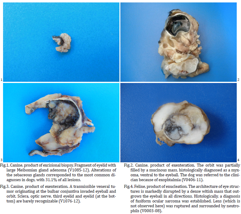
Eyelid was the most prevalent site of lesion in each of the species studied, accounting for 55.1% of all samples submitted (248/450), followed by third eyelid (73/450; 16.2%), and conjunctiva (27/450; 6.0%). Regarding the type of disease, neoplasia was the most numerous process, with 79.1% of all samples (356/450) (Fig.1-4), followed by inflammatory lesions of uncertain cause (50/450; 11.1%) (Fig.5), and congenital anomalies (12/450; 2.7%) (Fig.6).
Overall, squamous cell carcinoma (SCC) was the most common entity, with a prevalence of 30.8% (139/450) (Fig.7).
There were 241 samples from 239 individual dogs. One dog had phthisis bulbi and a SCC in the ipsilateral third eyelid, and another one had a carcinoma of unknown origin occupying the orbit and a sebaceous epithelioma in the ipsilateral eyelid. Lesions in the eyelids, especially in the cutaneous side, accounted for most part of samples in dogs (204/241; 84.6%). When all sites were considered, alterations of the sebaceous glands (including Meibomian glands) corresponded to the most common diagnoses in dogs (75/241; 31.1%), followed by melanocytic tumors (52/241; 21.6%), and nonspecific conjunctivitis (13/241; 5.4%). Squamous cell neoplasms were relatively common, with eight benign occurrences at the skin of eyelid and five malignant counterparts affecting conjunctiva or third eyelid. In one of these dogs, the third eyelid SCC was bilateral, and in another dog, it invaded the eyeball. In one situation, the dog had SCC affecting the conjunctiva and five other cutaneous sites along the body, as well as one cutaneous fibrosarcoma and one cutaneous mast cell tumor.
In cattle, sites affected by ocular and periocular lesions, in decreasing order, were eyelid (cutaneous side), cornea and third eyelid. SCC alone accounted for 80.3% of all diagnoses, while neoplastic lesions in total made 85.0%. Extensive invasion of the eyeball and orbit by SCC, with involvement of periocular tissues, was reported in seven cases. In five other cases, regional lymph nodes that were submitted with the ocular lesion had metastasis of SCC.
Neoplasia accounted for the major part of feline diagnosis, with 39/50 (78%) cases. All neoplasms were malignant. SCC, hemangiosarcoma and fibrosarcoma were the most common diagnoses, with 16 (32.0%), 10 (20.0%), and three (6.0%) cases, respectively. Nine of sixteen (56.2%) cats diagnosed with SCC had similar lesions in other parts of the head (ears; lips; nose; and/or contralateral eyelid), and in two occasions the neoplasm had already been removed before. In another two cats, clinicians reported invasion of eyeballs by cutaneous SCC with origin in the eyelids.
In horses, there were 24 submissions from 23 animals. Two out of 24 were samples from the same lesion, taken within a few weeks interval, and both were diagnosed as sarcoid, so they were condensed as a single diagnosis. When considering all samples, 19/23 (82.6%) accounted for neoplasms. Sarcoid and SCC were the most prevalent lesions, with 34.7% (8/23) each. There were six submissions from sheep, all of which represented SCC in the eyelid (5/6) or third eyelid (1/6). In all cases, animals had non-pigmented skin. In three sheep, there were SCC in another site (ear; third eyelid; and inferior eyelid).
Details about the distribution of lesions according to species, site (anatomical location), type of disease and diagnosis are presented in Tables 1-5 .


DISCUSSION
Out of 33,075 histopathologic exams, four hundred and fifty cases of ocular and periocular diseases were identified. Dogs corresponded to more than half of the exams, followed by cattle, cats, horses and others. This distribution according to species is, to a certain extent, proportional to the total number exams at LPV-UFSM. The State where LPV is located has strong tradition in meat and wool production, specially from cattle and sheep, with a large population of working horses and dogs, and local veterinarians used to specialize in large rather than small animal practice. Because of that, this lab used to handle primarily samples from cattle along its first decade of actuation. As the decades went by, there was a gradual change in the profile of samples submitted for pathologic examination in the region drained by the LPV-UFSM, when dogs rapidly became the most import species in terms of number of exams. This change probably relates to socio-cultural changes, such as the dramatic increase in the market of pets, but no study has been conducted to confirm that.
When considering anatomical location of lesions, eyelids accounted for the major part of submissions, with 55.1% of all samples, 86.3% of which were neoplasms. Third eyelid and conjunctiva accounted for 16.2% and 6.0% of the total, respectively, also with large proportions of neoplastic processes. One of the possible reasons why these sites are overrepresented is that from a clinical perspective, the diseases of the eyelid and conjunctiva form a major part of what the primary care veterinarian is likely to diagnose and treat (Njaa & Wilcock 2010). When it comes to neoplasms, surgery is the typical therapy, and although each tumor has some characteristic or suggestive clinical features, only histological or cytological examination of the specimen is definitively diagnostic (Maggs 2008). This might explain why 79.1% (356/450) of all samples in this study corresponded to neoplastic disease.
Neoplasms of the eyelids are quite common in dogs, horses, and cattle and less common or rare in cats (Stades et al. 2007), with prevalence varying by species (Maggs 2008). In dogs, sebaceous or Meibomian gland adenomas and epitheliomas, papillomas, and melanocytic tumors constitute more than 80% of canine eyelid neoplasms (Roberts et al. 1986). A substantial majority (75% to 90%) of these tumors are histologically benign, and even histologically malignant eyelid tumors in dogs rarely metastasize, although they are more likely to be locally invasive and recur following surgery (Krehbiel & Langham 1975).
In this study, eyelid neoplasms accounted for 58.9% of all canine samples (142/241), half of which (52.1%; 74/142) were due to alterations of the sebaceous and modified (Meibomian) sebaceous glands. That makes eyelid sebaceous neoplasms the most common canine lesion in our study, representing a third (30.7%) of all lesions, followed by eyelid melanocytic tumors (49/241; 20.3%), and nonspecific conjunctivitis (13/241; 5.4%). There were originally 31 and 18 diagnoses of eyelid (malignant) melanomas and melacytomas, respectively, something in large disagreement with the literature (Njaa & Wilcock 2010). Cutaneous melanocytic tumors of dogs, including the eyelid, are usually benign melanocytomas, unless they exhibit compelling anaplastic features and evidence of aggressive infiltration (Dubielzig et al. 2010). After reading the macroscopic and histologic descriptions, it was clear that in most cases, presence of junctional activity was used as the primary criterium of malignancy, usually regardless of absence of mitotic figures and stromal invasion. Based on a study about a comparative approach to melanocytic neoplasms (Smith et al. 2002), 25/31 melanomas were reclassified as melanocytomas.
In cattle, cats, and horses, differently than dogs, eyelid neoplasms are usually malignant (Stades et al. 2007). This corroborates the results obtained in this study, where eyelid tumors were malignant in 93.3% of cattle, 100% of cats, sheep and rabbit, and 90% of horses.
SCC was the predominant type of eyelid neoplasm in cattle, cats and sheep, as has been described (Maggs 2008). The cause of ocular SCC is still poorly understood; however, there are several factors including genetic susceptibility, nutrition levels, age, UV light, circumocular apigmentation and viruses that may contribute to its development (Tsujita & Plummer 2010). Besides the eyelids, SCC was observed in the third eyelid in cattle, horses, dogs, cats and a sheep; cornea, in cattle; and bulbar conjunctiva in cattle and one dog. Occasionally, the tumor invaded eyeball and orbit (especially in cattle and cats), was present in the skin in other parts of the head (ears; lips; nose; and/or contralateral eyelid) (cats and dogs) or body (dog), or draining lymph nodes (cattle); and/or had been removed before (especially cats). Indeed, SCC was the most common single entity in this study overall, with a prevalence of 30.8% (139/450).
Sarcoid and SCC were the the major lesions observed in horses, with 34.7% (8/23) of all diagnoses each. When considering eyelids only, sarcoid corresponded to 80% of submissions, whereas SCC was mainly observed in the third eyelid. It has been previously reported that approximately 10% of all equine neoplasms affect the eye or periocular structures, especially the eyelids, and that the most common periocular masses include sarcoid, SCC, papilloma, lymphoma and melanoma (Giuliano 2010), the former two being the most important. There is difference in prevalence of equine sarcoid and cutaneous and mucocutaneous (including ocular mucosa) SCC according to geographic areas (Valentine 2006).
Sarcoid is believed to be a bovine papillomavirus-associated tumor with a genetic predisposition, where Quarter horses and Arabians appear to be at higher risk for development of this neoplams. The higher prevalence of sarcoid in some areas is apparently related to a higher exposure to bovine papillomavirus, where large number of beef cattle are in close proximity to horses and other equids, and to a higher density of predisposed animals or viral vectors (Giuliano 2010), whereas the increased incidence of equine ocular and cutaneous SCC may reflect increased exposure to solar radiation, especially of horses on pasture and of horses in high desert areas, non-pigmented ocular adnexa (particularly of the nictitating membrane), and pale periocular pigmentation (Valentine 2006).
There were six submissions from ovine, and all corresponded to SCC in the eyelid (5/6) or third eyelid (1/6). Ovine neoplasms are uncommon in general. SCC is the most common ocular and periocular neoplasm in sheep, but it tends to occur in this location only in animals that have non-pigmented skin or white colored head (Ahmed & Hassanein 2012). Other periocular tumors that have been reported in sheep are Meibomian gland adenoma (Rezaie et al. 2012), and basal cell tumor (Gorham et al. 1990).
CONCLUSIONS
From 1964 to 2013, eyelid neoplasms were the most common pathologic diagnosis obtained from ocular and periocular lesions of domestic mammals submitted to LPV-UFSM. Squamous cell carcinoma was the most numerous entity in this study.
REFERENCES
Ahmed A.F. & Hassanein K.M.A. 2012. Ovine and caprine cutaneous and ocular neoplasms. Small Rum. Res. 106:189-200.
Dubielzig R.R., Ketring K.L., McLellan G.J. & Albert D.M. 2010. Diseases of the eyelids and conjunctiva, p.143-200. In: Ibid (Eds), Veterinary Ocular Pathology: A Comparative Review. Elsevier, Edinburgh.
Fernald R.D. 1997. The evolution of eyes. Brain Behav. Evol. 50:253-259.
Gelatt P.K.N. 2008. Veterinary ophthalmology: our past, present and future. Bull. Acad. Vét. France 161:299-306.
Giuliano E.A. 2010. Equine ocular adnexal and nasolacrimal disease, p.133-180. In: Gilger B. (Ed.), Equine Ophthalmology. 2nd ed. Saunders Elsevier, Maryland Heights.
Gorham S.L., Penney B.E. & Bradley L.D. 1990. Basal cell tumor in a sheep. Vet. Pathol. 27:466-467.
Krehbiel J.D. & Langham R.F. 1975. Eyelid neoplasms of dogs. Am. J. Vet. Res. 36:115-119.
Maggs D.J. 2008. Eyelids, p.107-134. In: Maggs D., Miller P. & Ofri R. (Eds), Slatter’s Fundamentals of Veterinary Ophthalmology. 4th ed. Saunders Elsevier, St Louis.
Njaa B.L. & Wilcock B.P. 2010. The ear and eye, p.1153-1244. In: Zachary J. & McGavin D. (Eds), Pathologic Basis of Veterinary Disease. 5th ed. Elsevier Mosby, St Louis.
Orellana M.E. & Pifano I.A. 2006. Patología ocular para el patólogo general. Revta Oftalmol. Venez. 62:16-31.
Rezaie A., Golshahi H. & Naddaf H. 2012. Coincidence of Meibomian adenoma and squamous cell carcinoma in the upper eyelid of a sheep: histopathological and immunohistochemical studies. Iranian J. Vet. Res. 13:343-346.
Roberts S.M., Severin G.A. & Lavach J.D. 1986. Prevalence and treatment of palpebral neoplasms in the dog: 200 cases (1975-1983). J. Am. Vet. Med. Assoc. 189:1355-1359.
Smith S.H., Goldschmidt M.H. & Mcmanus P.M. 2002. A comparative review of melanocytic neoplasms. Vet. Pathol. 39:651-678.
Stades F.C., Wyman M., Boevé M.H., Neumann W. & Spiess B. 2007. Eyelids, p.73-104. In: Ibid. (Eds), Ophthalmology for the Veterinary Practitioner. 2nd ed. Schluetersche, Hannover.
Tsujita H. & Plummer C.E. 2010. Bovine ocular squamous cell carcinoma. Vet. Clin. Food Anim. 26:511-529.
Valentine B.A. 2006. Survey of equine cutaneous neoplasia in the Pacific Northwest. J. Vet. Diagn. Invest. 18:123-126.
Vorobyev M., Marshall J., Osorio D., Ibarra N.H. & Menzel R. 2001. Colourful objects through animal eyes. Col. Res. Appl. 26:214-217.
Williams D.L. 2010. Welfare issues in farm animal ophthalmology. Vet. Clin. Food. Anim. 26:427-435.
Received on December 2, 2014
Accepted for publication on December 15, 2014
- Ahmed A.F. & Hassanein K.M.A. 2012. Ovine and caprine cutaneous and ocular neoplasms. Small Rum. Res. 106:189-200.
- Dubielzig R.R., Ketring K.L., McLellan G.J. & Albert D.M. 2010. Diseases of the eyelids and conjunctiva, p.143-200. In: Ibid (Eds), Veterinary Ocular Pathology: A Comparative Review. Elsevier, Edinburgh.
- Fernald R.D. 1997. The evolution of eyes. Brain Behav. Evol. 50:253-259.
- Gelatt P.K.N. 2008. Veterinary ophthalmology: our past, present and future. Bull. Acad. Vét. France 161:299-306.
- Giuliano E.A. 2010. Equine ocular adnexal and nasolacrimal disease, p.133-180. In: Gilger B. (Ed.), Equine Ophthalmology. 2nd ed. Saunders Elsevier, Maryland Heights.
- Gorham S.L., Penney B.E. & Bradley L.D. 1990. Basal cell tumor in a sheep. Vet. Pathol. 27:466-467.
- Krehbiel J.D. & Langham R.F. 1975. Eyelid neoplasms of dogs. Am. J. Vet. Res. 36:115-119.
- Njaa B.L. & Wilcock B.P. 2010. The ear and eye, p.1153-1244. In: Zachary J. & McGavin D. (Eds), Pathologic Basis of Veterinary Disease. 5th ed. Elsevier Mosby, St Louis.
- Orellana M.E. & Pifano I.A. 2006. Patología ocular para el patólogo general. Revta Oftalmol. Venez. 62:16-31.
- Rezaie A., Golshahi H. & Naddaf H. 2012. Coincidence of Meibomian adenoma and squamous cell carcinoma in the upper eyelid of a sheep: histopathological and immunohistochemical studies. Iranian J. Vet. Res. 13:343-346.
- Roberts S.M., Severin G.A. & Lavach J.D. 1986. Prevalence and treatment of palpebral neoplasms in the dog: 200 cases (1975-1983). J. Am. Vet. Med. Assoc. 189:1355-1359.
- Smith S.H., Goldschmidt M.H. & Mcmanus P.M. 2002. A comparative review of melanocytic neoplasms. Vet. Pathol. 39:651-678.
- Stades F.C., Wyman M., Boevé M.H., Neumann W. & Spiess B. 2007. Eyelids, p.73-104. In: Ibid. (Eds), Ophthalmology for the Veterinary Practitioner. 2nd ed. Schluetersche, Hannover.
- Tsujita H. & Plummer C.E. 2010. Bovine ocular squamous cell carcinoma. Vet. Clin. Food Anim. 26:511-529.
- Valentine B.A. 2006. Survey of equine cutaneous neoplasia in the Pacific Northwest. J. Vet. Diagn. Invest. 18:123-126.
- Vorobyev M., Marshall J., Osorio D., Ibarra N.H. & Menzel R. 2001. Colourful objects through animal eyes. Col. Res. Appl. 26:214-217.
- Williams D.L. 2010. Welfare issues in farm animal ophthalmology. Vet. Clin. Food. Anim. 26:427-435.

Publication Dates
-
Publication in this collection
29 Jan 2015 -
Date of issue
Dec 2014
History
-
Received
02 Dec 2014 -
Accepted
15 Dec 2014