Abstract:
Annually hundreds of crab-eating foxes (Cerdocyon thous) are referred to rehabilitation centers and zoos in Brazil. The ultrasonographic study of wildlife species is an important tool for a non-invasive and accurate anatomical description and provides important information for wildlife veterinary care. The aim of the present study was to determine the characteristics of the main abdominal organs as well as the vascular indexes of the abdominal aorta and renal arteries of crab-eating foxes (Cerdocyon thous) using mode B ultrasonography and Doppler ultrasonography, respectively. Ultrasonographic features of the main abdominal organs were described and slight differences were noticed between ultrasound imaging of abdominal organs of crab-eating foxes and other species. The bladder presented wall thickness of 12±0.01mm, with three defined layers. Both, the right and left kidneys presented corticomedullary ratio of 1:1 and similarly to the adrenals and the liver, they were homogeneous and hypoechoic compared to the spleen. The spleen was homogeneous and hyperechoic compared to the kidneys. The stomach presented 3 to 5 peristaltic movements per minute, wall thickness of 39±0.05mm and lumen and mucosa with hyperechoic and hypoechoic features, respectively. Small and large intestines presented 2 to 3 peristaltic movements per minute, wall thickness of 34±0.03mm and three defined layers with hyperechogenic (submucosa and serosa) and hypoechogenic (muscular) features. Ovaries of the female crab-eating fox were hypoechoic compared to the spleen and with heterogeneous parenchyma due to the presence of 2x2mm ovarian follicles. Prostates of the six males were regular and with a well defined boundary, with a homogeneous and hyperechoic parenchyma compared to the spleen. Vascular indexes of the abdominal aorta (PSV: 25.60±0.32cm/s; EDV: 6.96±1.68cm/s; PI: 1.15±0.07 e RI: 0.73±0.07) and right (PSV: 23.08±3.34cm/s; EDV: 9.33±2.36cm/s; PI: 1.01±0.65 e RI: 0.65±0.16) and left renal arteries (PSV: 23.74±3.94cm/s; EDV: 9.07±3.02cm/s; PI: 1.04±0.31 e RI: 0.64±0.10) were determined. Thus, conventional and Doppler ultrasonographic imaging provides basic information that can be used as reference for the species as well for other wild canids and it is a precise and non-invasive method that can be safely used to evaluate and diagnose abdominal injuries in these patients.
Index Terms:
Wild mammals; Cerdocyon thous; abdominal ultrasonography; Doppler.
Resumo:
O objetivo desse estudo foi descrever os achados ultrassonográficos convencionais dos principais órgãos abdominais e determinar com a utilização da ultrassonografia Doppler os índices vasculares da aorta abdominal e artérias renais em cachorros-do-mato (Cerdocyon thous). Foram avaliados nove cachorros-do-mato (Cerdocyon thous). Por meio da ultrassonografia convencional do abdômen dos animais, estudaram-se os achados ultrassonográficos normais e a biometria dos principais órgãos dessa cavidade como fígado, baço, bexiga, estômago, intestinos, adrenais, rins, ovários, próstata e tecido linfoide dos animais, obtendo-se resultados importantes para a caracterização sonográfica desses tecidos. A bexiga apresentou espessura da parede de 12 ± 0,01 mm, com três camadas bem definidas. O rim direito e esquerdo apresentaram relação corticomedular de 1:1 e com ecogenicidade semelhante as adrenais e fígado, homogêneas e hipoecoica em comparação ao baço. O baço apresentou-se homogêneo e hiperecoica em relação aos rins. O estômago apresentou de 3 a 5 movimentos peristálticos por minuto, espessura da parede de 39 ± 0,05mm e lúmen e mucosa com características hiperecoicas e hipoecoicas, respectivamente. O intestino delgado e grosso apresentaram de 2 a 3 movimentos peristálticos por minuto, espessura da parede de 34 ± 0,03mm e três camadas definidas, hiperecogênica (submucosa e serosa) e hipoecogênicas (musculares). Os ovários de uma fêmea se apresentaram hipoecoica, em comparação com o baço, e heterogêneos, devido à presença de folículos ovarianos de 2x2mm de diâmetro. As prostatas de seis machos eram regulares e com contornos definidos, parênquima homogêneo e hiperecoico em relação ao baço. Ao exame Doppler foram determinados os índices vasculares da aorta abdominal (PSV: 25,60±0,32cm/s; EDV: 6,96±1,68cm/s; PI: 1,15±0,07 e RI: 0,73±0,07) e das artérias renais direitas (PSV: 23,08±3,34cm/s; EDV: 9,33±2,36cm/s; PI: 1,01±0,65 e RI: 0,65±0,16) e esquerdas (PSV: 23,74±3,94cm/s; EDV: 9,07±3,02cm/s; PI: 1,04±0,31 e RI: 0,64±0,10). Pode-se concluir que a ultrassonografia convencional e Doppler podem ser ferramentas importantes no estudo morfofisiológico de órgãos abdominais em cachorros-do-mato, possibilitando o diagnóstico de alterações abdominais nesses animais e a utilização desses achados como referências para outros caninos silvestres.
Termos de Indexação:
Mamíferos selvagens; Cerdocyon thous; ultrassonografia abdominal; Doppler.
Introduction
The crab-eating fox (Cerdocyon thous) is a medium-size neotropical canid. It is a generalist species and occupies most habitats including marshland, savanna, cerrado, caatinga, chaco-cerrado-caatinga transitions, scrubland, woodlands, dry and semi-deciduous forests, gallery forest, Atlantic forest, Araucaria forest, isolated savanna within lowland, Amazon forest, and montane forest and readily adapts to deforestation, agricultural and horticultural development (e.g., sugarcane, eucalyptus, melon, pineapples) and habitats in regeneration (De Thoisy et al. 2013De Thoisy B., Vergara M., Silvestro P. & Vasconcelos I. 2013. Northern extension of records of the crab-eating fox in Brazil. Canid Biol. Conservation 16:1-3.).
No precise estimates of population sizes are available, however generally they are considered stable. Despite the stability of this species, annually hundreds of individuals are referred to rehabilitation centers and zoos in Brazil, victims of hunting, road-kills and wildfires (Martins 2004Martins I.A. 2004. Identificação dos canídeos brasileiros através dos seus pêlos guarda. Anais 25º Congresso Brasileiro de Zoologia, Brasília, DF, p.220. (Resumo)).
Determining anatomic features as well as morphophysiological basis of wildlife species represents an important scientific contribution, once it provides essential data to perform and improve management, diagnosis, clinical and surgical treatments whether in free-living or captive animals (Hofmann 1989Hofmann R. 1989. Evolutionary steps of ecophysiological adaptation and diversification of ruminants: a comparative view of their digestive system. Oecologia 78:443-457.).
The ultrasonographic study of wildlife species is an important tool for a non-invasive and accurate anatomical description (Mattoon et al. 2005Mattoon J.S., Auld D.M. & Nyland T.G. 2005. Técnica de varredura abdominal por ultra-som, p.53-84. In: Mattoon J.S. & Nyland T.G. (Eds), Ultra-Som Diagnóstico em Pequenos Animais. 2ª ed. Roca, São Paulo., Alves et al 2007Alves F.R., Costa F.B., Arouche M.M.S., Barros A.C.E., Miglino M.A., Vulcano L.C. & Guerra P.C. 2007. Ultrasonographic evaluation of the urinary system, liver and uterus of Cebus apella monkey. Pesq. Vet. Bras 27:377-382.). This technique allows the attainment of important and relevant information for wildlife veterinary care. Thus, here we describe the conventional ultrasonographic findings of the main abdominal organs of crab-eating foxes and the use of Doppler ultrasound to determine vascular indexes of the abdominal aorta and renal arteries in this species.
Materials and Methods
The study was conducted according to the principles of ethics of the European Commission for experiments involving animals (directive 83/609EEC) and the approval of the Animal Ethics and Welfare Committee of the College of Agriculture and Veterinary Sciences of São Paulo State University (FCAVJ-Unesp Jaboticabal, protocol 023704/12).
Nine clinically healthy crab-eating foxes (one female and eight males) aged between eight months and one year- old and with average weight of 6 kg were evaluated in this study. All animals were previously fastened for twelve hours and subjected to physical examination, complete blood count and serum biochemical assays, such as urea, creatinine, alanine aminotransferase and alkaline phosphatase.
The animals were sedated with 10mg/kg of Zoletil® (125mg of tiletamine hydrochloride and 125mg of zolazepam hydrochloride, in 5mL isotonic solution - VIRBAC, France) intramuscularly. Shortly after, a wide clipping was performed between the xiphoid process and the inguinal region, extending laterally to the ventral portion of the lumbar muscles close to the last pair of ribs on the left and the right sides (Feliciano et al. 2007Feliciano M.A.R., Muzzi L.A.L., Leite C.A.L. & Junqueira M.A. 2007. Two-dimensional conventional, high resolution two-dimensional and three-dimensional ultrasonography in the evaluation of pregnant bitch. Arq. Bras. Med. Vet. Zootec 59:1333-1337.).
All patients were positioned in dorsal or lateral recumbency and transabdominal ultrasound using a My Lab VET 30 ultrasound equipment (ESAOTE, Italy ) and a 7.5 and 10.0 MHz linear transducers was performed by a single experienced evaluator. Anatomic, biometric and ultrasonographic (ecogenicity, echotexture and wall thickness) features of the main abdominal organs (bladder, kidneys, adrenals, spleen, liver, digestive system, prostate, ovaries and lymphoid tissue) were assessed during the abdominal ultrasound scan.
Doppler mode ultrasound was used to determine the peak of systolic velocity (PSV), end-diastolic velocity (EDV), resistance index (RI = [PSV-EDV]/PSV) and pulsatility index (PI = [PSV-EDV]/mean velocity) of the abdominal aorta and renal arteries (Feliciano et al. 2013Feliciano M.A.R., Nepomuceno A.C., Crivelaro R.M., Oliveira M.E.F., Coutinho L.N. & Vicente W.R.R. 2013. Foetal echoencephalography and Doppler ultrasonography of the middle cerebral artery in canine fetuses. J. Small Anim. Pract. 54:149-152.).
For vascular indexes, Doppler was used to determine vessel volume using the uniform insonation method. The calliper was positioned in an area of the vessel with apertures to measure the spectral trace of the flow spectral curve and the vascular index, which were obtained following identification of the ultrasonic scanner for each waveform. Power Doppler was used to increase the sensitivity in detecting the blood flow of the vessels and to transform the examination from angle-independent to insonation or incident angle (Feliciano et al. 2012Feliciano M.A.R., Vicente W.R.R. & Silva M.A.M. 2012. Conventional and Doppler ultrasound for the differentiation of benign and malignant canine mammary tumours. J. Small Anim. Pract. 53:332-337.).
Results
All crab-eating foxes were healthy on physical examination and presented complete blood count, as well as biochemical profiles on the range considered normal for captive individuals of the species (Mattoso et al. 2012Mattoso C.R.S., Catenacci L.S., Beier S.L., Lopes R.S. & Takahira R.K. 2012. Hematologic, serum biochemistry and urinary values for captive crab-eating fox (Cerdocyon thous) in São Paulo state. Brazil. Pesq. Vet. Bras. 32:559-566.).
Mode B abdominal ultrasound technique was easy to perform however few animals presented behavioral changes due to stress. Biometric values of the main abdominal organs are described in Table 1, and conventional ultrasonographic evaluations of abdominal structures are shown in Figure 1.

Abdominal ultrasonographic images of crab-eating fox. Conventional ultrasonographic evaluation of abdominal structures: (A) bladder, (B) kidney, (C) liver, (D) spleen, (E) adrenal, (F) prostate, (G) stomach, (H) small intestine, and (I) ovaries with follicular structures (arrows).
Mode-B ultrasonography of the main abdominal organs of crab-eating foxes revealed the urinary bladder in the caudal abdomen, with average wall thickness of 12±0.01mm and with evident and highly defined layers (serosa: hyperechoic; muscular layer: hipoechoic; submucous coat (lamina propria): hyperechoic; mucosa: hipoechoic). The right kidney was anatomically positioned in the renal fossa of the caudate lobe of the liver and the left kidney medial to the spleen. Both were homogeneous and hypoechoic compared to the spleen and with a corticomedullary ratio of 1:1. The adrenals were visualized cranial to the kidneys, and similarly to these organs, they were also homogeneous and hypoechoic compared to the spleen. The spleen was homogeneous and hyperechoic compared to the kidney and located in the left hypochondrium. The liver was delimited in the cranial abdomen and presented homogeneous and hypoechogenic parenchyma when compared to the spleen. Also in the cranial abdomen, the stomach presented the lumen and mucosa with hyperechoic and hypoechoic characteristics, respectively. The average wall thickness of the stomach was 39±0.05mm and this structure presented 3 to 5 peristaltic movements per minute. The duodenum was visualized in the cranioventral part of the abdomen and the other parts of the small intestines in the medial region of the abdominal cavity. Large intestines were located in the medial and caudal part of the abdomen and were filled with fecal and/or gaseous luminal content. Both small and large intestines presented average wall thickness of 34±0.03mm with three well defined layers with hyperechogenic (submucosa and serosa) and hypoechogenic (muscular) features and 2 to 3 peristaltic movements per minute. A well developed, homogeneous and hyperechoic linfoid tissue was noticed in the cranio-medial abdomen close to the small intestine. The ovaries of the single female crab-eating fox were next to the caudal poles of each kidney and were hypoechoic compared to the spleen. Also they presented a heterogeneous parenchyma due to the presence of 2 x 2mm ovarian follicles. The prostates of the eight males were anatomically positioned in the caudal abdomen, caudal to the urinary bladder. The prostatic glands presented a regular a well defined boundary, with a homogeneous and hyperechoic parenchyma compared to the spleen.
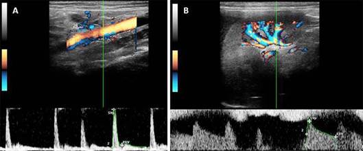
With the aid of the color flow and spectral Doppler vascular indexes of the abdominal aorta, left and right renal arteries were determined as shown in Table 2 and Figure 2.

Doppler ultrasonography images of abdominal aorta and renal arteries of a crab-eating fox. (A) Doppler ultrasonography of abdominal aorta. (B) Renal arteries with the presence of traces of the method.
Discussion
In wild canids, ultrasonographic studies are restricted to the reproductive tract imaging (Souza et al 2012Souza N.P., Furtado P.V. & Paz R.C. 2012. Non-invasive monitoring of the estrous cycle in captive crab- eating foxes (Cerdocyon thous). Theriogenology, 77:233-239.). No reference related to the normal ultrasonographic findings and biometry of the main abdominal organs, such as liver, spleen, bladder, stomach, intestines, adrenals, kidneys, prostate, linfoid tissue and vascular indexes of the abdominal aorta and renal arteries were described in crab-eating foxes.
The use of a non-invasive technique such as the ultrasonographic evaluation of abdominal organs and structures, proved to be easy to perform in this species. The use of a 7.5 to 10 MHz linear transdutor in the abdominal region allowed the visualization of most abdominal structures. Additionally, ultrasonographic imaging proved to be effective to correlate anatomic and ultrasonographic features of abdominal organs, highlighting important characteristics considered normal for the species. Nevertheless, this method demonstrated to be a standard tool for diagnosis and clinical evaluation of crab-eating foxes, similarly to the described for ferrets (Mustela furo) (Neuwirth et al. 1997Neuwirth L., Collins B., Calderwood-Mays M. & Tran T. 1997. Adrenal ultrasonography correlated with histopathology in ferrets. Vet. Radiol. Ultrasound 38:69-74.), common marmoset (Callithrix jacchus) (Wagner & Kirberger 2005Wagner W.M. & Kirberger R.M. 2005. Transcutaneous ultrasonography of the normal common marmoset (Callithrix jacchus). Vet. Radiol. Ultrasound 46:251-258.), cheetahs (Acinonyx jubatus) (Carstens et al. 2006Carstens A., Kirberger R.M., Spotswood T., Wagner W.M. & Grimbeek R.J. 2006. Ultrasonography of the liver, spleen, and urinary tract of the cheetah (Acinonyx jubatus). Vet. Radiol. Ultrasound 47:376-383.), tufted capuchin (Cebus apela) (Alves et al. 2007Alves F.R., Costa F.B., Arouche M.M.S., Barros A.C.E., Miglino M.A., Vulcano L.C. & Guerra P.C. 2007. Ultrasonographic evaluation of the urinary system, liver and uterus of Cebus apella monkey. Pesq. Vet. Bras 27:377-382.) and coatis (Nasua nasua) (Ribeiro et al. 2013Ribeiro R.G., Costa A.P.A., Bragato N., Fonseca A.M., Duque J.C.M., Prado T.D., Silva A.C.R. & Borges N.C. 2013. Normal sonographic anatomy of the abdomen of coatis (Nasua nasua Linnaeus, 1766). BMC Vet. Res. 9:124.).
As described in cats and dogs (Santos 2009Santos I.F.C. 2009. Ultrassonografia abdominal de cães e gatos hígidos, adultos e filhotes. Dissertação de Mestrado em Medicina Veterinária, Faculdade de Medicina Veterinária e Zootecnia, Universidade Estadual Paulista, Botucatu, SP. 180p.), in crab-eating foxes ultrasonographic imaging of the bladder revealed a quite empty structure, with small amount of anechoic content with absence of sediment, and anatomically located in the caudal portion of the abdomen. Mean value (1.2mm) of the bladder wall thickness was similar to the described for companion animals (1 to 5mm) (Vac 2004Vac M.H. 2004. Sistema urinário: Rins, ureteres, bexiga urinária e uretra, p.111-114. In: Carvalho C.F. (Ed.), Ultrassonografia em Pequenos Animais. Roca, São Paulo.), especially in dogs with average weight of 5 kg (1.43mm) (Santos 2009Santos I.F.C. 2009. Ultrassonografia abdominal de cães e gatos hígidos, adultos e filhotes. Dissertação de Mestrado em Medicina Veterinária, Faculdade de Medicina Veterinária e Zootecnia, Universidade Estadual Paulista, Botucatu, SP. 180p.).
Both, right and left kidneys showed homogenous echogenicity and were hyperechoic when compared to the spleen. Similarly to the reported in marmosets (Wagner & Kirberger 2005Wagner W.M. & Kirberger R.M. 2005. Transcutaneous ultrasonography of the normal common marmoset (Callithrix jacchus). Vet. Radiol. Ultrasound 46:251-258.), dogs, cats (Santos 2009Santos I.F.C. 2009. Ultrassonografia abdominal de cães e gatos hígidos, adultos e filhotes. Dissertação de Mestrado em Medicina Veterinária, Faculdade de Medicina Veterinária e Zootecnia, Universidade Estadual Paulista, Botucatu, SP. 180p.) and coatis (Ribeiro et al. 2013Ribeiro R.G., Costa A.P.A., Bragato N., Fonseca A.M., Duque J.C.M., Prado T.D., Silva A.C.R. & Borges N.C. 2013. Normal sonographic anatomy of the abdomen of coatis (Nasua nasua Linnaeus, 1766). BMC Vet. Res. 9:124.) the right kidney was located in renal fossa of the caudate lobe of the liver and the left kidney was caudal to the greater curvature of the stomach and medial to the spleen. The kidneys were approximately 4.6 cm long and 2.4 cm wide, slightly smaller when compared to adult dogs weighing 5 to 15 kg, which presented average lengths of 5 cm (Barr et al. 1990Barr F.J., Holt P.E. & Gibbs C. 1990. Ultrasonographic measurement of normal renal parameters. Conventional and Doppler ultrasound for the differentiation of benign and malignant canine mammary tumors. J. Small Anim. Pract. 31:180-184.) and width of 2.97cm (Santos 2009Santos I.F.C. 2009. Ultrassonografia abdominal de cães e gatos hígidos, adultos e filhotes. Dissertação de Mestrado em Medicina Veterinária, Faculdade de Medicina Veterinária e Zootecnia, Universidade Estadual Paulista, Botucatu, SP. 180p.).
Unlikely to the described in marmosets that exhibit a poor corticomedullary definition (Wagner & Kirberger 2005Wagner W.M. & Kirberger R.M. 2005. Transcutaneous ultrasonography of the normal common marmoset (Callithrix jacchus). Vet. Radiol. Ultrasound 46:251-258.), in crab-eating foxes this feature is well defined and the kidney presents a corticomedullary ratio of 1:1, similarly to the reported in cheetahs (Carstens et al. 2006Carstens A., Kirberger R.M., Spotswood T., Wagner W.M. & Grimbeek R.J. 2006. Ultrasonography of the liver, spleen, and urinary tract of the cheetah (Acinonyx jubatus). Vet. Radiol. Ultrasound 47:376-383.), dogs and cats (Silva et al. 2008Silva V.C., Mamprim M.J. & Vulcano L.C. 2008. Ultrassonografia no diagnóstico das doenças renais em pequenos animais. Vet. Zootec. 15:435-444.) and coatis (Ribeiro et al. 2013Ribeiro R.G., Costa A.P.A., Bragato N., Fonseca A.M., Duque J.C.M., Prado T.D., Silva A.C.R. & Borges N.C. 2013. Normal sonographic anatomy of the abdomen of coatis (Nasua nasua Linnaeus, 1766). BMC Vet. Res. 9:124.). In Veterinary Medicine this proportion is important for the diagnosis of kidney diseases that may present an increased cortical echogenicity with enhanced or loss of corticomedullary definition (Nyland et al. 2002Nyland T.G. 2002. Trato urinário, p.161-183. In: Nyland T.G. & Matton J.S. (Eds), Ultra-som: diagnóstico em pequenos animais. Roca, São Paulo.). Furthermore, parasites that affect the kidneys of domestic and wildlife animals such as the Dioctophyme renale can cause loss of definition of the renal corticomedullary junction (Veiga et al. 2011Veiga C.C.P., Azevedo F.D. & Scott F.B. 2011. Ultrassonografia e doppler velocimetria na avaliação renal de cães parasitados por Dioctophyma renale: relato de caso. Revta Bras. Med. Vet. 33:151-154.). Thus, determining the renal corticomedullary ratio of the crab-eating fox presents great importance for the diagnosis of renal pathologies in this species.
Similarly to the described for the spleen of dogs, cats (Silva et al. 2008Silva V.C., Mamprim M.J. & Vulcano L.C. 2008. Ultrassonografia no diagnóstico das doenças renais em pequenos animais. Vet. Zootec. 15:435-444., Santos 2009Santos I.F.C. 2009. Ultrassonografia abdominal de cães e gatos hígidos, adultos e filhotes. Dissertação de Mestrado em Medicina Veterinária, Faculdade de Medicina Veterinária e Zootecnia, Universidade Estadual Paulista, Botucatu, SP. 180p.) and coatis (Ribeiro et al. 2013Ribeiro R.G., Costa A.P.A., Bragato N., Fonseca A.M., Duque J.C.M., Prado T.D., Silva A.C.R. & Borges N.C. 2013. Normal sonographic anatomy of the abdomen of coatis (Nasua nasua Linnaeus, 1766). BMC Vet. Res. 9:124.), in the crab-eating fox this organ presented a homogeneous echogenicity and hyperechoic when compared to the renal parenchyma.
Corroborating with the findings in raccoons, dogs and cats, the hepatic echogenicity presented a homogeneous echogenicity and hypoechoic when compared to the spleen (Mamprim 2004Mamprim M.J. 2004. Fígado e vesícula biliar, p.51-70. In: Carvalho C.F. (Ed.), Ultrassonografia em Pequenos Animais. Roca, São Paulo., Larson 2009Larson M.M. 2009. Ultrasound of the thorax (Non cardiac). Vet. Clin. North Am. Small Anim. Pract. 3:733-745., Santos 2009Santos I.F.C. 2009. Ultrassonografia abdominal de cães e gatos hígidos, adultos e filhotes. Dissertação de Mestrado em Medicina Veterinária, Faculdade de Medicina Veterinária e Zootecnia, Universidade Estadual Paulista, Botucatu, SP. 180p., Ribeiro et al. 2013Ribeiro R.G., Costa A.P.A., Bragato N., Fonseca A.M., Duque J.C.M., Prado T.D., Silva A.C.R. & Borges N.C. 2013. Normal sonographic anatomy of the abdomen of coatis (Nasua nasua Linnaeus, 1766). BMC Vet. Res. 9:124.).
The stomach of the crab-eating foxes presented ultrasonographic characteristics similar to the ones reported in dogs, however the thickness of the small intestines wall was similar to the findings described by Ramos et al. (2011)Ramos A.H., Santos L.M., Miglino M.A., Peres J.A. & Guerra R.R. 2011. Biometria, histologia e morfometria do sistema digestório do cachorro-do-mato (Cerdocyon thous) de vida livre. Biotemas 24:111-119. for these wild canids and smaller when compared to the canine small intestine. Additionally, the peristaltic movements of the stomach (3 to 5 minutes) and the small intestines (2 to 3 minutes) presented similar frequency when compared to the domestic dogs (Ramos et al. 2011Ramos A.H., Santos L.M., Miglino M.A., Peres J.A. & Guerra R.R. 2011. Biometria, histologia e morfometria do sistema digestório do cachorro-do-mato (Cerdocyon thous) de vida livre. Biotemas 24:111-119.). Despite the hydric and food fasting and all the precautions taken for the ultrasound examination, the visualization and acquisition of reliable measures of the large intestines of crab-eating foxes were compromised due to the gas content present on its lumen.
Ultrasonographic imaging of the right and left adrenals was easy to perform differently from previous studies in companion animals that mentioned that these structures were difficult to visualize once they presented small dimensions and were involved by the retroperitoneal fat (Santos 2009Santos I.F.C. 2009. Ultrassonografia abdominal de cães e gatos hígidos, adultos e filhotes. Dissertação de Mestrado em Medicina Veterinária, Faculdade de Medicina Veterinária e Zootecnia, Universidade Estadual Paulista, Botucatu, SP. 180p.). Additionally, as described in dogs (Santos 2009Santos I.F.C. 2009. Ultrassonografia abdominal de cães e gatos hígidos, adultos e filhotes. Dissertação de Mestrado em Medicina Veterinária, Faculdade de Medicina Veterinária e Zootecnia, Universidade Estadual Paulista, Botucatu, SP. 180p.), ferrets (Neuwirth et al. 1997Neuwirth L., Collins B., Calderwood-Mays M. & Tran T. 1997. Adrenal ultrasonography correlated with histopathology in ferrets. Vet. Radiol. Ultrasound 38:69-74.) and coatis (Ribeiro et al. 2013Ribeiro R.G., Costa A.P.A., Bragato N., Fonseca A.M., Duque J.C.M., Prado T.D., Silva A.C.R. & Borges N.C. 2013. Normal sonographic anatomy of the abdomen of coatis (Nasua nasua Linnaeus, 1766). BMC Vet. Res. 9:124.) these glands were cranial to the kidneys and the left adrenal was slightly bigger than the one in the right side. Similar echobiometric findings of the adrenals were noticed between crab-eating foxes and the domestic dog. In the crab-eating foxes mean values of length (left adrenal: 18.7mm and right adrenal: 13.4mm) and width (left adrenal: 3.7mm and right adrenal: 4mm) were in the normal range reported for length and width in dogs. In this domestic species the left adrenal may range for length and width from 10.7 to 50.2mm and 1.9 to 12.4mm, respectively, and the right adrenal can measure from10 to 39.3mm in length and from 3.1 to 12mm in width.
In companion animals, the ovaries are located caudal and slightly lateral to the caudal poles of the ipsilateral kidneys (Davidson & Baker 2009Davidson A.P. & Baker T.W. 2009. Reproductive ultrasound of the bitch and queen. Topics Companion Anim. Med. 24:55-63.). Their visualization is considered quite difficult in the bitch and queen once these structures are hypoecogenic and can be confused to the mesenterium (Mattoon et al. 2005Mattoon J.S., Auld D.M. & Nyland T.G. 2005. Técnica de varredura abdominal por ultra-som, p.53-84. In: Mattoon J.S. & Nyland T.G. (Eds), Ultra-Som Diagnóstico em Pequenos Animais. 2ª ed. Roca, São Paulo.). In the present study, the ovaries were exactly in the same anatomic position described for the bitch and queen, however the visualization of multiple anechoic or hypoechoic cyst-like structures with average size of 2x2mm in the ovarian parenchyma probably helped to locate these structures. Additionally, the presence of ovarian follicles indicates the onset of folliculogenesis what lead us to suggest that in crab-eating foxes females, puberty occurs with less than a year of age. However further studies determining the endocrinological patterns of reproductive hormones and male effect in this species should be carried out to confirm this data.
Ultrasonographic imaging of prostate gland of crab-eating foxes was characterized by symmetric prostatic lobes, smooth edges, homogeneous parenchyma and discreetly higher or similar echogenicity when compared to the spleen, renal cortex and liver, findings that corroborates with the ones described for the normal prostate of domestic dogs (Nyland 2002Nyland T.G. 2002. Trato urinário, p.161-183. In: Nyland T.G. & Matton J.S. (Eds), Ultra-som: diagnóstico em pequenos animais. Roca, São Paulo., Monteiro 2009Monteiro S.C. 2009. Avaliação interobservador do aprendizado em ultrassonografia abdominal do sistema urogenital, adrenais e espaço retroperitoneal de cães. Dissertação de Mestrado em Ciências, Universidade Federal do Paraná, Curitiba. 113p.).
Abdominal scanning of the animals showed presence of a well developed lymphoid tissue, homogenous and hyperechoic compared to the spleen. In crab-eating foxes a malignant insulinoma with invasion by neoplasic cells of the lymphoid vessels of the pancreas was reported by Malta et al. (2008)Malta M.C.C., Luppi M.M., Oliveira R.G., Langohr I.M., Ecco R. & Santos R.L. 2008. Malignant insulinoma in a crab-eating fox (Cerdocyon thous). Braz. J. Vet. Pathol1(1):25-27., therefore the ultrasonographic description of the normal lymphoid tissue is important to distinguish it from altered and pathological status.
Regarding Doppler ultrasound of the abdominal aorta and renal arteries of crab-eating foxes, it is worth emphasizing the uniqueness of this study, once there is a lack of information describing the vascular indexes of these vessels in this species. Thus, the use of this imaging technique can be considered a standard tool for physiological studies, as well as the detection of hemodynamic abnormalities in these animals.
Comparing RI values of the right and left renal arteries of these wild canids and healthy sedated dogs, we noticed that our data was very similar to the ones described by Melo et al. (2006)Melo M.B., Veado J.C.C. & Silva E.F. 2006. Renal arteries Dopplerfluxometry: normal systolic and diastolic flow velocities and resistive index values in the main renal arteries. Arq. Bras. Med. Vet. Zootec 58:691-693. and slightly higher when compared to the RI values reported by Rivers et al. (1997)Rivers B.J., Walter P.A., Polzin D.J. & King V.L. 1997. Duplex Doppler estimation of intrarenal Pourcelot resistive index in dogs and cats with renal disease. J. Vet. Intern. Med. 11:250-260.. Despite the use of an association of tiletamine and zolazepam, the sedation protocol did not cause any significant effect on pressure, heart and respiratory rates of crab-eating foxes. Nevertheless, these differences among studies can be credited to the different sedation protocol, species, individual variation and the branch of the renal artery assessed.
The mean values for vascular indexes of the abdominal aorta, left and right renal arteries of crab-eating foxes were different from those described in literature for the domestic dog. This difference can be noticed when comparing our data with the values reported by Carvalho et al. (2008)Carvalho C.F., Chammas M.C., Sterman F.A., Barros N. & Cerri G.G. 2008. Ultra- sonografia dúplex-Doppler na avaliação morfológica e hemodinâmica das artérias aorta e mesentérica cranial em cães. Braz. J. Vet. Res. Anim. Sci. 45:24-31. for the peak systolic velocity (95.49±35.43cm/s), end diastolic velocity (76.20 to 84.40cm/s) the abdominal aorta and Melo et al. (2006)Melo M.B., Veado J.C.C. & Silva E.F. 2006. Renal arteries Dopplerfluxometry: normal systolic and diastolic flow velocities and resistive index values in the main renal arteries. Arq. Bras. Med. Vet. Zootec 58:691-693. for systolic velocities of the right (79.9cm/s), left kidneys (80.22cm/s) and diastolic velocity of the right (28.86cm/s) and the left kidneys (29.62cm/s).
Conclusion
In our study conventional and Doppler ultrasonographic imaging provided basic information that can be used as reference for the species as well for other wild canids. It is a precise and non-invasive method that can be safely used to evaluate and diagnose abdominal injuries in these patients.
References
- Alves F.R., Costa F.B., Arouche M.M.S., Barros A.C.E., Miglino M.A., Vulcano L.C. & Guerra P.C. 2007. Ultrasonographic evaluation of the urinary system, liver and uterus of Cebus apella monkey. Pesq. Vet. Bras 27:377-382.
- Barr F.J., Holt P.E. & Gibbs C. 1990. Ultrasonographic measurement of normal renal parameters. Conventional and Doppler ultrasound for the differentiation of benign and malignant canine mammary tumors. J. Small Anim. Pract. 31:180-184.
- Carstens A., Kirberger R.M., Spotswood T., Wagner W.M. & Grimbeek R.J. 2006. Ultrasonography of the liver, spleen, and urinary tract of the cheetah (Acinonyx jubatus). Vet. Radiol. Ultrasound 47:376-383.
- Carvalho C.F., Chammas M.C., Sterman F.A., Barros N. & Cerri G.G. 2008. Ultra- sonografia dúplex-Doppler na avaliação morfológica e hemodinâmica das artérias aorta e mesentérica cranial em cães. Braz. J. Vet. Res. Anim. Sci. 45:24-31.
- Davidson A.P. & Baker T.W. 2009. Reproductive ultrasound of the bitch and queen. Topics Companion Anim. Med. 24:55-63.
- De Thoisy B., Vergara M., Silvestro P. & Vasconcelos I. 2013. Northern extension of records of the crab-eating fox in Brazil. Canid Biol. Conservation 16:1-3.
- Feliciano M.A.R., Muzzi L.A.L., Leite C.A.L. & Junqueira M.A. 2007. Two-dimensional conventional, high resolution two-dimensional and three-dimensional ultrasonography in the evaluation of pregnant bitch. Arq. Bras. Med. Vet. Zootec 59:1333-1337.
- Feliciano M.A.R., Vicente W.R.R. & Silva M.A.M. 2012. Conventional and Doppler ultrasound for the differentiation of benign and malignant canine mammary tumours. J. Small Anim. Pract. 53:332-337.
- Feliciano M.A.R., Nepomuceno A.C., Crivelaro R.M., Oliveira M.E.F., Coutinho L.N. & Vicente W.R.R. 2013. Foetal echoencephalography and Doppler ultrasonography of the middle cerebral artery in canine fetuses. J. Small Anim. Pract. 54:149-152.
- Hofmann R. 1989. Evolutionary steps of ecophysiological adaptation and diversification of ruminants: a comparative view of their digestive system. Oecologia 78:443-457.
- Larson M.M. 2009. Ultrasound of the thorax (Non cardiac). Vet. Clin. North Am. Small Anim. Pract. 3:733-745.
- Malta M.C.C., Luppi M.M., Oliveira R.G., Langohr I.M., Ecco R. & Santos R.L. 2008. Malignant insulinoma in a crab-eating fox (Cerdocyon thous). Braz. J. Vet. Pathol1(1):25-27.
- Mamprim M.J. 2004. Fígado e vesícula biliar, p.51-70. In: Carvalho C.F. (Ed.), Ultrassonografia em Pequenos Animais. Roca, São Paulo.
- Mattoon J.S., Auld D.M. & Nyland T.G. 2005. Técnica de varredura abdominal por ultra-som, p.53-84. In: Mattoon J.S. & Nyland T.G. (Eds), Ultra-Som Diagnóstico em Pequenos Animais. 2ª ed. Roca, São Paulo.
- Mattoso C.R.S., Catenacci L.S., Beier S.L., Lopes R.S. & Takahira R.K. 2012. Hematologic, serum biochemistry and urinary values for captive crab-eating fox (Cerdocyon thous) in São Paulo state. Brazil. Pesq. Vet. Bras. 32:559-566.
- Melo M.B., Veado J.C.C. & Silva E.F. 2006. Renal arteries Dopplerfluxometry: normal systolic and diastolic flow velocities and resistive index values in the main renal arteries. Arq. Bras. Med. Vet. Zootec 58:691-693.
- Martins I.A. 2004. Identificação dos canídeos brasileiros através dos seus pêlos guarda. Anais 25º Congresso Brasileiro de Zoologia, Brasília, DF, p.220. (Resumo)
- Monteiro S.C. 2009. Avaliação interobservador do aprendizado em ultrassonografia abdominal do sistema urogenital, adrenais e espaço retroperitoneal de cães. Dissertação de Mestrado em Ciências, Universidade Federal do Paraná, Curitiba. 113p.
- Neuwirth L., Collins B., Calderwood-Mays M. & Tran T. 1997. Adrenal ultrasonography correlated with histopathology in ferrets. Vet. Radiol. Ultrasound 38:69-74.
- Nyland T.G. 2002. Trato urinário, p.161-183. In: Nyland T.G. & Matton J.S. (Eds), Ultra-som: diagnóstico em pequenos animais. Roca, São Paulo.
- Ramos A.H., Santos L.M., Miglino M.A., Peres J.A. & Guerra R.R. 2011. Biometria, histologia e morfometria do sistema digestório do cachorro-do-mato (Cerdocyon thous) de vida livre. Biotemas 24:111-119.
- Ribeiro R.G., Costa A.P.A., Bragato N., Fonseca A.M., Duque J.C.M., Prado T.D., Silva A.C.R. & Borges N.C. 2013. Normal sonographic anatomy of the abdomen of coatis (Nasua nasua Linnaeus, 1766). BMC Vet. Res. 9:124.
- Rivers B.J., Walter P.A., Polzin D.J. & King V.L. 1997. Duplex Doppler estimation of intrarenal Pourcelot resistive index in dogs and cats with renal disease. J. Vet. Intern. Med. 11:250-260.
- Santos I.F.C. 2009. Ultrassonografia abdominal de cães e gatos hígidos, adultos e filhotes. Dissertação de Mestrado em Medicina Veterinária, Faculdade de Medicina Veterinária e Zootecnia, Universidade Estadual Paulista, Botucatu, SP. 180p.
- Silva V.C., Mamprim M.J. & Vulcano L.C. 2008. Ultrassonografia no diagnóstico das doenças renais em pequenos animais. Vet. Zootec. 15:435-444.
- Souza N.P., Furtado P.V. & Paz R.C. 2012. Non-invasive monitoring of the estrous cycle in captive crab- eating foxes (Cerdocyon thous). Theriogenology, 77:233-239.
- Vac M.H. 2004. Sistema urinário: Rins, ureteres, bexiga urinária e uretra, p.111-114. In: Carvalho C.F. (Ed.), Ultrassonografia em Pequenos Animais. Roca, São Paulo.
- Veiga C.C.P., Azevedo F.D. & Scott F.B. 2011. Ultrassonografia e doppler velocimetria na avaliação renal de cães parasitados por Dioctophyma renale: relato de caso. Revta Bras. Med. Vet. 33:151-154.
- Wagner W.M. & Kirberger R.M. 2005. Transcutaneous ultrasonography of the normal common marmoset (Callithrix jacchus). Vet. Radiol. Ultrasound 46:251-258.
Publication Dates
-
Publication in this collection
Dec 2014
History
-
Received
08 Mar 2014 -
Accepted
13 Dec 2014