Abstract:
Mycoplasma haemofelis is the agent of feline infectious anemia, although Candidatus M. haemominutum can also be associated. This study evaluated the frequency and hematological alterations caused by hemoplasma infections and co-infections with FeLV, FIV and Toxoplasma gondii in domestic cats from two distinct areas (urban - G1 and periurban - G2) of Brasília, Brazil. One hundred cats were evaluated, 51 from the G1 area and 49 from G2. No cats were positive for T. gondii. Hemoplasma infection was diagnosed in 33% cats from G1 and 32.6% from G2 (p>0.05). In G1 35.3% of the positive cats were infected with Mycoplasma haemofelis, 47.06% with Candidatus Mycoplasma haemominutum and 17.64% with mixed hemoplasma species infection; 12.5% of the cats identified as PCR positive in G2 were infected with Mycoplasma haemofelis, 18.75% with Candidatus Mycoplasma haemominutum and 68.75% with mixed infection. Cats from the periurban area had higher mixed hemoplasmas infection rates than those from urban area, and most of them were asymptomatic carriers. Cytology results were positive in only 5% of cats from G1. Mycoplasma haemofelis infected cats had normocytic normochromic anemia while the cats infected with Candidatus Mycoplasma haemominutum or with both species did not. 37.2% of G1 cats were co-infected with Mycoplasma haemofelis and FeLV, and presented lower PCV and hemoglobin concentration than those infected only with Mycoplasma haemofelis. The co-infection with Candidatus Mycoplasma haemominutum and FeLV produced lower WBC, segmented cells and platelets, and increased total protein concentration.
Index Terms:
Anemia; Mycoplasma spp.; diagnosis; PCR; cats.
Resumo:
Mycoplasma haemofelis é o principal agente causador da anemia infecciosa felina, apesar de outras espécies de hemoplasmas, Candidatus M. haemominutum e Candidatus M. turicensis, também estarem associadas à hemoplasmose felina. O presente estudo avaliou a frequência de hemoplasmas, as principais alterações hematológicas associadas com a infecção e co-infecções com FeLV, FIV e Toxoplasma gondii em gatos domésticos de duas áreas diferentes (urbana - G1 e periurbana - G2) de Brasília, Brasil. 100 gatos foram avaliados, 51 de G1 e 49 de G2. Nenhum gato foi positivo para T. gondii. Hemoplasmose foi diagnosticada em 33% dos gatos de G1 e 32,6% de G2 (p>0,05). Em G1, 35.3% dos gatos hemoplasma positivos estavam infectados por Mycoplasma haemofelis, 47,06% por Candidatus Mycoplasma haemominutum e 17,64% apresentaram infecção concomitante por Mycoplasma haemofelis e Candidatus Mycoplasma haemominutum. Em G2, 12,5% dos gatos hemoplasma positivos estavam infectados por Mycoplasma haemofelis, 18,75% por Candidatus Mycoplasma haemominutum e 68,75% apresentaram infecção concomitante por Mycoplasma haemofelis e Candidatus Mycoplasma haemominutum. Gatos da região periurbana eram, em sua maioria, assintomáticos e apresentaram mais infecção concomitante por duas espécies de hemoplasmas do que gatos da região urbana. Os resultados da citologia foram positivos apenas em 5% dos gatos de G1. Gatos com infectados apenas com Mycoplasma haemofelis apresentaram anemia normocítica normocrômica e gatos com infecção única com Candidatus Mycoplasma haemominutum ou em combinação com Mycoplasma haemofelis não apresentaram alterações hematológicas. 37,2% dos gatos de G1 estavam co-infectados por Mycoplasma haemofelis e FeLV, e apresentaram VG e concentração de hemoglobina menores do que os gatos infectados apenas por Mycoplasma haemofelis. A co-infecção por Candidatus Mycoplasma haemominutum e FeLV resultou em baixo número de leucócitos, segmentados e plaquetes, além de aumento das concentrações de proteínas totais.
Termos de Indexação:
Anemia; Mycoplasma spp.; diagn; smai; PCR; felinos.
Introduction
Hemoplasmosis is caused by haemotropic Mycoplasma spp. and can lead to hemolytic anemia in different mammals. Three species of this genus are known to cause infection in cats: Mycoplasma haemofelis, Candidatus Mycoplasma haemominutum and Candidatus Mycoplasma turicensis (Peters et al. 2008Peters I.R., Helps C.R., McAuliffe L., Neimark H., Lappin M.R., Gruffydd-Jones T.J., Day M.J., Hoelzle L.E., Willi B., Meli M., Hofmann-Lehmann R. & Tasker S. 2008. RNase P RNA gene (rnpB) phylogeny of Hemoplasmas and other Mycoplasma species. J. Clin. Microbiol.46:1873-1877.). Experimental infections with Mycoplasma haemofelis resulted in more severe anemia than other feline hemoplasma species, although it may be considered an opportunist agent, causing anemia only when the animals are immunosuppressed by other diseases or after surgical intervention (Willi et al. 2005Willi B., Boretti F.S., Cattori V., Tasker S., Meli M.L., Reusch C., Lutz H. & Hofmann-Lehmann R. 2005. Identification, molecular characterisation and experimental transmission of a new hemoplasma isolate from a cat with hemolytic anaemia in Switzerland. J. Clin. Microbiol.43:2581-2585.). Candidatus Mycoplasma haemominutum and Candidatus Mycoplasma turicensis can lead to hematological alterations during co-infections with others hemoplasma species or retroviruses. Cats with ready access to streets and high exposure to disease vectors have increased risk of becoming infected with hemotropic mycoplasmas (Ishak et al. 2007Ishak A.M., Radecki S. & Lappin M.R. 2007. Prevalence of Mycoplasma haemofelis, 'Candidatus Mycoplasma haemominutum', Bartonella species, Ehrlichia species, and Anaplasma phagocytophilum DNA in the blood of cats with anemia. J. Feline Med. Surg.9:1-7.).
Since hemoplasmas haven't been successfully cultivated in vitro to date, the diagnosis of the infection was mainly based on cytological identification of organisms on the surface of red blood cells. However, this method may result in false positive and false negative results in the presence of Howell-Jolly bodies and during cyclic parasitemia, respectively. Nowadays, the polymerase chain reaction (PCR) has become the test of choice for the diagnosis of hemoplasma infections (Messick et al. 1998Messick J.B., Berent L.M. & Cooper S.K. 1998. Development and evaluation of a PCR-based assay for detection of Haemobartonella felis in cats and differentiation of H. felis from related bacteria by restriction fragment length polymorphism analysis. J. Clin. Microbiol.36:462-466., Inokuma et al. 2004Inokuma H., Taroura S., Okuda M., Hisasue M., Itamoto K., Une S., Nakaichi M. & Taura Y. 2004. Molecular survey of Mycoplasma haemofelis and 'Candidatus Mycoplasma haemominutum' infection in cats in Yamaguchi and srrounding areas. J. Vet. Med. Sci.66:1017-1020.).
This study used a conventional and accessible PCR method to evaluate the frequency of hemoplasma infections in domestic cats presenting different clinical conditions from two distinct regions of Brasília, Brazil. The study also aimed to evaluate the main hematological alterations present in hemoplasma positive cats and the effects of retrovirus (FeLV and FIV) infection on blood parameters of cats infected by hemoplasma.
Materials and Methods
One hundred cats were evaluated, 51 from the urban area (G1) and 49 from a periurban area (G2). The cats from G1 were evaluated at the Veterinary Hospital of Brasília University, whilst the cats from G2 were evaluated at their homes. All cats were submitted to clinical examination and blood collection by venipuncture of cephalic or jugular veins which were then transferred to ethylenediamine tetra-acetic acid-coated tubes for complete blood cell count (CBC), hemoparasite search (cytology) and PCR for Mycoplasma spp., FeLV and FIV. Hemoglobin concentration, erythrocytes (RBC) and leukocytes (WBC) numbers were determined using a semi-automatic veterinary blood cell counter (CC-550, Celm(tm), Brazil) whereas the platelets number was obtained by manual counting on a hemocytometer. The packed cell volume (PCV) was determined by microhematocrit centrifugation followed by plasma protein concentration analysis performed by refractometry. Differential leukocyte count, cell morphology evaluation and hemoparasite searches were performed on Giemsa-stained blood smears using a light microscope (CX40RF200, Olympus, Japan). Mean corpuscular volume (MCV) and mean corpuscular hemoglobin concentration (MCHC) were calculated from hemoglobin, PCV and red blood cell count.
A DNA extraction from whole blood samples was performed using commercial kit (QIAmp DNA blood mini kit, Qiagen, USA) according to manufacture's instructions.
A restriction fragment length polymorphism PCR - RFLP-PCR - (PCR1) was used to identify Mycoplasma spp. infection as previously described (Criado-Fornelio et al. 2003Criado-Fornelio A., Martinez-Marcos A., Buling-Sarana A. & Barba-Carretero J.C. 2003. Presence of Mycoplasma haemofelis, Mycoplasma haemominutum and piroplasmids in cats from southern Europe: a molecular study. Vet. Microbiol.93:307-317.), in which a DNA template (2μL) was amplified using 0.5mM of each primer, 1.0U Taq DNA polymerase, 0.25mM deoxynucleoside triphosphate, 1X PCR buffer and 2.5mM MgCl2 in a 25μL reaction. Amplification was performed in a thermal-cycler (FTGene5D - Techgene, UK) with an initial denaturation step at 94°C for 10 min, followed by a 40-cycle program with denaturation (94°C; 30 s), annealing (50°C; 30 s), primer extension (72°C; 30 s), and a final extension at 72°C for 10 min. In each set of reactions, water was used as negative control and a hemoplasma positive cat previously diagnosed by cytology and PCR was the positive control. Amplicons of expected size were detected after electrophoresis in a 1.5% agarose gel stained with ethidium bromide. The positive samples were submitted to digestion with restriction endonuclease EcoRI (REact± - Invitrogen, EUA), which produce two fragments (269 and 349bp) in case of Candidatus M. haemominutum DNA and no fragments if it was M. haemofelis DNA.
A second PCR (PCR2) with a different set of primers (Fl: GACTTTGGTTTCGGCCAAGG and R3: CAGAGTACTATCATAATTATCCCTC) specific to Mycoplasma haemofelis, was used to confirm the presence of this species in the positive samples (Berent et al. 1998Berent L.M., Messick J.B. & Cooper S.K. 1998. Detection of Haemobartonella felis in cats with experimentally induced acute and chronic infections, using a polymerase chain reaction assay. Am. J. Vet. Res.59:1215-1220.). In M. haemofelis PCR, template DNA (2μL) was amplified within a 25μL reaction containing 0.6mM of each primer, 1.5U Taq DNA polymerase, 0.25mM deoxynucleoside triphosphate, 1X PCR buffer, 3.0 mM MgCl2, and a final volume of water. Amplification was performed in a thermal-cycler (FTGene5D - Techgene, UK) with an initial denaturation step at 94°C for 10 min, followed by a 40-cycle program with denaturation (94°C; 45 s), annealing (54°C; 45 s), primer extension (72°C; 45 s), and a final extension at 72°C for 7 min. The 393bp amplicons were detected after electrophoresis in a 1.5% agarose gel stained with ethidium bromide.
A positive result for the blood smear and/or PCR characterized a hemoplasma positive cat. Sequencing of RFLP-PCR and Mycoplasma haemofelis PCR positive products were performed and compared with available cat hemoplasmas sequences on GeneBank through BLAST tool to confirm the primer specificity.
FeLV and FIV infections were evaluated through PCR as previously published (Hohdatsu et al. 1998Hohdatsu T., Motokawa K., Usami M., Amioka M., Okada S. & Koyama H. 1998. Genetic subtyping and epidemiological study of feline immunodeficiency virus by nested polymerase chain reaction-restriction fragment length polymorphism analysis of the gag gene. J. Virol. Methods70:107-111., Miyazawa & Jarrett 1997Miyazawa T. & Jarrett O. 1997. Feline leukaemia virus proviral DNA detected by polymerase chain reaction in antigenaemic but non-viraemic ('discordant') cats. Archs Virol.142:323-332., Tzavaras et al. 1990Tzavaras T., Stewart M., McDougall A., Fulton R., Testa N., Onions D.E. & Neil J.C. 1990. Molecular cloning and characterization of a defective recombinant feline leukaemia virus associated with myeloid leukaemia. J. General Virol.71(Pt 2):343-354.). Plasmids containing full-length p61E FeLV-A provirus and samples from persistently infected cats according to ELISA commercial test (Snap± - CITEcombo FeLV/FIV test kit, IDEXX Systems, Portland, USA) were used as positive controls for FeLV and FIV respectively. Water was used as negative control in each PCR test set.
Statistical analyses were carried out using the SAS± version 9.1 and Microsoft± Excel. Test t was used to compare means, test Z to compare proportions and test F for equality of variances. Comparisons between sexes (male and female), origin of cat (G1 or G2), test result (positive or negative) and type (infecting species: Mycoplasma haemofelis, Candidatus M. haemominutum, Mycoplasma spp. and co-infection) for hematological data were carried out using analysis of variance and mean comparison test (Duncan at 5% significance level). Non-parametric data such as percentage of infected cats were tested using chi-squared test (X2 ).
Results and Discussion
The main clinical signs found in cats from G1 during clinical inspection were apathy, partial or total anorexia and pale and/or icteric mucous membranes, in contrast to the cats from G2, which had ectoparasites, skin lesions and poor body condition.
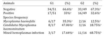
Table 1 summarizes the results of hemoplasma infections and the distribution of hemoplasmas species identified in cats from G1 and G2. No difference was observed in the hemoplasma frequency rates according to the sex gender (p>0.05).
Number and percentage of infected (positive) and non-infected (negative) cats by PCR from urban (G1) and periurban (G2) areas of the Federal District, Brazil
These clinical differences may be related with the cats' origin: cat samples from G1 were obtained when they were admitted to the veterinary hospital with suspicion of disease or behavior alteration. The clinical signs observed could be either related to hemoplasma infection or another debilitating condition of the cat. The G2 cats were from a region with low socioeconomic status and many of the cats had street access. Moreover, the samples from the G2 cats were collected in their homes after clinical evaluation, independent of their clinical condition. None of the cats showed clinical signs related to hemoplasma infection.
The frequency of hemoplasma infections observed in both groups wasn't different, but higher than the prevalence previously reported in North America (19.7 to 22.7%) (Gary et al. 2006Gary A.T., Richmond H.L., Tasker S., Hackett T.B. & Lappin M.R. 2006. Survival of Mycoplasma haemofelis and 'Candidatus Mycoplasma haemominutum' in blood of cats used for transfusions. J. Feline Med. Surg.8:321-326.) and similar to the rates detected in European cats (28 to 30%)(Criado-Fornelio et al. 2003Criado-Fornelio A., Martinez-Marcos A., Buling-Sarana A. & Barba-Carretero J.C. 2003. Presence of Mycoplasma haemofelis, Mycoplasma haemominutum and piroplasmids in cats from southern Europe: a molecular study. Vet. Microbiol.93:307-317.). However, the higher frequency of Candidatus Mycoplasma haemominutum (47%) over Mycoplasma haemofelis (35.3%) in cats from urban areas was the opposite found in Great Britain, where 66% of hemoplasma positive cats were infected by Mycoplasma haemofelis and 33% by Candidatus Mycoplasma haemominutum. This difference could be associated with different forms of transmission other than vectors and the weather conditions associated with it (Criado-Fornelio et al. 2003Criado-Fornelio A., Martinez-Marcos A., Buling-Sarana A. & Barba-Carretero J.C. 2003. Presence of Mycoplasma haemofelis, Mycoplasma haemominutum and piroplasmids in cats from southern Europe: a molecular study. Vet. Microbiol.93:307-317.). It is postulated that transmissions can also occur via contact with contaminated saliva, feces and/or urine. Furthermore, it's possible that each feline hemoplasma may have a different transmission route (Dean et al. 2008Dean R.S., Helps C.R., Gruffydd Jones T.J. & Tasker S. 2008. Use of real-time PCR to detect Mycoplasma haemofelis and 'Candidatus Mycoplasma haemominutum' in the saliva and salivary glands of haemoplasma-infected cats. J. Feline Med. Surg.10:413-417.).
No difference was observed in the hemoplasma frequency rates according to the sex gender (p>0.05), even though hemoplasma infection rates are usually higher in male cats than female cats (Willi et al. 2005Willi B., Boretti F.S., Cattori V., Tasker S., Meli M.L., Reusch C., Lutz H. & Hofmann-Lehmann R. 2005. Identification, molecular characterisation and experimental transmission of a new hemoplasma isolate from a cat with hemolytic anaemia in Switzerland. J. Clin. Microbiol.43:2581-2585.). We consider that a similar level of exposure to risk factors and/or the low number of studied animals could be related with this finding.
Cats living in periurban areas (G2) had higher number of mixed hemoplasma infections (Table 1), but these were asymptomatic animals. Given that the G2 cats were assessed independent of their clinical condition, it's plausible that the high frequency of mixed hemoplasma infection without clinical signs or hematological changes (as discussed further) can be related with longer exposure to hemoplasma species and no contribution of retroviruses. Therefore, hemoplasma infections detected by either PCR or cytology don´t necessarily indicate clinical disease (Macieira et al. 2008Macieira D.B., De Menezes R.D., Damico C.B., Almosny N.R., McLane H.L., Daggy J.K. & Messick J.B. 2008. Prevalence and risk factors for hemoplasmas in domestic cats naturally infected with feline immunodeficiency virus and/or feline leukemia virus in Rio de Janeiro - Brazil. J. Feline Med. Surg.10:120-129.) but points out the role of asymptomatic carrier cats as sources of transmission.
The hemoparasite search of the blood smear revealed only 5% hemoplasma positive cats, all from G1 and with characteristic clinical signs of hemoplasmosis, such as intense anemia, jaundice, apathy and anorexia (Willi et al. 2006Willi B., Boretti F.S., Baumgartner C., Tasker S., Wenger B., Cattori V., Meli M.L., Reusch C.E., Lutz H. & Hofmann-Lehmann R. 2006. Prevalence, risk factor analysis, and follow-up of infections caused by three feline hemoplasma species in cats in Switzerland. J. Clin. Microbiol.44:961-969.). Even though the positive samples from the blood smear were also positive on PCR, the difference in PCR results (33%) confirm the higher sensibility of PCR compared to cytology in the diagnosis of uncultivable hemoplasmas, especially in asymptomatic carrier cats, as those found in G2 (Watanabe et al. 2008Watanabe M., Hisasue M., Souma T., Ohshiro S., Yamada T. & Tsuchiya R. 2008. Molecular detection of Mycoplasma haemofelis and 'Candidatus Mycoplasma haemominutum' Infection in cats by direct PCR using whole blood without DNA extraction. J. Vet. Med. Sci.70:1095-1099.).
The sequences from PCR1 products had 83% similarity with Candidatus Mycoplasma haemominutum, which confirmed the results from enzymatic digestion, while PCR2 products sequences had 99% similarity with Mycoplasma haemofelis.
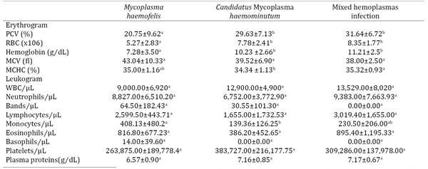
The cats from G1 had lower (p<0.05) PCV, RBC and hemoglobin concentration than the G2 cats (Table 2), even though both groups had mean values within reference values. These observations are also commonly noticed in hemoplasma infected cats, suggesting the low pathogenicity of some hemoplasma infections (Willi et al. 2006Willi B., Boretti F.S., Baumgartner C., Tasker S., Wenger B., Cattori V., Meli M.L., Reusch C.E., Lutz H. & Hofmann-Lehmann R. 2006. Prevalence, risk factor analysis, and follow-up of infections caused by three feline hemoplasma species in cats in Switzerland. J. Clin. Microbiol.44:961-969.). As G1 animals had a higher infection rate of Mycoplasma haemofelis (Table 1), known to be more pathogenic (Criado-Fornelio et al., 2003Criado-Fornelio A., Martinez-Marcos A., Buling-Sarana A. & Barba-Carretero J.C. 2003. Presence of Mycoplasma haemofelis, Mycoplasma haemominutum and piroplasmids in cats from southern Europe: a molecular study. Vet. Microbiol.93:307-317.), the reduced values observed in the erythrograms from these cats may be caused by hemoparasite infections. Also, 37.2% of G1 cats were FeLV positive, in opposition to G2, where none of the cats had retroviruses. The effect of FeLV on bone marrow, as discussed below, could have contributed to these results. It can be assumed that periurban cats have a higher resistance to hemoplasma infections, as indicated by their normal erythrograms (Table 2), due to intense or constant contact with different sources of infection. This condition makes the cats themselves important sources of transmission.
The hematological analysis of cats infected with hemoplasma and those not infected didn't reveal significant differences (data not shown). This finding is mainly attributed to the mild effects of Candidatus Mycoplasma haemominutum alone or in co-infection with Mycoplasma haemofelis (Table 3).
Cats infected with Mycoplasma haemofelis presented mild, normocytic and normochromic anemia, as previously described (Table 3) (Sykes et al. 2007Sykes J.E., Drazenovich N.L., Kyles A.E., Ball L.M. & Leutenegger C.M. 2007. Detection of mixed infections with "Candidatus Mycoplasma haemominutum" and Mycoplasma haemofelis using real-time TaqMan polymerase chain reaction. J. Vet. Diagn. Invest.19:250-255.), in alignment with the higher pathogenicity of this hemoplasma (Willi et al. 2005Willi B., Boretti F.S., Cattori V., Tasker S., Meli M.L., Reusch C., Lutz H. & Hofmann-Lehmann R. 2005. Identification, molecular characterisation and experimental transmission of a new hemoplasma isolate from a cat with hemolytic anaemia in Switzerland. J. Clin. Microbiol.43:2581-2585.). These results strongly reinforce that the infectious agent species, Mycoplasma haemofelis, is responsible for reducing erythrogram values in G1 cats.
The previously related association between mixed hemoplasma species infection and anemia (Willi et al. 2006Willi B., Boretti F.S., Baumgartner C., Tasker S., Wenger B., Cattori V., Meli M.L., Reusch C.E., Lutz H. & Hofmann-Lehmann R. 2006. Prevalence, risk factor analysis, and follow-up of infections caused by three feline hemoplasma species in cats in Switzerland. J. Clin. Microbiol.44:961-969.) wasn't observed in this study, in fact the mixed hemoplasmas infected cats had higher PCV than cats with single species of hemoplasma infection (Table 3). These results may indicate a long duration of exposure to hemoplasmas, resulting in a balance between host and parasite. Furthermore, no cat co-infected with multiple hemoplasma species had retrovirus infection, but 6 out of 19 cats infected with a single hemoplasma species were also infected with FeLV, which may have contributed to the lower hematological parameters.
In G1, 37.2% cats were diagnosed as FeLV positive and one was FIV positive. In G2 none of the animals tested were infected by retroviruses. Cats infected with hemoplasmas didn't have higher frequency of FeLV compared with those not infected with hemoplasmas (p>0.05), even when considering the species of hemoplasmas individually. However, there was a significant reduction (p<0.05) in the PCV, RBC, hemoglobin concentration and monocytes in the cats infected with hemoplasma and FeLV compared with those negative for both infections, indicating a strong pathological effect of both agents on bone marrow of co-infected animals.
Cats infected with Mycoplasma haemofelis and FeLV had lower PCV and hemoglobin concentration than those only infected by Mycoplasma haemofelis (Table 4). The co-infection by Candidatus Mycoplasma haemominutum and FeLV produced lower WBC, segmented cells and platelets, and increased total protein concentration (Table 4). These findings are supported by the well known effect of FeLV on hematopoietic precursors cells (Hofmann-Lehmann et al. 1997Hofmann-Lehmann R., Holznagel E., Ossent P. & Lutz H. 1997. Parameters of disease progression in long-term experimental feline retrovirus (feline immunodeficiency virus and feline leukemia virus) infections: hematology, clinical chemistry, and lymphocyte subsets. Clin. Diagn. Lab. Immunol.4:33-42.). Although the exact mechanism isn't clear, there is evidence of a suppressive effect on bone marrow, increased hemolytic effect of hemoplasmas in the presence of FeLV, opportunistic infections inducing iron sequestration, metastasis to bone marrow, myelodisplasic disorders or nutritional deficiencies (Gleich & Hartmann 2009Gleich S. & Hartmann K. 2009. Hematology and serum biochemistry of feline immunodeficiency virus-infected and feline leukemia virus-infected cats. J. Vet. Intern. Med.23:552-558.).
Only one cat had FIV infection, which was expected considering the low frequency of such retrovirus in the region studied (Marcola et al. 2013Marcola T.G., Gomes C.P., Silva P.A., Fernandes G.R., Paludo G.R. & Pereira R.W. 2013. Identification of a novel subtype of feline immunodeficiency virus in a population of naturally infected felines in the Brazilian Federal District. Virus Genes46:546-550.). This cat had a co-infection of Candidatus Mycoplasma haemominutum and FeLV with typical clinical signs from FeLV such as low PCV, erythrocytes and hemoglobin, that could have been caused by the associated infection between Candidatus Mycoplasma haemominutum and FeLV, and changes associated with FIV like PPT above reference values and low number of platelets (data not show).
Conclusions
This study showed a high number of cats infected with hemoplasmas in Brasília, Brazil.
Cats from periurban area had higher mixed hemoplasmas infection rates than those from the urban area, and most of them were asymptomatic carriers.
The major hematological changes observed were in erythrogram of animals infected by Mycoplasma haemofelis and presenting FeLV co-infection, which reinforces the strong pathogenic effect of this hemoplasma species and the importance of a concomitant agent.
Therefore, it is important to consider other common infectious agents in cats when dealing with hemoplasma infections.
Hemoplasma positive cats without hematological changes may be in the chronic phase of the infection or infected by less pathogenic hemoplasma species, as Candidatus Mycoplasma haemominutum.
Acknowledgements
Thanks to Capes for scholarships and Finatec to financial support, to Prof. Márcio Pimentel for logistical support and Dr Sajal K. Ghosh, Cancer Research Center, Boston University School of Medicine, Boston, USA, that kindly donated the positive controls for FeLV PCR.
References
- Berent L.M., Messick J.B. & Cooper S.K. 1998. Detection of Haemobartonella felis in cats with experimentally induced acute and chronic infections, using a polymerase chain reaction assay. Am. J. Vet. Res.59:1215-1220.
- Criado-Fornelio A., Martinez-Marcos A., Buling-Sarana A. & Barba-Carretero J.C. 2003. Presence of Mycoplasma haemofelis, Mycoplasma haemominutum and piroplasmids in cats from southern Europe: a molecular study. Vet. Microbiol.93:307-317.
- Dean R.S., Helps C.R., Gruffydd Jones T.J. & Tasker S. 2008. Use of real-time PCR to detect Mycoplasma haemofelis and 'Candidatus Mycoplasma haemominutum' in the saliva and salivary glands of haemoplasma-infected cats. J. Feline Med. Surg.10:413-417.
- Gary A.T., Richmond H.L., Tasker S., Hackett T.B. & Lappin M.R. 2006. Survival of Mycoplasma haemofelis and 'Candidatus Mycoplasma haemominutum' in blood of cats used for transfusions. J. Feline Med. Surg.8:321-326.
- Gleich S. & Hartmann K. 2009. Hematology and serum biochemistry of feline immunodeficiency virus-infected and feline leukemia virus-infected cats. J. Vet. Intern. Med.23:552-558.
- Hofmann-Lehmann R., Holznagel E., Ossent P. & Lutz H. 1997. Parameters of disease progression in long-term experimental feline retrovirus (feline immunodeficiency virus and feline leukemia virus) infections: hematology, clinical chemistry, and lymphocyte subsets. Clin. Diagn. Lab. Immunol.4:33-42.
- Hohdatsu T., Motokawa K., Usami M., Amioka M., Okada S. & Koyama H. 1998. Genetic subtyping and epidemiological study of feline immunodeficiency virus by nested polymerase chain reaction-restriction fragment length polymorphism analysis of the gag gene. J. Virol. Methods70:107-111.
- Inokuma H., Taroura S., Okuda M., Hisasue M., Itamoto K., Une S., Nakaichi M. & Taura Y. 2004. Molecular survey of Mycoplasma haemofelis and 'Candidatus Mycoplasma haemominutum' infection in cats in Yamaguchi and srrounding areas. J. Vet. Med. Sci.66:1017-1020.
- Ishak A.M., Radecki S. & Lappin M.R. 2007. Prevalence of Mycoplasma haemofelis, 'Candidatus Mycoplasma haemominutum', Bartonella species, Ehrlichia species, and Anaplasma phagocytophilum DNA in the blood of cats with anemia. J. Feline Med. Surg.9:1-7.
- Macieira D.B., De Menezes R.D., Damico C.B., Almosny N.R., McLane H.L., Daggy J.K. & Messick J.B. 2008. Prevalence and risk factors for hemoplasmas in domestic cats naturally infected with feline immunodeficiency virus and/or feline leukemia virus in Rio de Janeiro - Brazil. J. Feline Med. Surg.10:120-129.
- Marcola T.G., Gomes C.P., Silva P.A., Fernandes G.R., Paludo G.R. & Pereira R.W. 2013. Identification of a novel subtype of feline immunodeficiency virus in a population of naturally infected felines in the Brazilian Federal District. Virus Genes46:546-550.
- Messick J.B., Berent L.M. & Cooper S.K. 1998. Development and evaluation of a PCR-based assay for detection of Haemobartonella felis in cats and differentiation of H. felis from related bacteria by restriction fragment length polymorphism analysis. J. Clin. Microbiol.36:462-466.
- Miyazawa T. & Jarrett O. 1997. Feline leukaemia virus proviral DNA detected by polymerase chain reaction in antigenaemic but non-viraemic ('discordant') cats. Archs Virol.142:323-332.
- Peters I.R., Helps C.R., McAuliffe L., Neimark H., Lappin M.R., Gruffydd-Jones T.J., Day M.J., Hoelzle L.E., Willi B., Meli M., Hofmann-Lehmann R. & Tasker S. 2008. RNase P RNA gene (rnpB) phylogeny of Hemoplasmas and other Mycoplasma species. J. Clin. Microbiol.46:1873-1877.
- Sykes J.E., Drazenovich N.L., Kyles A.E., Ball L.M. & Leutenegger C.M. 2007. Detection of mixed infections with "Candidatus Mycoplasma haemominutum" and Mycoplasma haemofelis using real-time TaqMan polymerase chain reaction. J. Vet. Diagn. Invest.19:250-255.
- Tzavaras T., Stewart M., McDougall A., Fulton R., Testa N., Onions D.E. & Neil J.C. 1990. Molecular cloning and characterization of a defective recombinant feline leukaemia virus associated with myeloid leukaemia. J. General Virol.71(Pt 2):343-354.
- Watanabe M., Hisasue M., Souma T., Ohshiro S., Yamada T. & Tsuchiya R. 2008. Molecular detection of Mycoplasma haemofelis and 'Candidatus Mycoplasma haemominutum' Infection in cats by direct PCR using whole blood without DNA extraction. J. Vet. Med. Sci.70:1095-1099.
- Willi B., Boretti F.S., Baumgartner C., Tasker S., Wenger B., Cattori V., Meli M.L., Reusch C.E., Lutz H. & Hofmann-Lehmann R. 2006. Prevalence, risk factor analysis, and follow-up of infections caused by three feline hemoplasma species in cats in Switzerland. J. Clin. Microbiol.44:961-969.
- Willi B., Boretti F.S., Cattori V., Tasker S., Meli M.L., Reusch C., Lutz H. & Hofmann-Lehmann R. 2005. Identification, molecular characterisation and experimental transmission of a new hemoplasma isolate from a cat with hemolytic anaemia in Switzerland. J. Clin. Microbiol.43:2581-2585.
Publication Dates
-
Publication in this collection
Aug 2016
History
-
Received
11 June 2015 -
Accepted
13 Apr 2016