ABSTRACT:
Sida carpinifolia is a plant responsible for poisoning several species of animals. This paper describes Hypomyelinogenesis in fetuses and neonates of cattle that consumed S. carpinifolia. Neonates manifested ataxia and muscle tremors. Two bovine newborns and four fetuses were necropsied and showed no significant gross changes. Histopathologic findings included vacuolation of pancreatic acinar cells, thyroid follicular cells, hepatocytes, cells of renal tubules and neurons of the fetus and the white matter of the telencephalic frontal lobe of the neonates and also revealed axonal spheroids in the brain of the fetuses and neonates. The lectin-histochemical evaluation shoved staining for the lectins Con-A, WGA and s-WGA. The Luxol Fast Blue staining revealed a marked decrease of myelin in the brain of all the fetuses and a moderate decrease in the neonates. Histologic and lectin-histochemic findings indicate that the consumption of S. carpinifolia by pregnant bovine females can cause hypomyelinogenesis in fetuses and neonates.
INDEX TERMS:
Poisonous plants; hypomyelinogenesis; Sida carpinifolia; Malvaceae; calves; cattle; swainsonine; alfa-manosidosis; lysosomal storage disease; plant poisoning; toxicoses
RESUMO:
Sida carpinifolia é uma planta responsável por intoxicar várias espécies animais. Este artigo descreve hipomielinogênese em fetos e neonatos de bovinos que consumiram S. carpinifolia. Os neonatos manifestaram ataxia e tremores musculares. Dois neonatos e quatro fetos bovinos foram necropsiados e não havia alterações macroscópicas significativas. Os achados histopatológicos incluíram vacuolização de células acinares do pâncreas, células foliculares da tireoide, hepatócitos, células renais tubulares e neurônios nos fetos. Nos neonatos havia vacuolização na substância branca do lobo frontal telencefálico, além de esferoides axonais no encéfalo dos fetos e dos recém-nascidos. A avaliação lectino-histoquímica demonstrou marcação para as lectinas Con-A, WGA e s-WGA. A coloração de Luxol Fast Blue revelou diminuição acentuada da mielina no telencéfalo de todos os fetos e diminuição moderada nos neonatos. Os achados histológicos e lectina-histoquímicos indicam que o consumo de S. carpinifolia por fêmeas bovinas gestantes pode causar hipomielinogênese em fetos e neonatos.
TERMOS DE INDEXAÇÃO:
Plantas tóxicas; Hipomielinogênese; Sida carpinifolia; Malvaceae; bezerros; bovinos; swainsonina; alfa-manosidose; doença do acúmulo lisossomal; intoxicação por plantas; toxicoses
Introduction
Sida carpinifolia (L.f.) is a small sub-shrub plant of tropical and subtropical regions. It grows throughout Brazil but mainly in the humid and shaded areas of the South, South-East and West-Central regions (Matos et al. 2011Matos F.J.A., Lorenzi H., Santos L.F.L., Matos M.E.O., Silva M.G.V. & Sousa M.P. 2011. Plantas Tóxicas: estudo da fitotoxicologia química de plantas brasileiras. Instituto Plantarum de Estudos da Flora, São Paulo, p.123-124., Pedroso et al. 2012Pedroso P.M.O., Colodel E.M., Seitz A.L., Correa G.L.F., Soares M.P. & Driemeier D. 2012. Pathological findings in fetuses of goats and cattle poisoned by Sida carpinifolia (Malvaceae). Pesq. Vet. Bras. 32(3):227-230. <http://dx.doi.org/10.1590/S0100-736X2012000300008>
https://doi.org/10.1590/S0100-736X201200...
). Cases of intoxication by this plant have already been described in goats (Driemeier et al. 2000Driemeier D., Colodel E.M., Gimeno E.J. & Barros S.S. 2000. Lysosomal storage disease caused by Sida carpinifolia poisoning in goats. Vet. Pathol. 37(2):153-159. <http://dx.doi.org/10.1354/vp.37-2-153> <PMid:10714644>
https://doi.org/10.1354/vp.37-2-153...
), ponies (Loretti et al. 2003Loretti A.P., Colodel E.M., Gimeno E.J. & Driemeier D. 2003. Lysossomal storage disease in Sida carpinifolia toxicosis: an induced mannosidosis in horses. Equine Vet. J. 35(5):434-438. <http://dx.doi.org/10.2746/042516403775600523> <PMid:12875319>
https://doi.org/10.2746/0425164037756005...
), sheep (Seitz et al. 2005Seitz A.L., Colodel E.M., Barros S.S. & Driemeier D. 2005. Experimental poisoning by Sida carpinifolia (Malvaceae) in sheep. Pesq. Vet. Bras. 25:15-20. <http://dx.doi.org/10.1590/S0100-736X2005000100004>
https://doi.org/10.1590/S0100-736X200500...
), cattle (Furlan et al. 2009Furlan F.H., Lucioli J., Veronezi L.O., Medeiros A.L., Barros S.S., Traverso S.D. & Gava A. 2009. Spontaneous lysosomal storage disease caused by Sida carpinifolia (Malvaceae) poisoning in cattle. Vet. Pathol. 46(2):343-347. <http://dx.doi.org/10.1354/vp.46-2-343> <PMid:19261649>
https://doi.org/10.1354/vp.46-2-343...
) and in Fallow Deer (Dama dama) (Pedroso et al. 2009Pedroso P.M., Von Hohendorf R., de Oliveira L.G., Schmitz M., da Cruz C.E. & Driemeier D. 2009. Sida carpinifolia (Malvaceae) poisoning in fallow deer (Dama dama). J. Zoo Wildl. Med. 40(3):583-585. <http://dx.doi.org/10.1638/2009-0029.1> <PMid:19746879>
https://doi.org/10.1638/2009-0029.1...
), as well as changes in fetus of goats and cattle with transplacental intoxication by S. carpinifolia (Pedroso et al. 2012Pedroso P.M.O., Colodel E.M., Seitz A.L., Correa G.L.F., Soares M.P. & Driemeier D. 2012. Pathological findings in fetuses of goats and cattle poisoned by Sida carpinifolia (Malvaceae). Pesq. Vet. Bras. 32(3):227-230. <http://dx.doi.org/10.1590/S0100-736X2012000300008>
https://doi.org/10.1590/S0100-736X201200...
), in addition to stillbirth (Driemeier et al. 2000Driemeier D., Colodel E.M., Gimeno E.J. & Barros S.S. 2000. Lysosomal storage disease caused by Sida carpinifolia poisoning in goats. Vet. Pathol. 37(2):153-159. <http://dx.doi.org/10.1354/vp.37-2-153> <PMid:10714644>
https://doi.org/10.1354/vp.37-2-153...
). Other plants containing swainsonin, such as Astragalus spp. and Oxytropis spp., affect reproduction by reducing libido and spermatogenesis in males, as well as reduced fetal development, reproductive maturity, and neonatal/maternal behavior (Panter et al. 2013Panter K.E., Welch K.D., Gardner D.R. & Green B.T. 2013. Poisonous plants: effects on embryo and fetal development. Birth Defects Res. C, Embryo Today 99(4):223-234. <http://dx.doi.org/10.1002/bdrc.21053> <PMid:24339034>
https://doi.org/10.1002/bdrc.21053...
). Ipomea carnea, another plant that causes a lysosomal accumulation disease, has been associated with poor survival of goats at birth, as well as poor performance of survivors (Gotardo et al. 2011Gotardo A.T., Pfister J.A., Ferreira M.B. & Górniak S.L. 2011. Effects of prepartum ingestion of Ipomoea carnea on postpartum maternal and neonate behavior in goats. Birth Defects Res. B, Dev. Reprod. Toxicol. 92(2):131-138. <http://dx.doi.org/10.1002/bdrb.20291> <PMid:21465638>
https://doi.org/10.1002/bdrb.20291...
).
In goats and cattle fetuses poisoned via transplacental exposure to S. carpinifolia, the main histologic findings were vacuolation of renal tubular cells, follicular cells of the thyroid and the Purkinje neurons (Pedroso et al. 2012Pedroso P.M.O., Colodel E.M., Seitz A.L., Correa G.L.F., Soares M.P. & Driemeier D. 2012. Pathological findings in fetuses of goats and cattle poisoned by Sida carpinifolia (Malvaceae). Pesq. Vet. Bras. 32(3):227-230. <http://dx.doi.org/10.1590/S0100-736X2012000300008>
https://doi.org/10.1590/S0100-736X201200...
). Hypomyelinogenesis has been associated with congenital α-mannosidosis in Anglo-Nubian goats (Hueza et al. 2007Hueza I.M., Guerra J.L., Haraguchi M., Gardner D.R., Asano N., Ikeda K. & Górniak S.L. 2007. Assessment of the perinatal effects of maternal ingestion of Ipomoea carnea in rats. Exp. Toxicol. Pathol. 58(6):439-446. <http://dx.doi.org/10.1016/j.etp.2007.01.001> <PMid:17418550>
https://doi.org/10.1016/j.etp.2007.01.00...
) and in Persian cats (Vandevelde et al. 1982Vandevelde M., Fankhauser R., Bichsel P., Wiesmann U. & Herschkowitz N. 1982. Hereditary neurovisceral mannosidosis associated with α-mannosidase deficiency in a family of persian cats. Acta Neuropathol., Berlin 58(1):64-68. <http://dx.doi.org/10.1007/BF00692699> <PMid:7136518>
https://doi.org/10.1007/BF00692699...
). This paper describes Hypomyelinogenesis in fetus and newborn calves transplacentally poisoned by S. carpinifolia.
Materials and Methods
Four fetuses and two newborns from cows diagnosed with Sida carpinifolia poisoning, necropsied from January 2008 to December 2015 were evaluated. Clinical data were obtained from the owners and the veterinarians. Samples of various organs and tissues were collected at necropsy, fixed in 10% formalin, and routinely processed for histology, embedded in paraffin and sectioned in 3μm slices. Histochemical staining with hematoxylin and eosin (HE) was performed for all samples, and Luxol Fast Blue (LFB) was used for the frontal lobe and cerebellum sections. Central nervous system (CNS) samples were submitted to lectin-histochemistry staining using Con-A (Canavalia ensiformis), SBA (Glycine max), DBA (Dolichos biflorus), UEA-I (Ulex europaeus I), WGA (Triticum vulgaris), sWGA (succinyl-Triticum vulgaris), PNA (Arachis hypogaea), RCA-I (Ricinus communis I) and BS-I (Bandeiraea simplicifolia I) lectins, as described by Pedroso et al. (2012)Pedroso P.M.O., Colodel E.M., Seitz A.L., Correa G.L.F., Soares M.P. & Driemeier D. 2012. Pathological findings in fetuses of goats and cattle poisoned by Sida carpinifolia (Malvaceae). Pesq. Vet. Bras. 32(3):227-230. <http://dx.doi.org/10.1590/S0100-736X2012000300008>
https://doi.org/10.1590/S0100-736X201200...
. Fetuses and newborns of similar age from non-poisoned cows were used as negative controls. Cerebellar sections from adult cattle poisoned by S. carpinifolia were used as positive control. Immunohistochemichal evaluation was performed using the polymer method (EnVision, DAKO®). Heat-induced antigen retrieval was obtained in microwave in maximum power for 6 minutes, placing the samples in citrate buffer (pH 6.0). Anti-neurofilament monoclonal antibody (Clone 2F11, DAKO®) were applied at 1:1000 dilution and the slides incubated overnight at 4°C. The reaction was revealed with DAB (DAKO®) and the slides counterstained with Harris hematoxylin. Brain samples from non-poisoned fetuses and newborns of similar ages were included in all tests.
Serum samples (Newborns 1 and 2) and fragments of liver, spleen and heart (Newborns 1, 2 and Fetuses 1, 2) were submitted to reverse transcriptase polymerase chain reaction (RT-PCR) for Pestivirus and Bluetongue virus, respectively, as described by Vilcek et al. (1994)Vilcek S., Herring A.J., Herring J.A., Nettleton P.F., Lowings J.P. & Paton D.J. 1994. Pestiviruses isolated from pigs, cattle and sheep can be allocated into at least three genogroups using polymerase chain reaction and restriction endonuclease analysis. Arch. Virol. 136(3-4):309-323. <http://dx.doi.org/10.1007/BF01321060> <PMid:8031236>
https://doi.org/10.1007/BF01321060...
and Maan et al. (2012)Maan N.S., Maan S., Belaganahalli M.N., Ostlund E.N., Johnson D.J., Nomikou K. & Mertens P.P.C. 2012. Identification and differentiation of the twenty six Bluetongue virus serotypes by RT-PCR amplification of the serotype-specific Genome Segment 2. Plos One 7(2):1-9. <http://dx.doi.org/10.1371/journal.pone.0032601> <PMid:22389711>
https://doi.org/10.1371/journal.pone.003...
. Fragments of liver (Newborns 1, 2 and Fetuses 1, 2) fixed in 10% formalin were submitted to atomic absorption spectrometry to determine copper levels.
Results
From January 2008 to December 2015 were performed 4,188 necropsies of cattle, and diagnosed 11 cases of Sida carpinifolia poisoning in the species. Four of these cases were pregnant cows and two newborns. The remaining cases were non-pregnant cattle. Data about the origin, age, breed and sex of the animals are shown in Table 1. All cases were from farms (A, B, C, D and E) of the county of Triunfo, Rio Grande do Sul, Brazil.
On farm A, pregnant cows were kept on pasture infested by S. carpinifolia, and in the last third of gestation these cows were transferred to a S. carpinifolia free paddock. At the time, two calves (Newborn 1 and 2) were born with clinical signs of ataxia, muscle tremors, pleurothotonus and difficulty standing (Fig.1), with no clinical changes being observed in the mothers.

Hypomyelinogenesis in bovine fetuses and newborns associated with transplacental poisoning by Sida carpinifolia. Difficulty standing of Neonate 2.
On farm B, 10 abortions were reported after heifers kept in a pasture infested with S. carpinifolia. A heifer at 5 months gestation (Fetus 1) presented ataxia, posteriorly decubitus and, due to an unfavourable prognosis, was euthanized. The other heifers of this establishment were kept in pastures free of S. carpinifolia and showed no reproductive or neurological alterations. On farm C, a 6 months pregnant cow (Fetus 2) who presented ataxia, anorexia, and progression to decubitus was euthanized. On D farm, infested by the plant, a cow at 9 months gestation (Fetus 3) presented ataxia, difficulty standing and progressed to sternal decubitus and death. On farm E, a 5 months pregnant cow (Fetus 4) showed clinical signs of incoordination, generalized muscle tremors and difficulty standing and was euthanized. No significant gross changes were observed in the fetuses and neonates, with the exception of Fetus 3, which presented hydronephrosis.
At histopathologic evaluation of Newborn 1 and 2 was observed mild vacuolation in the telencephalic white matter of frontal lobe, and axonal spheroids in the telencephalon and cerebellum, but there was no vacuolation or cellular swelling in the other organs. In Fetus 1 and 2 there were observed accentuated diffuse swelling and vacuolation in the acinar cells of the pancreas, follicular cells of the thyroid, hepatocytes and renal tubular cells. Moderate to accentuated swelling and vacuolation were also observed in the cytoplasm of the Purkinje neurons, and in neurons of the telencephalic cortex, obex (Fig.2A), thalamus and hippocampus. In addition, hyperplasia was observed in the Bergmann astrocytes in the cerebellum, and axonal spheroids in sections of cerebellum, obex, thalamus and hippocampus. Fetus 3 exhibited moderate cytoplasmic swelling and vacuolation in the Purkinje neurons and in the neurons from the telencephalic cortex, thalamus and hippocampus; in addition, axonal spheroids were observed in the cerebellum, thalamus and hippocampus. In Fetus 4 were observed moderate swelling and vacuolation in the follicular thyroid cells and in the renal tubular cells, with no alterations in the other organs.

Hypomyelinogenesis in bovine fetuses and newborns associated with transplacental poisoning by Sida carpinifolia. (A) Accentuated swelling and vacuolation of the cytoplasm of neurons in the obex of Fetus 1. HE, obj.20x. (B) Labelling on Purkinje neurons for Con A in cerebellum of Neonate 2. Lectin-histochemistry, obj.20x.
The anti-neurofilament immunostaining revealed no differences between the axons of the affected cattle and their respective controls. The lectin-histochemical exam in all fetus and newborns showed discrete to accentuated positive staining for Con A, WGA and sWGA in the cerebellum, frontal cortex and obex neurons (Fig.2B). The LFB histochemical staining revealed a moderate decrease of myelin in the brain of newborns (Fig.3A); that were accentuated in the brain of all fetus (Fig.3B), and discrete in the cerebellum of all cases. There was normal staining in the CNS sections of the control calves (Fig.3C).
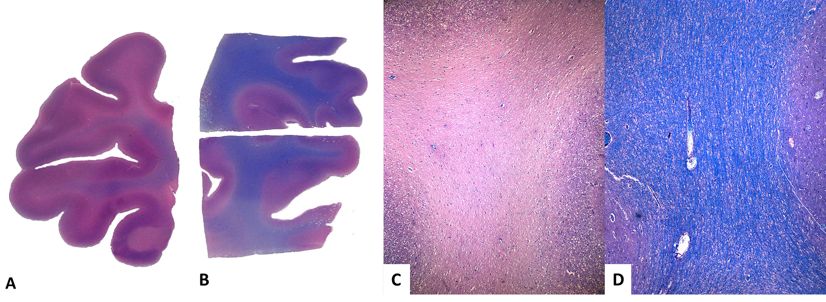
Hypomyelinogenesis in bovine fetuses and newborns associated with transplacental poisoning by Sida carpinifolia. (A) Moderate decrease of myelin in frontal lobe of Neonate 2. Luxol Fast Blue, submacroscopic aspect of stained sections. (B) Normal myelination of a control newborn. Luxol Fast Blue, submacroscopic aspect of stained sections. (C) Accentuated decrease of myelin in frontal lobe of Fetus 1. Luxol Fast Blue, obj.20x. (D) Normal myelination in frontal lobe of control fetus. Luxol Fast Blue, obj.20x.
No gross changes were observed in the cows. The histologic findings of the cows consisted mainly of vacuolation and moderate to severe swelling of the neurons of the cerebral cortex, thalamus, cerebellum and spinal cord and vacuolation of the hepatocytes and renal tubule epithelial cells.
For the other lectins used there were discrete or absent marking. The RT-PCR for Pestivirus and Bluetongue virus results negative. The hepatic copper levels of two newborn calves tested were 243 and 170μg/g (reference value = higher than 50μg/g).
Discussion
Diagnoses of transplacental Sida carpinifolia poisoning in bovine fetuses and newborns were performed through epidemiological, clinical and pathological findings, most of which were also observed in cows and confirmed by lectin-histochemistry. The invasion of grazing areas by S. carpinifolia is frequently observed in the region from Rio Grande do Sul where the poisoning occurred, mainly intermixed with eucalyptus plantations.
Indolizidine alkaloid, called swainsonine, is the toxic principle of S. carpinifolia, and inhibits α-mannosidase, a lysosomal enzyme (Colodel et al. 2002Colodel E.M., Driemeier D., Loretti A.P., Gimeno E.J., Traverso S.D., Seitz A.L. & Zlotowski P. 2002. Aspectos clínicos e patológicos da intoxicação por Sida carpinifolia (Malvaceae) em caprinos no Rio Grande do Sul. Pesq. Vet. Bras. 22(2):51-57. <http://dx.doi.org/10.1590/S0100-736X2002000200004>
https://doi.org/10.1590/S0100-736X200200...
). Inhibition of this enzyme induces the accumulation of mannose-rich oligosaccharides in the lysosomes of several cells, particularly in neurons, hepatocytes and acinar pancreatic cells. Swaisonine also inhibits the α-mannosidase II of the Golgi complex, an enzyme that participates in the glycosylation of many glycoproteins, a process that favours the increase of mannose accumulation in the lysosomes (Driemeier et al. 2000Driemeier D., Colodel E.M., Gimeno E.J. & Barros S.S. 2000. Lysosomal storage disease caused by Sida carpinifolia poisoning in goats. Vet. Pathol. 37(2):153-159. <http://dx.doi.org/10.1354/vp.37-2-153> <PMid:10714644>
https://doi.org/10.1354/vp.37-2-153...
). These cellular accumulations were observed in some organs of all fetuses, but were not observed in the newborn calves. In these no swelling or cell vacuolization was observed, suggesting that the decrease or absence of exposure to the toxic principle leads to regression of this lesion, since the affected cows were removed from the plant-infested pasture in the last third of gestation. Similar results were observed in a study on adult goats that were removed from infested pastures and showed a lower intensity or absence of neuronal vacuolization. This is probably due to the interruption in the exposure to swaisonine, with a return to normal α-mannosidase levels (Colodel et al. 2002Colodel E.M., Driemeier D., Loretti A.P., Gimeno E.J., Traverso S.D., Seitz A.L. & Zlotowski P. 2002. Aspectos clínicos e patológicos da intoxicação por Sida carpinifolia (Malvaceae) em caprinos no Rio Grande do Sul. Pesq. Vet. Bras. 22(2):51-57. <http://dx.doi.org/10.1590/S0100-736X2002000200004>
https://doi.org/10.1590/S0100-736X200200...
).
The mothers of the newborns did not present clinical disease. In pregnant rats, swainsonine has been detected in the amniotic fluid through amniocentesis, which indicates transplacental passage of the toxic principle (Hueza et al. 2007Hueza I.M., Guerra J.L., Haraguchi M., Gardner D.R., Asano N., Ikeda K. & Górniak S.L. 2007. Assessment of the perinatal effects of maternal ingestion of Ipomoea carnea in rats. Exp. Toxicol. Pathol. 58(6):439-446. <http://dx.doi.org/10.1016/j.etp.2007.01.001> <PMid:17418550>
https://doi.org/10.1016/j.etp.2007.01.00...
). In fetuses, probably, the toxic principle is not eliminated as it is in adults, because the glomeruli of the fetus are not yet fully developed, and high levels of the toxic principle remain in the body during the synthesis of myelin, which occurs during an important stage of development.
The pathogenesis involving the decrease of the myelin is uncertain and needs to be better elucidated. A possible cause for hypomyelinogenesis in cases of poisoning of bovine fetuses and neonates by S. carpinifolia is the interference in the production of thyroid hormones, due to the loss of function of the thyroid epithelial cells, which appear vacuolated at histopathological examination. Thyroid hormones are involved in the development of the CNS, acting on neuronal maturation and myelinogenesis (Calzá et al. 2015Calzà L., Fernández M. & Giardino L. 2015. Role of the thyroid system in myelinationand neural connectivity. Comp. Physiol. 5(3):1405-1421. <http://dx.doi.org/10.1002/cphy.c140035> <PMid:26140723>
https://doi.org/10.1002/cphy.c140035...
). Another possibility for myelin deficiency is the occurrence of abnormal substrate deposition in the oligodendrocytes, interfering with their normal metabolic function (Vandevelde et al. 1982Vandevelde M., Fankhauser R., Bichsel P., Wiesmann U. & Herschkowitz N. 1982. Hereditary neurovisceral mannosidosis associated with α-mannosidase deficiency in a family of persian cats. Acta Neuropathol., Berlin 58(1):64-68. <http://dx.doi.org/10.1007/BF00692699> <PMid:7136518>
https://doi.org/10.1007/BF00692699...
). As no differences were found between the axons of the poisoned and the control calves, the myelin deficiency was considered primary and not secondary to axonal destruction.
Some diseases that induce hypomyelinogenesis in cattle in Brazil should be included in the differential diagnosis. These diseases include bovine viral diarrhoea, caused by a Pestivirus, copper deficiency and bluetongue. The previously mentioned viral diseases were discarded in the etiology of hypomyelinogenesis in this study through molecular tests, and the copper deficiency through hepatic copper dosage.
Lysosomal storage diseases of hereditary origin, such as β-mannosidosis, have already been described as inducers of hypomyelinogenesis in cattle (Bryan et al. 1993Bryan L., Schmutz S., Hodges S.D. & Snyder F.F. 1993. Bovine β-mannosidosis: pathologic and genetic findings in salers calves. Vet. Pathol. 30(2):130-139. <http://dx.doi.org/10.1177/030098589303000205> <PMid:8470335>
https://doi.org/10.1177/0300985893030002...
). As the lectin-histochemistry detects accumulated residue inside the cells, the technique served as an important tool in the diagnosis of lysosomal accumulation disease (Driemeier et al. 2000Driemeier D., Colodel E.M., Gimeno E.J. & Barros S.S. 2000. Lysosomal storage disease caused by Sida carpinifolia poisoning in goats. Vet. Pathol. 37(2):153-159. <http://dx.doi.org/10.1354/vp.37-2-153> <PMid:10714644>
https://doi.org/10.1354/vp.37-2-153...
, Pedroso et al. 2012Pedroso P.M.O., Colodel E.M., Seitz A.L., Correa G.L.F., Soares M.P. & Driemeier D. 2012. Pathological findings in fetuses of goats and cattle poisoned by Sida carpinifolia (Malvaceae). Pesq. Vet. Bras. 32(3):227-230. <http://dx.doi.org/10.1590/S0100-736X2012000300008>
https://doi.org/10.1590/S0100-736X201200...
). Staining was evident for sWGA and WGA, indicating the expression of D-N-acetyl-glucosaminase and acetyl neuraminic acid, and for Con-A, which is specific for D-mannose and D-glucose (Driemeier et al. 2000Driemeier D., Colodel E.M., Gimeno E.J. & Barros S.S. 2000. Lysosomal storage disease caused by Sida carpinifolia poisoning in goats. Vet. Pathol. 37(2):153-159. <http://dx.doi.org/10.1354/vp.37-2-153> <PMid:10714644>
https://doi.org/10.1354/vp.37-2-153...
).
Conclusion
The epidemiological, pathological, histochemical and lectin-histochemical findings indicate that the α-mannosidosis acquired by the consumption of Sida carpinifolia by pregnant cows is responsible for hypomyelinogenesis in bovine foetuses and neonates.
Acknowledgements
The authors thank all colleagues and who contributed in some way for this paper.
References
- Bryan L., Schmutz S., Hodges S.D. & Snyder F.F. 1993. Bovine β-mannosidosis: pathologic and genetic findings in salers calves. Vet. Pathol. 30(2):130-139. <http://dx.doi.org/10.1177/030098589303000205> <PMid:8470335>
» https://doi.org/10.1177/030098589303000205 - Calzà L., Fernández M. & Giardino L. 2015. Role of the thyroid system in myelinationand neural connectivity. Comp. Physiol. 5(3):1405-1421. <http://dx.doi.org/10.1002/cphy.c140035> <PMid:26140723>
» https://doi.org/10.1002/cphy.c140035 - Colodel E.M., Driemeier D., Loretti A.P., Gimeno E.J., Traverso S.D., Seitz A.L. & Zlotowski P. 2002. Aspectos clínicos e patológicos da intoxicação por Sida carpinifolia (Malvaceae) em caprinos no Rio Grande do Sul. Pesq. Vet. Bras. 22(2):51-57. <http://dx.doi.org/10.1590/S0100-736X2002000200004>
» https://doi.org/10.1590/S0100-736X2002000200004 - Driemeier D., Colodel E.M., Gimeno E.J. & Barros S.S. 2000. Lysosomal storage disease caused by Sida carpinifolia poisoning in goats. Vet. Pathol. 37(2):153-159. <http://dx.doi.org/10.1354/vp.37-2-153> <PMid:10714644>
» https://doi.org/10.1354/vp.37-2-153 - Furlan F.H., Lucioli J., Veronezi L.O., Medeiros A.L., Barros S.S., Traverso S.D. & Gava A. 2009. Spontaneous lysosomal storage disease caused by Sida carpinifolia (Malvaceae) poisoning in cattle. Vet. Pathol. 46(2):343-347. <http://dx.doi.org/10.1354/vp.46-2-343> <PMid:19261649>
» https://doi.org/10.1354/vp.46-2-343 - Gotardo A.T., Pfister J.A., Ferreira M.B. & Górniak S.L. 2011. Effects of prepartum ingestion of Ipomoea carnea on postpartum maternal and neonate behavior in goats. Birth Defects Res. B, Dev. Reprod. Toxicol. 92(2):131-138. <http://dx.doi.org/10.1002/bdrb.20291> <PMid:21465638>
» https://doi.org/10.1002/bdrb.20291 - Hueza I.M., Guerra J.L., Haraguchi M., Gardner D.R., Asano N., Ikeda K. & Górniak S.L. 2007. Assessment of the perinatal effects of maternal ingestion of Ipomoea carnea in rats. Exp. Toxicol. Pathol. 58(6):439-446. <http://dx.doi.org/10.1016/j.etp.2007.01.001> <PMid:17418550>
» https://doi.org/10.1016/j.etp.2007.01.001 - Loretti A.P., Colodel E.M., Gimeno E.J. & Driemeier D. 2003. Lysossomal storage disease in Sida carpinifolia toxicosis: an induced mannosidosis in horses. Equine Vet. J. 35(5):434-438. <http://dx.doi.org/10.2746/042516403775600523> <PMid:12875319>
» https://doi.org/10.2746/042516403775600523 - Maan N.S., Maan S., Belaganahalli M.N., Ostlund E.N., Johnson D.J., Nomikou K. & Mertens P.P.C. 2012. Identification and differentiation of the twenty six Bluetongue virus serotypes by RT-PCR amplification of the serotype-specific Genome Segment 2. Plos One 7(2):1-9. <http://dx.doi.org/10.1371/journal.pone.0032601> <PMid:22389711>
» https://doi.org/10.1371/journal.pone.0032601 - Matos F.J.A., Lorenzi H., Santos L.F.L., Matos M.E.O., Silva M.G.V. & Sousa M.P. 2011. Plantas Tóxicas: estudo da fitotoxicologia química de plantas brasileiras. Instituto Plantarum de Estudos da Flora, São Paulo, p.123-124.
- Panter K.E., Welch K.D., Gardner D.R. & Green B.T. 2013. Poisonous plants: effects on embryo and fetal development. Birth Defects Res. C, Embryo Today 99(4):223-234. <http://dx.doi.org/10.1002/bdrc.21053> <PMid:24339034>
» https://doi.org/10.1002/bdrc.21053 - Pedroso P.M., Von Hohendorf R., de Oliveira L.G., Schmitz M., da Cruz C.E. & Driemeier D. 2009. Sida carpinifolia (Malvaceae) poisoning in fallow deer (Dama dama). J. Zoo Wildl. Med. 40(3):583-585. <http://dx.doi.org/10.1638/2009-0029.1> <PMid:19746879>
» https://doi.org/10.1638/2009-0029.1 - Pedroso P.M.O., Colodel E.M., Seitz A.L., Correa G.L.F., Soares M.P. & Driemeier D. 2012. Pathological findings in fetuses of goats and cattle poisoned by Sida carpinifolia (Malvaceae). Pesq. Vet. Bras. 32(3):227-230. <http://dx.doi.org/10.1590/S0100-736X2012000300008>
» https://doi.org/10.1590/S0100-736X2012000300008 - Seitz A.L., Colodel E.M., Barros S.S. & Driemeier D. 2005. Experimental poisoning by Sida carpinifolia (Malvaceae) in sheep. Pesq. Vet. Bras. 25:15-20. <http://dx.doi.org/10.1590/S0100-736X2005000100004>
» https://doi.org/10.1590/S0100-736X2005000100004 - Vandevelde M., Fankhauser R., Bichsel P., Wiesmann U. & Herschkowitz N. 1982. Hereditary neurovisceral mannosidosis associated with α-mannosidase deficiency in a family of persian cats. Acta Neuropathol., Berlin 58(1):64-68. <http://dx.doi.org/10.1007/BF00692699> <PMid:7136518>
» https://doi.org/10.1007/BF00692699 - Vilcek S., Herring A.J., Herring J.A., Nettleton P.F., Lowings J.P. & Paton D.J. 1994. Pestiviruses isolated from pigs, cattle and sheep can be allocated into at least three genogroups using polymerase chain reaction and restriction endonuclease analysis. Arch. Virol. 136(3-4):309-323. <http://dx.doi.org/10.1007/BF01321060> <PMid:8031236>
» https://doi.org/10.1007/BF01321060
Publication Dates
-
Publication in this collection
July 2018
History
-
Received
17 Nov 2017 -
Accepted
04 Dec 2017