
ABSTRACT:
Wild species, especially those threatened with extinction, are increasingly being investigated to obtain information that can be useful for their preservation. The objective of the present study was to standardize the vertebral heart scale (VHS) and cardiothoracic ratio (CTR) of the collared peccary (Tayassu tajacu Linnaeus, 1758) sedated with ketamine and midazolam. Fourteen clinically healthy collared peccaries were examined in the two-year age group weighing 15-22kg. The animals were submitted to digital radiography of the thorax in lateral and dorsal ventral projections to calculate the VHS and CTR. The VHS mean values for males and females was 8.88±0.51v for right recumbency and 8.84±0.39v for left decubitus, and there were no significant between-gender differences regarding recumbency (p>0.05). The CTR showed mean values of 0.50±0.05 (males) and 0.45±0.04 (females), but the gender-differences were not significant (p>0.05). A positive correlation was shown between VHS and CTR (r=0.98, right decubitus; r=0.96, left decubitus). Establishing reference values for heart measurements in collared peccaries using digital radiography of the thorax permitted standardization of the VHS and CTR values for this wild species. In the studied wild animal model, the VHS and CTR heart assessment indexes were shown to be essential diagnostic tools for investigations of alterations in the size of the cardiac silhouette.
INDEX TERMS:
Vertebral heart scale; cardiothoracic ratio; heart size; collared peccaries; Tayassu tajacu; ketamine; midazolam; cardiac silhouette; wild animals; cardiology; morphology
RESUMO:
Espécies silvestres têm sido cada vez mais exploradas como forma de obter informações que favoreçam sua conservação, especialmente aquelas ameaçadas de extinção. Este trabalho teve por objetivo a padronização dos valores de VHS (vertebral heart scale) e índice cardiotorácico (ICT) de catetos (Tayassu tajacu Linnaeus, 1758) contidos com Cetamina e Midazolam. Foram avaliados 14 catetos clinicamente saudáveis, com faixa etária de 2 anos e variação média de peso entre 15 a 22Kg. Os animais foram submetidos a radiografia digital de tórax em projeções laterolaterais e dorsoventrais para o cálculo do vertebral heart scale (VHS) e Índice Cardiotorácico (ICT). O VHS evidenciou valores médios entre machos e fêmeas de 8,88±0,51v para decúbito direito e 8,84±0,39v para decúbito esquerdo, não ocorrendo diferença estatística entre os decúbitos (p>0,05). O ICT revelou valores médios de 0,50±0,05 (machos) e de 0,45±0,04 (fêmeas), não sendo verificada diferença estatística significativa entre os sexos (p>0,05). Verificou-se correlação positiva entre VHS e o ICT (r=0,98, decúbito direito, r= 0,96, decúbito esquerdo). O estabelecimento dos valores de referência para mensurações cardíacas em catetos, a partir de radiografias digitais do tórax, permitiu a padronização do VHS e ICT para esta espécie silvestre. No modelo animal silvestre estudado, os índices de avaliação cardíaca VHS e ICT mostraram-se como uma ferramenta diagnóstica imprescindível para investigações sobre as alterações do tamanho da silhueta cardíaca.
TERMOS DE INDEXAÇÃO:
Vertebral heart scale; índice cardiotorácico; tamanho cardíaco; caititus; Tayassu tajacu; cetamina; midazolam; pecari; silhueta cardíaca; animais silvestres; cardiologia; morfologia
Introduction
Radiographic examination is an image diagnosis method that is very important in veterinary medicine. The values obtained from VHS can help in the prediction of cardiac alterations that may reflect in clinical or surgical procedures (Carvalho 2004Carvalho C.F. 2004. Ultrassonografia em Pequenos Animais. Roca, São Paulo. 365p.).
Thoracic radiographs are routinely used in human and animal medicine to assess alterations in cardiac silhouette conformation. In addition, this tool provides digital impressions of the appearance of the lung parenchyma, upper intrathoracic airways, anatomic integrity of the thoracic cavity walls, pleural space and lung vascularization (Lamb & Boswood 2002Lamb C.R. & Boswood A. 2002. Role of survey radiography in diagnosis canine cardiac disease. Comp. Contin. Educ. Pract. Vet. 24:316-326., Gabay 2003Gabay A. 2003. Radiologia cardiovascular, p.40-45. In: Blerenian G.C., Mucha C.J. & Camacho A.A. (Eds), Afecções Cardiovasculares em Pequenos Animais. Interbook, São Paulo., Soares et al. 2004Soares E.C., Larsson M.H.M.A. & Pinto A.C.B.C.F. 2004. Aspectos radiográficos da doença valvar crônica. Ciência Rural 34(1):119-124. <http://dx.doi.org/10.1590/S0103-84782004000100018>
https://doi.org/10.1590/S0103-8478200400...
, Kealy & McAllister 2005Kealy J.K. & McAllister H. 2005. Radiologia e Ultrassonografia do Cão e do Gato. 3ª ed. Manole, São Paulo. 436p.)
Studies of the heart in radiographic images include the left and right decubitus positions; the dorsal ventral incidence (DV) is preferred to the ventral dorsal (VD) because the former better shows the appearance of the cardiac silhouette, avoiding outline distortions in the image. Furthermore, in the DV incidence, the caudal lung vessels present better radiographic definition, favoring diagnoses of lung congestion and permitting indirect assessment of heart performance (Ruehl & Thrall 1981Ruehl Junior W.W. & Thrall D.E. 1981. The effect of dorsal versus ventral recumbency on the radiography appearance of the canine thorax. Vet. Radiol. Ultrasound 22(1):10-16. <http://dx.doi.org/10.1111/j.1740-8261.1981.tb00602.x>
https://doi.org/10.1111/j.1740-8261.1981...
, Lamb & Boswood 2002Lamb C.R. & Boswood A. 2002. Role of survey radiography in diagnosis canine cardiac disease. Comp. Contin. Educ. Pract. Vet. 24:316-326., Kealy & McAllister 2005Kealy J.K. & McAllister H. 2005. Radiologia e Ultrassonografia do Cão e do Gato. 3ª ed. Manole, São Paulo. 436p.).
The qualitative radiographic method is the one used most for heart assessments, where the veterinary or human radiologist relates anatomic knowledge to possible alterations in the radiographic image inside the thoracic cavity. The advantages of this method are convenience and speed, but it should be adopted only by experienced professionals who know the normal variations that the image can present (Suter & Lord 1971Suter P.F. & Lord P.F. 1971. A critical evaluation of radiographic findings in canine cardiovascular diseases. J. Am. Vet. Med. Assoc. 158(3):358-371. <PMid:4250978>, Lamb & Boswood 2002Lamb C.R. & Boswood A. 2002. Role of survey radiography in diagnosis canine cardiac disease. Comp. Contin. Educ. Pract. Vet. 24:316-326., Soares et al. 2004Soares E.C., Larsson M.H.M.A. & Pinto A.C.B.C.F. 2004. Aspectos radiográficos da doença valvar crônica. Ciência Rural 34(1):119-124. <http://dx.doi.org/10.1590/S0103-84782004000100018>
https://doi.org/10.1590/S0103-8478200400...
).
Among the methods proposed, biometric measuring methods aim to assess heart size quantitatively to increase accuracy and decrease the subjectivity present in qualitative assessments (Buchanan & Bücheler 1995Buchanan J.W. & Bücheler J. 1995. Vertebral scale system to measure canine heart size in radiographs. J. Am. Vet. Med. Assoc. 206(2):194-199. <PMid:7751220>). In biometric methods, measurements of the heart dimensions and cardiothoracic relationships are necessary. In lateral projection, the heart length between the base and the apex should be approximately 70% of the dorsal ventral distance of the thoracic cavity (Schelling 2002Schelling C.G. 2002. Exame radiográfico do coração, p.15-38. In: Tilley L.P. & Goodwin J.K. (Eds), Manual de Cardiologia para Cães e Gatos. 3ª ed. Roca, São Paulo .), while the width or cranial-caudal diameter of the heart can range from 2.5 (in deep-chested dogs) to 3.5 intercostal spaces (in wide-chested dogs) (Buchanan & Bücheler 1995Buchanan J.W. & Bücheler J. 1995. Vertebral scale system to measure canine heart size in radiographs. J. Am. Vet. Med. Assoc. 206(2):194-199. <PMid:7751220>, Schelling 2002Schelling C.G. 2002. Exame radiográfico do coração, p.15-38. In: Tilley L.P. & Goodwin J.K. (Eds), Manual de Cardiologia para Cães e Gatos. 3ª ed. Roca, São Paulo .).
Studies on Brazilian fauna have high priority because of their ecological importance and the potential for animal science exploitation presented by various species (Neves et al. 2013Neves L.R., Pereira F.B., Tavares-Dias M. & Luque J.L. 2013. Seasonal influence on the parasite fauna of a wild population of Astronotus ocellatus (Perciformes: Cichlidae) from the Brazilian Amazon. J. Parasitol. 99(4):718-721. <http://dx.doi.org/10.1645/12-84.1> <PMid:23421456>
https://doi.org/10.1645/12-84.1...
). Basic information on wild animal morphology regarding their physiology and biology in captivity is lacking. Collared peccaries (Tayassu tajacu) are wild Suiformes belonging to the genus Tayassu and the family Dicotylidae. These animals are subject to intense predatory pressure, which has increased the need for greater knowledge regarding their cardiovascular physiology (Lazure et al. 2010Lazure L., Bachand M., Ansseau C. & Almeida-Cortez J.S. 2010. Fate of native and introduced seeds consumed by captive white-lipped and collared peccaries (Tayassu pecari Link, 1795 and Pecari tajacu Linnaeus, 1758) in the Atlantic rainforest, Brazil. Braz. J. Biol. 70(1):47-53. <http://dx.doi.org/10.1590/S1519-69842010000100008> <PMid:20231959>
https://doi.org/10.1590/S1519-6984201000...
, Azevedo et al. 2012Azevedo C.S., Lima M.F.F., Silva V.C.A., Young R.J. & Rodrigues M. 2012. Visitor influence on the behavior of captive greater rheas (Rhea americana, Rheidae Aves). J. Appl. Anim. Welf. Sci. 15(2):113-125. <http://dx.doi.org/10.1080/10888705.2012.624895> <PMid:22458873>
https://doi.org/10.1080/10888705.2012.62...
).
The collared peccary is highly adaptable to different environments, extremely robust and resistant to various diseases. These characteristics greatly favor their use in scientific investigations, as observed by Bezerra et al. (2014)Bezerra D.O., Feitosa M.L.T., Almeida H.M., Costa F.A.L., Braga J.F.V., Souza F.A.L., Alves F.R., Pessoa G.T. & Carvalho M.A.M. 2014. Collared Pecary (Tayassu tajacu) as a new model of renal ischemic injury induced by clamping the renal artery. Acta. Cir. Bras. 29(9):560-572. <http://dx.doi.org/10.1590/S0102-8650201400150003> <PMid:25252202>
https://doi.org/10.1590/S0102-8650201400...
and Pessoa et al. (2014)Pessoa G.T., Feitosa M.L.T., Argolo-Neto N.M., Rocha A.R., Costa C.R.M., Silva G.C., Bezerra D.O., Coelho C.J.C., Sousa S.S. & Carvalho M.A.M. 2014. Isolation, culture and differentiation potential of collared peccary (Tayassu tajacu) adipose-derived stem cells. Acta. Scient. Vet. 42:1-10., which used these animals as models for renal ischemic lesions and the isolation of mesenchymal cells derived from adipose tissue. Thus, these animals are more accessible alternatives for scientific studies than is the domestic pig (Sus scrofa domesticus) (Monteiro et al. 2009Monteiro R., Brandau R., Gomes W.J. & Braile D.M. 2009. Trends in animal experimentation. Revta Bras. Cir. Cardiol. 24(4):506-513. <http://dx.doi.org/10.1590/S0102-76382009000500012> <PMid:20305924>
https://doi.org/10.1590/S0102-7638200900...
) or mini pigs (Vodička et al. 2005Vodička P., Smetana Junior K., Dvoránková B., Emerick T., Xu Y.Z., Ourednik J., Ourednik V. & Motlík J. 2005. The miniature pig as an animal model in biomedical research. Ann. N.Y. Acad. Sci. 1049(1):161-171. <http://dx.doi.org/10.1196/annals.1334.015> <PMid:15965115>
https://doi.org/10.1196/annals.1334.015...
).
Studies of heart measurements in domestic animals have been conducted for species such as dogs (Lamb et al. 2001Lamb C.R., Wikeley H., Boswood A. & Pfeiffer D.U. 2001. Use of breed-specific ranges for the vertebral heart scale as an aid to the radiographic diagnosis of cardiac disease in dogs. Vet. Rec. 148(23):707-711. <http://dx.doi.org/10.1136/vr.148.23.707> <PMid:11430680>
https://doi.org/10.1136/vr.148.23.707...
), cats (Litster & Buchanan 2000Litster A.L. & Buchanan J.W. 2000. Vertebral scale system to measure heart size in radiographs of cats. J. Vet. Med. Assoc. 216(2):210-214. <http://dx.doi.org/10.2460/javma.2000.216.210> <PMid:10649755>
https://doi.org/10.2460/javma.2000.216.2...
), horses (Patteson et al. 1995Patteson M.W., Gibbs C., Wotton P.R. & Cripps P.J. 1995. Echocardiographic measurements of cardiac dimensions and indices of cardiac function in normal adult thoroughbred horses. Equine Vet. J. 27(19):18-27. <PMid:8933065>), goats (Ukaha et al. 2013Ukaha R.O., Kene R.O.C. & Gboniko O.E. 2013. Vertebral scale system to measure heart size in thoracic radiographs of West African dwarf goats. Nig. Vet. J. 34(4):912-916.) and pigs. However, variables such as breed, animal size and thoracic shape have influenced the standardization of the mean vertebral scale of the normal heart, as has been observed in the domestic species already assessed (Azevedo et al. 2016Azevedo G.M., Pessoa G.T., Moura L.S., Sousa F.C.A., Rodrigues R.P.S., Sanches M.P., Fontenele R.D., Barbosa M.A.P.S., Neves W.C., Sousa J.M. & Alves F.R. 2016. Comparative study of the Vertebral Heart Scale (VHS) and the Cardiothoracic Ratio (CTR) in healthy poodle breed dogs. Acta. Scient. Vet. 44:1-7.).
Buchanan & Bücheler (1995)Buchanan J.W. & Bücheler J. 1995. Vertebral scale system to measure canine heart size in radiographs. J. Am. Vet. Med. Assoc. 206(2):194-199. <PMid:7751220> determined that values for dogs obtained in lateral projections were smaller or equal to 10.5 vertebra (v) in 98% of the radiography of clinically normal dogs. Corroborating the findings of these authors, Lamb et al. (2001)Lamb C.R., Wikeley H., Boswood A. & Pfeiffer D.U. 2001. Use of breed-specific ranges for the vertebral heart scale as an aid to the radiographic diagnosis of cardiac disease in dogs. Vet. Rec. 148(23):707-711. <http://dx.doi.org/10.1136/vr.148.23.707> <PMid:11430680>
https://doi.org/10.1136/vr.148.23.707...
analyzed the use of specific VHS values as a method to diagnose heart disease in dogs. The authors reported that significant differences exist between the various mean values for different breeds, and suggested standardizing this technique for each breed in question.
The VHS (vertebral heart scale) is a well-established routine in veterinary medicine (Jepsen-Grant et al. 2013Jepsen-Grant K., Pollard R.E. & Johnson L.R. 2013. Vertebral heart scores in eight dog breeds. Vet. Radiol. Ultrasound 54(1):3-8. <http://dx.doi.org/10.1111/j.1740-8261.2012.01976.x> <PMid:22994206>
https://doi.org/10.1111/j.1740-8261.2012...
), and it correlates positively with cardiomegaly development in companion animals (Guglielmini et al. 2012Guglielmini C., Diana A., Santarelli G., Torbidone A., Di Tommaso M., Toaldo M.B. & Cipone M. 2012. Accuracy of radiographic vertebral heart score and sphericity index in the detection of pericardial effusion in dogs. J. Am. Vet. Med. Assoc. 241(8):1048-1055. <http://dx.doi.org/10.2460/javma.241.8.1048> <PMid:23039979>
https://doi.org/10.2460/javma.241.8.1048...
). However, the VHS has been little used as a diagnostic tool in wild animals because standard values are that measure heart biometry available for only a few species, hindering the identification of determined abnormalities in wild animals. Felkai et al. (2014)Felkai A., Vogelnest L., Mcnabb S., Allan G. & Sangster C. 2014. Dilated cardiomyopathy in a De Brazza’s monkey (Cercopithecus neglectus). J. Med. Primatol. 43(3):209-212. <http://dx.doi.org/10.1111/jmp.12108> <PMid:24611814>
https://doi.org/10.1111/jmp.12108...
claimed that the presence of pleural residual leakage and cardiomegaly were associated with increased VHS values in the primate Cercopithecus neglectus, which was later confirmed by echocardiographic examination. In humans, a cardiothoracic ratio of >0.5 on a postero-anterior radiograph showed a good predictor of left ventricular dysfunction (Chana et al. 2015Chana H.S., Martin C.A., Cakebread H.E., Adjei F.D. & Gajendragadkar P.R. 2015. Diagnostic accuracy of cardiothoracic ratio on admission chest radiography to detect left or right ventricular systolic dysfunction: a retrospective study. J. R. Soc. Med. 108(8):317-324. <http://dx.doi.org/10.1177/0141076815588314> <PMid:26152673>
https://doi.org/10.1177/0141076815588314...
). Studies have aimed to standardize CTR values in nonhuman primates (Schillaci et al. 2009Schillaci M.A., Parish S. & Jones-Engel L. 2009. Radiographic measurement of the cardiothoracic ratio in pet macaques from Sulawesi, Indonesia. Radiography 15(4):29-33. <http://dx.doi.org/10.1016/j.radi.2009.05.005>
https://doi.org/10.1016/j.radi.2009.05.0...
) and wild rodents (Moura et al. 2015Moura C.R.C., Diniz A.N., Moura L.S., Sousa F.C.A., Baltazar P.I., Freire L.D., Guerra P.C., Sousa J.M., Giglio R.F., Pessoa G.T., Rodrigues R.P.S. & Alves F.R. 2015. Cardiothoracic ratio and vertebral heart scale in clinically normal black-rumped agoutis (Dasyprocta prymnolopha Wagler, 1831). J. Zoo. Wild. Med. 46(2):314-319. <http://dx.doi.org/10.1638/2014-0038R.1> <PMid:26056885>
https://doi.org/10.1638/2014-0038R.1...
), but are still scarce in several wild species, with no reports for species proposed in this research.
Rearing wild animals in captivity is a species management problem and requires safe and efficient restraint methods that ensure adequate immobilization to carry out necessary procedures such as biological material collections, morphometric measurements, physiological measurements and clinical surgical interventions (Mayor et al. 2007Mayor P., Guimarães D.A.A., Le-pendu I., Da Silva J., Jori F. & Lópezbéjar M. 2007. Reproductive performance of captive collared peccaries (Tayassu tajacu) in the eastern Amazon. Anim. Rep. Sci. 102(1/2):88-97. <http://dx.doi.org/10.1016/j.anireprosci.2006.10.015> <PMid:17101243>
https://doi.org/10.1016/j.anireprosci.20...
). Thus, the objective of the present study was to standardize the VHS (vertebral heart scale) and cardiothoracic ratio (CTR) in collared peccaries (Tayassu tajacu Linnaeus, 1758) sedated with ketamine and midazolam.
Materials and Methods
Animals. In this study, fourteen healthy adult collared peccaries (Tayassu tajacu Linnaeus, 1758) were used, seven males (20±1.19kg) and seven females (17.85±2.41kg), each approximately two years old, from the Nucleus for Wild Animal Study and Preservation (Núcleo de Estudos e Preservação de Animais Silvestres - NEPAS (Registro IBAMA nº 02/08-618) of the Agrarian Science Center (CCA) at the Federal University of Piauí (UFPI), Teresina, Piauí, Brazil).
The protocols used in the present study were approved by the Committee of Ethics in Animal Experimentation (Comitê de Ética em Experimentação Animal - CEEA/UFPI (nº 047/15) and authorized by the Ministry of the Environment through the Biodiversity Authorization and Information System (SISBIO) of the Brazilian Institute for the Environment and Renewable Natural Resources (IBAMA) (nº 48113-1).
Exclusion criteria. Subjects with an unremarkable clinical history and physical examination were included in the study. The cardiovascular and respiratory systems were inspected by auscultation of the heart valves and lung fields. The time of capillary perfusion and the diagnosis of dehydration were also evaluated by hemodynamic investigation. The systemic infectious processes were evaluated by palpating the peripheral lymph nodes (submandibular, cervical, and popliteal), abdominal palpation, and temperature measurement. In addition, all peccaries were subjected to blood, biochemical, and electrolyte tests. Animals with evidence of systemic diseases, cardiovascular abnormalities (murmurs or arrhythmias), or any degree of valve insufficiency observed on echocardiogram, or with excessive stress during the examination, were excluded from the study.
Anesthetic protocol. The peccaries were fasted (given no solid food) for 12 hours and unwatered for 6 hours and then captured in their enclosures with a capture net. For chemical sedation, a mixture of 15mg/kg ketamine hydrochloride (Vetanarcol) with 3mg/kg midazolam maleate (Dormonid®) was administered intramuscularly. The procedures were started when the anesthetic effect was observed, 15 minutes after application.
Radiographic examinations. To conduct the radiographic examinations, the animals were placed in sternal and lateral (right/left) decubitus to obtain radiographic images in right and left dorsal ventral (DV) and lateral projections of the thoracic region. A mobile INTECAL CR-7 X-ray apparatus was used. The images acquired in a sensitive radiographic chassis were analyzed in a CR 30-X (Agfa HealthCare) digital radiographic image-capture apparatus installed in the Image Diagnosis Sector of the University Veterinary Hospital (HVU/UFPI), saved on a computer hard drive, and coupled to the analysis system.
VHS execution. Means were collected for the VHS (vertebral heart scale) according to the protocol established by Buchanan & Bücheler (1995)Buchanan J.W. & Bücheler J. 1995. Vertebral scale system to measure canine heart size in radiographs. J. Am. Vet. Med. Assoc. 206(2):194-199. <PMid:7751220> and Litster & Buchanan (2000)Litster A.L. & Buchanan J.W. 2000. Vertebral scale system to measure heart size in radiographs of cats. J. Vet. Med. Assoc. 216(2):210-214. <http://dx.doi.org/10.2460/javma.2000.216.210> <PMid:10649755>
https://doi.org/10.2460/javma.2000.216.2...
, which directs sampling of the heart’s length and width at its greatest diameter and subsequent comparison with the vertebral bodies from the cranial endplate of the fourth thoracic vertebra (T4) (Fig.1). The measurements were taken first; later, the mean values were estimated to establish the normality criteria.
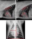
Digital radiographic image of the collared peccary thoracic cavity. (A) Right lateral projection. (B) Left lateral projection to obtain the vertebral heart scale (VHS) calculation. (C) Dorsal ventral projection to obtain the cardiothoracic ratio (CTR). Teresina, Piauí/Brazil, 2017.
Cardiothoracic ratio. The CTR was assessed by comparing the greatest widths of the cardiac silhouette and the distance between the thoracic walls at height T8, according to methodology described by Schillaci et al. (2009)Schillaci M.A., Parish S. & Jones-Engel L. 2009. Radiographic measurement of the cardiothoracic ratio in pet macaques from Sulawesi, Indonesia. Radiography 15(4):29-33. <http://dx.doi.org/10.1016/j.radi.2009.05.005>
https://doi.org/10.1016/j.radi.2009.05.0...
for Macaca fascicularis. Heart width was measured at its two greatest distances (C and D) from the vertical line that divides the limits between the right and left sides of the heart, at the point of its greatest diameter. Similarly, the thoracic width was measured at the point of greatest thoracic diameter (E) as follows:
Statistical analysis. The data were submitted to a normality test (Shapiro-Wilk and Kolmogorov-Smirnov test); then, group means were analyzed by Student’s t-tests and paired for parameter interpretation at a confidence interval of 5% (p<0.05).
Results
The animals assessed had cardiac silhouettes between the second and fourth intercostal spaces, situated on the mid-mediastinum in both decubitus (right lateral and left lateral) positions (Fig.1). The weight of the males studied averaged 20.0±1.19kg and the females averaged 17.85±2.41kg; there was no significant difference between the males and female weights (p>0.05), the mean weight of males and females combined was 19.06±2.04kg.
The VHS (vertebral heart scale) in the males showed a mean of 9.22±0.29v for the right decubitus and 8.87±0.42v for the left decubitus; there was no significant difference between them (p>0.05). For the females, the VHS showed a mean value of 8.55±0.47v for the right decubitus and 8.81±0.42v for the left decubitus; there was also no significant difference between them (p>0.05). When the VHS of males and females was compared for both of the decubitus positions, no difference was observed (p>0.05), and the estimated VHS was 8.85±0.39v (Table 1).
The cardiothoracic ratio (CTR) mean values were 0.50±0.05 for the males and 0.45±0.04 for the females, and there was no significant difference between the genders (p>0.05) (Table 1). Thus, the combined male and female CTR was 0.48±0.05. Positive correlation was observed between the VHS in both decubitus and the CTR (r=0.98, right decubitus; r=0.96, left decubitus), taking CTR as the dependent variable (Fig.2).
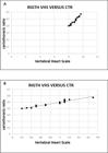
Correlation between the VHS and CTR of the peccary. (A) Linear correlation between vertebral heart scale (VHS) and cardiothoracic ratio (CTR) the in right lateral decubitus. (B) Linear correlation between VHS and CTR the in left lateral decubitus. Teresina, Piauí/Brazil, 2017.
Discussion
The mean of the combined VHS values of collared peccary males and females (8.88±0.51v) was lower than that observed in other wild animals, such as coatis (Nasua nasua, 9.36±0.75v; Martini et al. 2013Martini A.C., Meireles Y.S., Monzem S., Vasconcelos L.P., Monteiro N.C., Turbino R., Dahroug M.A.A., Farias D., Néspoli P.B., Gonçalves G.F., Souza R.L. & Guimarães R.D. 2013. Avaliação radiográfica da silhueta cardíaca, pelo método VHS (Vertebral Heart Size), de quatis (Nasua nasua Linnaeus, 1766) jovens e adultos mantidos em cativeiro. Semina, Ciênc. Agrárias 34(2):3823-3829.), robust capuchin monkeys (Cebus apella, 9.25±0.32v; Rocha-Neto et al. 2015Rocha-Neto H.J., Moura L.S., Pessoa G.T., Ambrósio C.E., Sousa F.C.A., Rodrigues R.P.S., Guerra P.C. & Alves F.C. 2015. Cardiothoracic ratio and vertebral heart size (VHS) to standardize the heart size of the tufted capuchin (Cebus paella, Linnaeus, 1758) in computerized radiographic images. Pesq. Vet. Bras. 35(10):853-858. <http://dx.doi.org/10.1590/S0100-736X2015001000006>
https://doi.org/10.1590/S0100-736X201500...
), marmosets (Callithrix jacchus, 9.42±0.44v; Wagner & Kirberger 2005Wagner W.M. & Kirberger R.M. 2005. Radiographic anatomy of the thorax and abdomen of the common marmoset (Callithrix jacchus). Vet. Radiol. Ultrasound 46(3):217-224. <http://dx.doi.org/10.1111/j.1740-8261.2005.00044.x> <PMid:16050279>
https://doi.org/10.1111/j.1740-8261.2005...
) and alpacas (Alpaca cria, 9.36±0.65v; Nelson et al. 2011Nelson N.C., Mattoon N.J.S. & Anderson D.E. 2011. Radiographic appearance of the thorax of clinically normal Alpaca crias. Am. J. Vet. Res. 72(11):1439-1448. <http://dx.doi.org/10.2460/ajvr.72.11.1439> <PMid:22023121>
https://doi.org/10.2460/ajvr.72.11.1439...
); however, it was higher compared with ferrets (Mustela putorius furo, 5.39±0.45v) and hedgehogs (Atelerix albiventris, 5.39±0.45v) (Black et al. 2011Black P.A., Marshall C., Seyfried A.W. & Bartin A.M. 2011. Cardiac assessment of African Hedgehogs, Atelerx albiventris. J. Zoo. Wild. Med. 24(1):49-53. <http://dx.doi.org/10.1638/2010-0012.1>
https://doi.org/10.1638/2010-0012.1...
).
There is a directly proportional correlation of the VHS (vertebral heart scale) values with the physical size of the animal, a fact that was observed when the collared peccary was compared with domestic Suiformes (Sus scrofa domesticus). The VHS values observed in the collared peccary were lower than conventional pigs (9.05±0.15v) and close to the values found for mini pigs (8.6±0.14v).
Thoracic conformation affects the VHS values, a fact observed in narrow- and wide-chested dogs, as in the American Pit Bull Terrier breed, which presents lower and higher VHS values at 10.5 and 11.8 vertebrae, respectively (Cardoso et al. 2011Cardoso M.J.L., Caludino J.L. & Melussi M. 2011. Mensuração do tamanho cardíaco pelo método VHS (vertebral heart size) em cães sadios da raça American pit bull terrier. Ciência Rural 41(1):127-131. <http://dx.doi.org/10.1590/S0103-84782011000100020>
https://doi.org/10.1590/S0103-8478201100...
). In contrast, in dogs with an intermediate-size thorax, such as the Indian Spitz, Labrador Retriever and crossbred dogs (SRD), the established VHS value is 9.7±0.5v. Lastly, Bavegems et al. (2005)Bavegems V., Van Caelenberg A., Duchateau L., Sys S.U., Van Bree H. & De Rick A. 2005. Vertebral heart size ranges specific for Whippets. Vet. Radiol. Ultrasound 46(5):400-403. <http://dx.doi.org/10.1111/j.1740-8261.2005.00073.x> <PMid:16250398>
https://doi.org/10.1111/j.1740-8261.2005...
standardized the VHS variable at 11.0±0.5v (right lateral) and 11.3±0.5v (left lateral) for Whippets, dogs with a narrow and deep thorax, and significant differences were observed between the decubitus positions.
The results showed that the thoracic cavity morphology in collared peccaries varies little, similar to that observed in non-human primates, such as Callithrix jacchus (Wagner & Kirberger 2005Wagner W.M. & Kirberger R.M. 2005. Radiographic anatomy of the thorax and abdomen of the common marmoset (Callithrix jacchus). Vet. Radiol. Ultrasound 46(3):217-224. <http://dx.doi.org/10.1111/j.1740-8261.2005.00044.x> <PMid:16050279>
https://doi.org/10.1111/j.1740-8261.2005...
), Chlorocebus sabaeus (Young et al. 2013Young A.N., Du Plessis W.M., Rodriguez D. & Beierschmitt A. 2013. Thoracic radiographic anatomy in vervet monkeys (Chlorocebus sabaeus). J. Med. Primatol. 42(6):310-317. <http://dx.doi.org/10.1111/jmp.12058> <PMid:23848259>
https://doi.org/10.1111/jmp.12058...
) and Macaca mulata (Ji et al. 2013Ji Y., Xie L., Liu S., Cheng K., Xu F., Li X., Wang T., Zhou Q., Fang L. & Xie P. 2013. Correlation of thoracic radiograph measurements with age in adolescent Chinese rhesus macaques (Macaca mulatta). J. Am. Assoc. Lab. Anim. Sci. 52(1):78-82. <PMid:23562037>). This characteristic is essential for applying biometric tests, enabling the detection of discrete increases in the cardiac silhouettes of these animals and permitting early diagnosis of cardiovascular diseases (Schillaci et al. 2010Schillaci M.A., Lischka A.R., Karamitsos A.A., Engel G.A., Paul N., Ramoul R., Rompis A., Putra A., Wandia I.N. & Jones-Engel L. 2010. Radiographic measurement of the cardiothoracic ratio in a feral population of long-tailed macaques (Macaca fascicularis). Radiography 16(2):163-166. <http://dx.doi.org/10.1016/j.radi.2010.01.003>
https://doi.org/10.1016/j.radi.2010.01.0...
).
In the collared peccary, no significant statistical difference was observed between males and females for the CTR, a characteristic also observed in tonkeana and cylopis monkeys by Schillaci et al. (2009)Schillaci M.A., Parish S. & Jones-Engel L. 2009. Radiographic measurement of the cardiothoracic ratio in pet macaques from Sulawesi, Indonesia. Radiography 15(4):29-33. <http://dx.doi.org/10.1016/j.radi.2009.05.005>
https://doi.org/10.1016/j.radi.2009.05.0...
, and capuchin monkeys (Cebus apella) by Rocha-Neto et al. (2015)Rocha-Neto H.J., Moura L.S., Pessoa G.T., Ambrósio C.E., Sousa F.C.A., Rodrigues R.P.S., Guerra P.C. & Alves F.C. 2015. Cardiothoracic ratio and vertebral heart size (VHS) to standardize the heart size of the tufted capuchin (Cebus paella, Linnaeus, 1758) in computerized radiographic images. Pesq. Vet. Bras. 35(10):853-858. <http://dx.doi.org/10.1590/S0100-736X2015001000006>
https://doi.org/10.1590/S0100-736X201500...
and in agoutis (Dasyprocta prymnolopha) by Moura et al. (2015)Moura C.R.C., Diniz A.N., Moura L.S., Sousa F.C.A., Baltazar P.I., Freire L.D., Guerra P.C., Sousa J.M., Giglio R.F., Pessoa G.T., Rodrigues R.P.S. & Alves F.R. 2015. Cardiothoracic ratio and vertebral heart scale in clinically normal black-rumped agoutis (Dasyprocta prymnolopha Wagler, 1831). J. Zoo. Wild. Med. 46(2):314-319. <http://dx.doi.org/10.1638/2014-0038R.1> <PMid:26056885>
https://doi.org/10.1638/2014-0038R.1...
. These results are probably due to behavioral characteristics in captivity, both for males and females; there were no marked differences in the cardio-hemodynamic profile in this wild species, and thus there was close conformation of the cardiac silhouette between the genders.
However, differences were observed in humans in CTR values between men and women (Giamouzis et al. 2008Giamouzis G., Sui X., Love T.E., Butler J., Young J.B. & Ahmed A.A. 2008. Propensity-matched study of the association of cardiothoracic ratio with morbidity and mortality in chronic heart failure. Am. J. Cardiol. 101(3):343-347. <http://dx.doi.org/10.1016/j.amjcard.2007.08.039> <PMid:18237597>
https://doi.org/10.1016/j.amjcard.2007.0...
, Dimopoulos et al. 2013Dimopoulos K., Giannakoulas G., Bendayan I., Liodakis E., Petraco R., Diller G.P., Piepoli M.F., Swan L., Mullen M., Best N., Poole-Wilson P.A., Francis D.P., Rubens M.B. & Gatzoulis M.A. 2013. Cardiothoracic ratio from postero-anterior chest radiographs: A simple, reproducible and independent marker of disease severity and outcome in adults with congenital heart disease. Int. J. Cardiol. 166(2):453-457. <http://dx.doi.org/10.1016/j.ijcard.2011.10.125> <PMid:22137450>
https://doi.org/10.1016/j.ijcard.2011.10...
), and women presented a risk 2.5 to 4 times higher for developing left ventricular hypertrophy than did men (Chen et al. 2011Chen K.H., Hung C.C., Lin-tan D.T., Huang W.H., Hsu C.W., Weng S.M. & Lin J.L. 2011. Cardiothoracic ratio association with mortality in patients on maintenance peritoneal dialysis. Ther. Apheresis Dialysis 15(1):81-88. <http://dx.doi.org/10.1111/j.1744-9987.2010.00860.x> <PMid:21272257>
https://doi.org/10.1111/j.1744-9987.2010...
).
The heart size ratios reflected in CTRs show a strong relationship with the health and youth of the animals. The collared peccaries studied were, at the physical examination, free from cardiorespiratory alterations and the lung fields were very audible. Studies of humans showed significant association between the CTR and malnutrition and anemia, afflictions that can lead to alterations in normal heart sounds (Chen et al. 2011Chen K.H., Hung C.C., Lin-tan D.T., Huang W.H., Hsu C.W., Weng S.M. & Lin J.L. 2011. Cardiothoracic ratio association with mortality in patients on maintenance peritoneal dialysis. Ther. Apheresis Dialysis 15(1):81-88. <http://dx.doi.org/10.1111/j.1744-9987.2010.00860.x> <PMid:21272257>
https://doi.org/10.1111/j.1744-9987.2010...
). Further, the females were not pregnant during the collection periods, a fact that could result in alterations in the thoracic conformation of this portion of the experiment (Edelstein et al. 2005Edelstein M., Feijó A.J.C., Preussler C.M. & Orengo P. 2005. Rx de tórax nas cardiopatias e gravidez. Revta Soc. Cardiol. 14(5):1-3.).
The CTR observed in the collared peccaries (0.50±0.05) presented values like those observed in humans. Although we have not studied animals with the presence of cardiopathies, the proximity between these values suggests that, potentially, the peccary may present cut-off points close to those reported in humans for the determination of cardiac pathological alterations. Screaton (2010)Screaton N. 2010. The cardiothoracic ratio an inaccurate and outdated measurement: new data from CT. Eur. Soc. Radiol. 20(7):1597-1598. <http://dx.doi.org/10.1007/s00330-010-1721-y> <PMid:20204648>
https://doi.org/10.1007/s00330-010-1721-...
demonstrated that the CTR was intimately related to the volume of the left ventricle. Rubens (1996)Rubens M. 1996. The chest x-ray in adult heart disease, p.253-283. In: Julian D., Camm A.J. & Fox K.M. (Eds), Diseases of the Heart. 2nd ed. Saunders, London. described CTR values of over 0.5 in humans as pathological. Hemingway et al. (1998)Hemingway H., Shipley M., Christie D. & Marmot M. 1998. Cardiothoracic ratio and relative heart volume as predictors of coronary heart disease mortality, the Whitehall Study 25 year follow-up. Eur. Heart. J. 19(6):859-869. <http://dx.doi.org/10.1053/euhj.1997.0862> <PMid:9651709>
https://doi.org/10.1053/euhj.1997.0862...
assessed the cardiothoracic ratio in adults with congenital cardiopathies and observed that patients with congenital heart diseases had a significantly larger CTR (52.0%±7.6%) than did the control group formed by patients without alterations (42.3%±4.0%), and concluded that patients with high CTR presented a significantly higher risk of death. Furthermore, the CTR was a predictor of 13% of the mortality in patients with heart diseases, indicating the value of early monitoring and reduction of the CTR (Gao et al. 2009Gao N., Kwan B.C., Chow K.M., Chung K.Y., Leung C.B., Li P.K. & Szeto C.C. 2009. Longitudinal changes of cardio thoracic ratio and vascular pedicle width as predictors of volume status during one year in Chinese peritoneal dialysis patients. Kidney Blood Pressure Res. 32(1):45-50. <http://dx.doi.org/10.1159/000203349> <PMid:19229117>
https://doi.org/10.1159/000203349...
).
Conclusions
Establishing reference values for heart measurements in the collared peccary from digital radiography of the thorax permitted standardization of the vertebral heart scale (VHS) and cardiothoracic ratio (CTR) for this wild species.
The CTR showed positive correlation compared to the VHS, and can meet the needs of clinical precision in its application.
In the wild animal model studied, the VHS and CTR heart assessment indexes were shown to be essential diagnostic tools for studying alterations in the size of the cardiac silhouette.
Acknowledgements
On behalf of the authors, Prof. Dr. João Macedo de Sousa thanks the team at the Image Diagnosis Sector of the University Veterinary Hospital (HVU-UFPI). They also thank the Coordination for the Improvement of Higher Education Personnel (CAPES) for a doctoral grant.
References
- Azevedo C.S., Lima M.F.F., Silva V.C.A., Young R.J. & Rodrigues M. 2012. Visitor influence on the behavior of captive greater rheas (Rhea americana, Rheidae Aves). J. Appl. Anim. Welf. Sci. 15(2):113-125. <http://dx.doi.org/10.1080/10888705.2012.624895> <PMid:22458873>
» https://doi.org/10.1080/10888705.2012.624895 - Azevedo G.M., Pessoa G.T., Moura L.S., Sousa F.C.A., Rodrigues R.P.S., Sanches M.P., Fontenele R.D., Barbosa M.A.P.S., Neves W.C., Sousa J.M. & Alves F.R. 2016. Comparative study of the Vertebral Heart Scale (VHS) and the Cardiothoracic Ratio (CTR) in healthy poodle breed dogs. Acta. Scient. Vet. 44:1-7.
- Bavegems V., Van Caelenberg A., Duchateau L., Sys S.U., Van Bree H. & De Rick A. 2005. Vertebral heart size ranges specific for Whippets. Vet. Radiol. Ultrasound 46(5):400-403. <http://dx.doi.org/10.1111/j.1740-8261.2005.00073.x> <PMid:16250398>
» https://doi.org/10.1111/j.1740-8261.2005.00073.x - Bezerra D.O., Feitosa M.L.T., Almeida H.M., Costa F.A.L., Braga J.F.V., Souza F.A.L., Alves F.R., Pessoa G.T. & Carvalho M.A.M. 2014. Collared Pecary (Tayassu tajacu) as a new model of renal ischemic injury induced by clamping the renal artery. Acta. Cir. Bras. 29(9):560-572. <http://dx.doi.org/10.1590/S0102-8650201400150003> <PMid:25252202>
» https://doi.org/10.1590/S0102-8650201400150003 - Black P.A., Marshall C., Seyfried A.W. & Bartin A.M. 2011. Cardiac assessment of African Hedgehogs, Atelerx albiventris J. Zoo. Wild. Med. 24(1):49-53. <http://dx.doi.org/10.1638/2010-0012.1>
» https://doi.org/10.1638/2010-0012.1 - Buchanan J.W. & Bücheler J. 1995. Vertebral scale system to measure canine heart size in radiographs. J. Am. Vet. Med. Assoc. 206(2):194-199. <PMid:7751220>
- Cardoso M.J.L., Caludino J.L. & Melussi M. 2011. Mensuração do tamanho cardíaco pelo método VHS (vertebral heart size) em cães sadios da raça American pit bull terrier. Ciência Rural 41(1):127-131. <http://dx.doi.org/10.1590/S0103-84782011000100020>
» https://doi.org/10.1590/S0103-84782011000100020 - Carvalho C.F. 2004. Ultrassonografia em Pequenos Animais. Roca, São Paulo. 365p.
- Chana H.S., Martin C.A., Cakebread H.E., Adjei F.D. & Gajendragadkar P.R. 2015. Diagnostic accuracy of cardiothoracic ratio on admission chest radiography to detect left or right ventricular systolic dysfunction: a retrospective study. J. R. Soc. Med. 108(8):317-324. <http://dx.doi.org/10.1177/0141076815588314> <PMid:26152673>
» https://doi.org/10.1177/0141076815588314 - Chen K.H., Hung C.C., Lin-tan D.T., Huang W.H., Hsu C.W., Weng S.M. & Lin J.L. 2011. Cardiothoracic ratio association with mortality in patients on maintenance peritoneal dialysis. Ther. Apheresis Dialysis 15(1):81-88. <http://dx.doi.org/10.1111/j.1744-9987.2010.00860.x> <PMid:21272257>
» https://doi.org/10.1111/j.1744-9987.2010.00860.x - Dimopoulos K., Giannakoulas G., Bendayan I., Liodakis E., Petraco R., Diller G.P., Piepoli M.F., Swan L., Mullen M., Best N., Poole-Wilson P.A., Francis D.P., Rubens M.B. & Gatzoulis M.A. 2013. Cardiothoracic ratio from postero-anterior chest radiographs: A simple, reproducible and independent marker of disease severity and outcome in adults with congenital heart disease. Int. J. Cardiol. 166(2):453-457. <http://dx.doi.org/10.1016/j.ijcard.2011.10.125> <PMid:22137450>
» https://doi.org/10.1016/j.ijcard.2011.10.125 - Edelstein M., Feijó A.J.C., Preussler C.M. & Orengo P. 2005. Rx de tórax nas cardiopatias e gravidez. Revta Soc. Cardiol. 14(5):1-3.
- Felkai A., Vogelnest L., Mcnabb S., Allan G. & Sangster C. 2014. Dilated cardiomyopathy in a De Brazza’s monkey (Cercopithecus neglectus). J. Med. Primatol. 43(3):209-212. <http://dx.doi.org/10.1111/jmp.12108> <PMid:24611814>
» https://doi.org/10.1111/jmp.12108 - Gabay A. 2003. Radiologia cardiovascular, p.40-45. In: Blerenian G.C., Mucha C.J. & Camacho A.A. (Eds), Afecções Cardiovasculares em Pequenos Animais. Interbook, São Paulo.
- Gao N., Kwan B.C., Chow K.M., Chung K.Y., Leung C.B., Li P.K. & Szeto C.C. 2009. Longitudinal changes of cardio thoracic ratio and vascular pedicle width as predictors of volume status during one year in Chinese peritoneal dialysis patients. Kidney Blood Pressure Res. 32(1):45-50. <http://dx.doi.org/10.1159/000203349> <PMid:19229117>
» https://doi.org/10.1159/000203349 - Giamouzis G., Sui X., Love T.E., Butler J., Young J.B. & Ahmed A.A. 2008. Propensity-matched study of the association of cardiothoracic ratio with morbidity and mortality in chronic heart failure. Am. J. Cardiol. 101(3):343-347. <http://dx.doi.org/10.1016/j.amjcard.2007.08.039> <PMid:18237597>
» https://doi.org/10.1016/j.amjcard.2007.08.039 - Guglielmini C., Diana A., Santarelli G., Torbidone A., Di Tommaso M., Toaldo M.B. & Cipone M. 2012. Accuracy of radiographic vertebral heart score and sphericity index in the detection of pericardial effusion in dogs. J. Am. Vet. Med. Assoc. 241(8):1048-1055. <http://dx.doi.org/10.2460/javma.241.8.1048> <PMid:23039979>
» https://doi.org/10.2460/javma.241.8.1048 - Hemingway H., Shipley M., Christie D. & Marmot M. 1998. Cardiothoracic ratio and relative heart volume as predictors of coronary heart disease mortality, the Whitehall Study 25 year follow-up. Eur. Heart. J. 19(6):859-869. <http://dx.doi.org/10.1053/euhj.1997.0862> <PMid:9651709>
» https://doi.org/10.1053/euhj.1997.0862 - Jepsen-Grant K., Pollard R.E. & Johnson L.R. 2013. Vertebral heart scores in eight dog breeds. Vet. Radiol. Ultrasound 54(1):3-8. <http://dx.doi.org/10.1111/j.1740-8261.2012.01976.x> <PMid:22994206>
» https://doi.org/10.1111/j.1740-8261.2012.01976.x - Ji Y., Xie L., Liu S., Cheng K., Xu F., Li X., Wang T., Zhou Q., Fang L. & Xie P. 2013. Correlation of thoracic radiograph measurements with age in adolescent Chinese rhesus macaques (Macaca mulatta). J. Am. Assoc. Lab. Anim. Sci. 52(1):78-82. <PMid:23562037>
- Kealy J.K. & McAllister H. 2005. Radiologia e Ultrassonografia do Cão e do Gato. 3ª ed. Manole, São Paulo. 436p.
- Lamb C.R. & Boswood A. 2002. Role of survey radiography in diagnosis canine cardiac disease. Comp. Contin. Educ. Pract. Vet. 24:316-326.
- Lamb C.R., Wikeley H., Boswood A. & Pfeiffer D.U. 2001. Use of breed-specific ranges for the vertebral heart scale as an aid to the radiographic diagnosis of cardiac disease in dogs. Vet. Rec. 148(23):707-711. <http://dx.doi.org/10.1136/vr.148.23.707> <PMid:11430680>
» https://doi.org/10.1136/vr.148.23.707 - Lazure L., Bachand M., Ansseau C. & Almeida-Cortez J.S. 2010. Fate of native and introduced seeds consumed by captive white-lipped and collared peccaries (Tayassu pecari Link, 1795 and Pecari tajacu Linnaeus, 1758) in the Atlantic rainforest, Brazil. Braz. J. Biol. 70(1):47-53. <http://dx.doi.org/10.1590/S1519-69842010000100008> <PMid:20231959>
» https://doi.org/10.1590/S1519-69842010000100008 - Litster A.L. & Buchanan J.W. 2000. Vertebral scale system to measure heart size in radiographs of cats. J. Vet. Med. Assoc. 216(2):210-214. <http://dx.doi.org/10.2460/javma.2000.216.210> <PMid:10649755>
» https://doi.org/10.2460/javma.2000.216.210 - Martini A.C., Meireles Y.S., Monzem S., Vasconcelos L.P., Monteiro N.C., Turbino R., Dahroug M.A.A., Farias D., Néspoli P.B., Gonçalves G.F., Souza R.L. & Guimarães R.D. 2013. Avaliação radiográfica da silhueta cardíaca, pelo método VHS (Vertebral Heart Size), de quatis (Nasua nasua Linnaeus, 1766) jovens e adultos mantidos em cativeiro. Semina, Ciênc. Agrárias 34(2):3823-3829.
- Mayor P., Guimarães D.A.A., Le-pendu I., Da Silva J., Jori F. & Lópezbéjar M. 2007. Reproductive performance of captive collared peccaries (Tayassu tajacu) in the eastern Amazon. Anim. Rep. Sci. 102(1/2):88-97. <http://dx.doi.org/10.1016/j.anireprosci.2006.10.015> <PMid:17101243>
» https://doi.org/10.1016/j.anireprosci.2006.10.015 - Monteiro R., Brandau R., Gomes W.J. & Braile D.M. 2009. Trends in animal experimentation. Revta Bras. Cir. Cardiol. 24(4):506-513. <http://dx.doi.org/10.1590/S0102-76382009000500012> <PMid:20305924>
» https://doi.org/10.1590/S0102-76382009000500012 - Moura C.R.C., Diniz A.N., Moura L.S., Sousa F.C.A., Baltazar P.I., Freire L.D., Guerra P.C., Sousa J.M., Giglio R.F., Pessoa G.T., Rodrigues R.P.S. & Alves F.R. 2015. Cardiothoracic ratio and vertebral heart scale in clinically normal black-rumped agoutis (Dasyprocta prymnolopha Wagler, 1831). J. Zoo. Wild. Med. 46(2):314-319. <http://dx.doi.org/10.1638/2014-0038R.1> <PMid:26056885>
» https://doi.org/10.1638/2014-0038R.1 - Nelson N.C., Mattoon N.J.S. & Anderson D.E. 2011. Radiographic appearance of the thorax of clinically normal Alpaca crias Am. J. Vet. Res. 72(11):1439-1448. <http://dx.doi.org/10.2460/ajvr.72.11.1439> <PMid:22023121>
» https://doi.org/10.2460/ajvr.72.11.1439 - Neves L.R., Pereira F.B., Tavares-Dias M. & Luque J.L. 2013. Seasonal influence on the parasite fauna of a wild population of Astronotus ocellatus (Perciformes: Cichlidae) from the Brazilian Amazon. J. Parasitol. 99(4):718-721. <http://dx.doi.org/10.1645/12-84.1> <PMid:23421456>
» https://doi.org/10.1645/12-84.1 - Patteson M.W., Gibbs C., Wotton P.R. & Cripps P.J. 1995. Echocardiographic measurements of cardiac dimensions and indices of cardiac function in normal adult thoroughbred horses. Equine Vet. J. 27(19):18-27. <PMid:8933065>
- Pessoa G.T., Feitosa M.L.T., Argolo-Neto N.M., Rocha A.R., Costa C.R.M., Silva G.C., Bezerra D.O., Coelho C.J.C., Sousa S.S. & Carvalho M.A.M. 2014. Isolation, culture and differentiation potential of collared peccary (Tayassu tajacu) adipose-derived stem cells. Acta. Scient. Vet. 42:1-10.
- Rocha-Neto H.J., Moura L.S., Pessoa G.T., Ambrósio C.E., Sousa F.C.A., Rodrigues R.P.S., Guerra P.C. & Alves F.C. 2015. Cardiothoracic ratio and vertebral heart size (VHS) to standardize the heart size of the tufted capuchin (Cebus paella, Linnaeus, 1758) in computerized radiographic images. Pesq. Vet. Bras. 35(10):853-858. <http://dx.doi.org/10.1590/S0100-736X2015001000006>
» https://doi.org/10.1590/S0100-736X2015001000006 - Rubens M. 1996. The chest x-ray in adult heart disease, p.253-283. In: Julian D., Camm A.J. & Fox K.M. (Eds), Diseases of the Heart. 2nd ed. Saunders, London.
- Ruehl Junior W.W. & Thrall D.E. 1981. The effect of dorsal versus ventral recumbency on the radiography appearance of the canine thorax. Vet. Radiol. Ultrasound 22(1):10-16. <http://dx.doi.org/10.1111/j.1740-8261.1981.tb00602.x>
» https://doi.org/10.1111/j.1740-8261.1981.tb00602.x - Schelling C.G. 2002. Exame radiográfico do coração, p.15-38. In: Tilley L.P. & Goodwin J.K. (Eds), Manual de Cardiologia para Cães e Gatos. 3ª ed. Roca, São Paulo .
- Schillaci M.A., Parish S. & Jones-Engel L. 2009. Radiographic measurement of the cardiothoracic ratio in pet macaques from Sulawesi, Indonesia. Radiography 15(4):29-33. <http://dx.doi.org/10.1016/j.radi.2009.05.005>
» https://doi.org/10.1016/j.radi.2009.05.005 - Schillaci M.A., Lischka A.R., Karamitsos A.A., Engel G.A., Paul N., Ramoul R., Rompis A., Putra A., Wandia I.N. & Jones-Engel L. 2010. Radiographic measurement of the cardiothoracic ratio in a feral population of long-tailed macaques (Macaca fascicularis). Radiography 16(2):163-166. <http://dx.doi.org/10.1016/j.radi.2010.01.003>
» https://doi.org/10.1016/j.radi.2010.01.003 - Screaton N. 2010. The cardiothoracic ratio an inaccurate and outdated measurement: new data from CT. Eur. Soc. Radiol. 20(7):1597-1598. <http://dx.doi.org/10.1007/s00330-010-1721-y> <PMid:20204648>
» https://doi.org/10.1007/s00330-010-1721-y - Soares E.C., Larsson M.H.M.A. & Pinto A.C.B.C.F. 2004. Aspectos radiográficos da doença valvar crônica. Ciência Rural 34(1):119-124. <http://dx.doi.org/10.1590/S0103-84782004000100018>
» https://doi.org/10.1590/S0103-84782004000100018 - Suter P.F. & Lord P.F. 1971. A critical evaluation of radiographic findings in canine cardiovascular diseases. J. Am. Vet. Med. Assoc. 158(3):358-371. <PMid:4250978>
- Ukaha R.O., Kene R.O.C. & Gboniko O.E. 2013. Vertebral scale system to measure heart size in thoracic radiographs of West African dwarf goats. Nig. Vet. J. 34(4):912-916.
- Vodička P., Smetana Junior K., Dvoránková B., Emerick T., Xu Y.Z., Ourednik J., Ourednik V. & Motlík J. 2005. The miniature pig as an animal model in biomedical research. Ann. N.Y. Acad. Sci. 1049(1):161-171. <http://dx.doi.org/10.1196/annals.1334.015> <PMid:15965115>
» https://doi.org/10.1196/annals.1334.015 - Wagner W.M. & Kirberger R.M. 2005. Radiographic anatomy of the thorax and abdomen of the common marmoset (Callithrix jacchus). Vet. Radiol. Ultrasound 46(3):217-224. <http://dx.doi.org/10.1111/j.1740-8261.2005.00044.x> <PMid:16050279>
» https://doi.org/10.1111/j.1740-8261.2005.00044.x - Young A.N., Du Plessis W.M., Rodriguez D. & Beierschmitt A. 2013. Thoracic radiographic anatomy in vervet monkeys (Chlorocebus sabaeus). J. Med. Primatol. 42(6):310-317. <http://dx.doi.org/10.1111/jmp.12058> <PMid:23848259>
» https://doi.org/10.1111/jmp.12058
Publication Dates
-
Publication in this collection
Aug 2018
History
-
Received
29 Aug 2017 -
Accepted
16 Oct 2017


