
ABSTRACT:
Avipoxvirus is the etiological agent of the avian pox, a well-known disease of captive and wild birds, and it has been associated with tumor-like lesions in some avian species. A white-faced whistling duck (Dendrocygna viduata) raised in captivity was referred to a Veterinary Teaching Hospital in Northeast due to cutaneous nodules present in both wings. A few days after the clinical examination, the animal died naturally. Once submitted to necropsy, histopathological evaluation of the lesions revealed clusters of proliferating epithelial cells expanding toward the dermis. Some of these cells had round, well-defined, intracytoplasmic eosinophilic material suggestive of poxvirus inclusion (Bollinger bodies). PCR performed on the DNA extracted from tissue samples amplified a fragment of the 4b core protein gene (fpv167), which was purified and sequenced. This fragment of Avipoxvirus DNA present in these tumor-like lesions showed high genetic homology (100.0%) with other poxviruses detected in different avian species in several countries, but none of them were related to tumor-like lesions or squamous cell carcinoma. This is the first report of Avipoxvirus detected in tumor-like lesions of a white-faced whistling duck with phylogenetic analysis of the virus.
INDEX TERMS:
Avipoxvirus; tumor-like lesions; white-faced whistling duck; Dendrocygna viduata; skin diseases; viral diseases; phylogenetic analysis; Anatidae
RESUMO:
Avipoxvirus é o agente etiológico da varíola (bouba) aviária, uma doença bem descrita em aves de cativeiro e selvagens, tendo sido associada a lesões semelhantes a tumores em algumas dessas espécies. Uma marreca piadeira (Dendrocygna viduata), criada em cativeiro, foi atendida em um Hospital Veterinário na região nordeste devido à presença de nódulos cutâneos em ambas as asas. Alguns dias após o exame clínico, o animal veio a óbito naturalmente. A ave foi submetida à necropsia e coletados fragmentos das lesões para análise histopatológica, que revelou proliferação de células epiteliais expandindo para a derme. Algumas dessas células possuíam material eosinofílico intracitoplasmático e bem definido, sugestivo de inclusão de poxvírus (corpúsculos de Bollinger). A PCR realizada a partir do DNA extraído de amostras das lesões amplificou um fragmento do gene da proteína do núcleo 4b (fpv 167), que foi purificado e sequenciado. Esse fragmento de DNA de Avipoxvirus presente nas lesões relevou alta homologia genética (100,0%) com outros poxvírus detectados em diferentes espécies de aves em vários países, mas nenhum deles estava relacionado a lesões tumorais ou carcinoma espinocelular. Este é o primeiro relato de Avipoxvirus detectado em lesões semelhantes a tumores em uma marreca piadeira com caracterização molecular do vírus.
TERMOS DE INDEXAÇÃO:
Avipoxvirus; lesões tumorais; marreca-irerê; Dendrocygna viduata; dermatopatias; doenças virais; análise filogenética; Anatidae
Introduction
Members of the family Poxviridae are double-stranded DNA viruses that infect and cause disease in many vertebrates. The genus Avipoxvirus is responsible for causing Avian pox, a well-known disease of captive and wild birds that affects a wide range of species worldwide (McFerran & McNulty 1993McFerran J. & McNulty M. 1993. Poxviridae, p.1-15. In: McFerran J. & McNulty M. (Eds), Virus Infections of Birds. Elsevier Scientific Publishing, Oxford., Friend & Franson 1999Friend M. & Franson J.C. 1999. Avian pox, p.163-170. In: Friend M. & Franson J.C. (Eds), Field Manual of Wildlife Diseases: General Field Procedures and Diseases of Birds. Biological Resources Division, Madison., van Riper & Forrester 2007Van Riper C. III. & Forrester D. 2007. Avian pox, p.131-176. In: Van Riper C. III & Forrester D. (Eds), Infectious Diseases of Wild Birds. Blackwell Publishing, Iowa.). Many species of domestic, pet and free-living birds can present various disease manifestations. The clinical signs in naturally infected birds vary depending on the virulence of the virus, susceptibility of the host, distribution and type of lesions and other complicating factors (Tripathy & Reed 2008Tripathy D. & Reed W. 2008. Pox, p.291-307. In: Saif Y.M., Fadly A.M., Glisson J.R., McDougald L.R., Nolan L.K. & Swayne D.E. (Eds), Diseases of Poultry. 12nd ed. Iowa State Press, Iowa.). The most common forms of the disease are cutaneous, characterized by nodular lesions on feather-free regions of the body (Bolte et al. 1999Bolte A.L., Meurer J. & Kaleta E.F. 1999. Avian host spectrum of avipoxviruses. Avian Pathol. 28(5):415-432. <https://dx.doi.org/10.1080/03079459994434> <PMid:26911595>
https://doi.org/10.1080/03079459994434...
), and/or diphtheritic form, with lesions in the digestive and upper-respiratory tracts mucosa (Gerlach 1994Gerlach H. 1994. Viruses, p.862-948. In Ritchie B.W., Harrison G.J. & Harrison L.R. (Eds), Avian Medicine: principles and application. Wingers publishing Lake Worth, Florida.).
Poxviridae members have oncogenic properties characterized by the expression of factors that are homologous to growth factors. These molecules can disturb the cell cycle and induce mitoses (Schafer 1998Schafer K. 1998. The cell cycle: a review. Vet. Pathol. 35(6):461-478. <https://dx.doi.org/10.1177/030098589803500601> <PMid:9823588>
https://doi.org/10.1177/0300985898035006...
, Bolte et al. 1999Bolte A.L., Meurer J. & Kaleta E.F. 1999. Avian host spectrum of avipoxviruses. Avian Pathol. 28(5):415-432. <https://dx.doi.org/10.1080/03079459994434> <PMid:26911595>
https://doi.org/10.1080/03079459994434...
) and tumor-like lesions, which have been reported in domestic and wild birds (Arai et al. 1991Arai S., Arai C., Fujimaki M., Iwamoto Y., Kawarada M., Saito Y., Nomura Y. & Suzuki T. 1991. Cutaneous tumour-like lesions due to poxvirus infection in Chilean flamingos. J. Comp. Pathol. 104(4):439-441. <https://dx.doi.org/10.1016/S0021-9975(08)80154-5>
https://doi.org/10.1016/S0021-9975(08)80...
, Ritchie & Carter 1995Ritchie B.W. & Carter K. 1995. Poxviridae, p.285-311. In: Ritchie B.W. (Ed.), Avian Viruses: function and control. Winger’s Publishing, Lake Worth, Florida., Tsai et al. 1997Tsai S.S., Chang T.C., Yang S.F., Chi Y.C., Cher R.S., Chien M.S. & Itakura C. 1997. Unusual lesions associated with avian poxvirus infection in rosy-faced lovebirds (Agapornis roseicollis). Avian Pathol. 26(1):75-82. <https://dx.doi.org/10.1080/03079459708419195> <PMid:18483891>
https://doi.org/10.1080/0307945970841919...
, Bolte et al. 1999Bolte A.L., Meurer J. & Kaleta E.F. 1999. Avian host spectrum of avipoxviruses. Avian Pathol. 28(5):415-432. <https://dx.doi.org/10.1080/03079459994434> <PMid:26911595>
https://doi.org/10.1080/03079459994434...
, Mete et al. 2001Mete A., Borst G.H. & Dorrestein G.M. 2001. Atypical poxvirus lesions in two Galapagos doves (Nesopelia g. galapagoensis). Avian Pathol. 30(2):159-162. <https://dx.doi.org/10.1080/03079450120044560> <PMid:19184890>
https://doi.org/10.1080/0307945012004456...
, Fallavena et al. 2002Fallavena L.C., Canal C.W., Salle C.T., Moraes H.L., Rocha S.L., Pereira R.A. & Silva A.B. 2002. Presence of avipoxvirus DNA in avian dermal squamous cell carcinoma. Avian Pathol. 31(3):241-246. <https://dx.doi.org/10.1080/03079450220136558> <PMid:12396347>
https://doi.org/10.1080/0307945022013655...
, Pesaro et al. 2009Pesaro S., Biancani B., Fabbrizi G. & Rossi G. 2009. Squamous cell carcinoma with presence of poxvirus-like inclusions in the foot of a pink-backed pelican (Pelecanus rufescens). Avian Pathol. 38(3):229-231. <https://dx.doi.org/10.1080/03079450902912176> <PMid:19468940>
https://doi.org/10.1080/0307945090291217...
). Although Avipoxvirus has also been associated with a dermal squamous cell carcinoma (SCC), (Fallavena et al. 1993Fallavena L.C., Rodrigues N.C., Scheufler W., Martins N.R., Braga A.C., Salle C.T. & Moraes H.L. 1993. Atypical fowlpox in broiler chickens in southern Brazil. Vet. Rec. 132(25):635. <https://dx.doi.org/10.1136/vr.132.25.635> <PMid:8394610>
https://doi.org/10.1136/vr.132.25.635...
, Back et al. 1995Back A., Soncini R.A., Ruthes O., Madureira Jr. S. & Flores R. 1995. An atypical fowl pox outbreak in broilers in southern Brazil. Avian Dis. 39(4):902-906. <https://dx.doi.org/10.2307/1592431>
https://doi.org/10.2307/1592431...
, Fallavena et al., 1997Fallavena L.C., Rodrigues N.C., Moraes H.L., Salle C.T., Silva A.B., Nascimento V.P. & Rodrigues O. 1997. Squamous cell carcinoma-like and pox lesions occurring simultaneously in chorioallantoic membranes of chicken embryos inoculated with materials from squamous cell carcinoma and pox lesions in broiler chickens. Avian Dis. 41(2):469-471. <PMid:9201417>, Hafner & Goodwin 1997Hafner S. & Goodwin M.A. 1997. Dermal squamous cell carcinoma, p.1044-1046. In: Calnek B.W., Barnes H.J., Beard C.W., McDougald L.R. & Saif Y.M. (Eds), Diseases of Poultry. 10th ed. Iowa State University Press, Iowa.), the etiology of naturally occurring cases of dermal SCC is still unknown.
In birds, the neoplastic changes induced by poxviruses are seen mainly in the skin of Passeriformes and Columbiformes (Gerlach 1994Gerlach H. 1994. Viruses, p.862-948. In Ritchie B.W., Harrison G.J. & Harrison L.R. (Eds), Avian Medicine: principles and application. Wingers publishing Lake Worth, Florida., Ritchie & Carter 1995Ritchie B.W. & Carter K. 1995. Poxviridae, p.285-311. In: Ritchie B.W. (Ed.), Avian Viruses: function and control. Winger’s Publishing, Lake Worth, Florida.). Tumor-like lesions were also reported in the skin of young Chilean flamingos (Arai et al. 1991Arai S., Arai C., Fujimaki M., Iwamoto Y., Kawarada M., Saito Y., Nomura Y. & Suzuki T. 1991. Cutaneous tumour-like lesions due to poxvirus infection in Chilean flamingos. J. Comp. Pathol. 104(4):439-441. <https://dx.doi.org/10.1016/S0021-9975(08)80154-5>
https://doi.org/10.1016/S0021-9975(08)80...
), in the cornea, bursa of Fabricius, in the lungs of Galapagos doves (Mete et al. 2001Mete A., Borst G.H. & Dorrestein G.M. 2001. Atypical poxvirus lesions in two Galapagos doves (Nesopelia g. galapagoensis). Avian Pathol. 30(2):159-162. <https://dx.doi.org/10.1080/03079450120044560> <PMid:19184890>
https://doi.org/10.1080/0307945012004456...
) and canaries (Ritchie & Carter 1995Ritchie B.W. & Carter K. 1995. Poxviridae, p.285-311. In: Ritchie B.W. (Ed.), Avian Viruses: function and control. Winger’s Publishing, Lake Worth, Florida.), and head bones of rosy-faced lovebirds (Tsai et al. 1997Tsai S.S., Chang T.C., Yang S.F., Chi Y.C., Cher R.S., Chien M.S. & Itakura C. 1997. Unusual lesions associated with avian poxvirus infection in rosy-faced lovebirds (Agapornis roseicollis). Avian Pathol. 26(1):75-82. <https://dx.doi.org/10.1080/03079459708419195> <PMid:18483891>
https://doi.org/10.1080/0307945970841919...
). In Anseriformes, poxvirus infection was reported to be associated (Pereira et al. 2014Pereira W.L.A., Gabriel A.L.M., Monger S.G.B., Moraes L.A., Queiroz D.K.S. & Souza A.J.S. 2014. Lesões cutâneas tipo tumorais associadas à infeção por Avipoxvirus em uma marreca-cabocla (Dendrocygna autumnalis). Ciênc. Anim. Bras. 15(2):234-238. <https://dx.doi.org/10.1590/1809-6891v15i217202>
https://doi.org/10.1590/1809-6891v15i217...
) and not associated (Morton & Dieterich 1979Morton J.K. & Dieterich R.A. 1979. Avian pox infection in an American green-winged teal (Anas crecca carolinensis) in Alaska. J. Wildl. Dis. 15(3):451-453. <https://dx.doi.org/10.7589/0090-3558-15.3.451>
https://doi.org/10.7589/0090-3558-15.3.4...
) with cutaneous tumor-like lesions.
Although, there are a few reports of tumor-like lesions and their possible association with Avipoxvirus, there is no description of the molecular characteristics of the viruses detected in these lesions. This paper describes the first report of tumor-like lesion in white-faced whistling duck (Dendrocygna viduata) and the phylogenetic analysis of Avipoxvirus present in the lesion of this bird.
Materials and Methods
Case history. A white-faced whistling duck (Dendrocygna viduata, family Anatidae, order Anseriformes), male, 15 years-old, 1.15kg, raised in captivity was admitted to Veterinary Teaching Hospital in Northeast Brazil, due to the presence of cutaneous nodules in the humerus-radio-ulnar joint region of both wings. These nodules were painful and the bird was restless and reluctant to allow physical examination. During the clinical examination, the bird presented lethargy and anorexia, but the cloacal temperature was normal (41°C).
The bird was sedated with tiletamine-zolazepam (18mg/kg, IM), and referred for radiographic examination, in dorsoventral and laterolateral positions. The radiographic exam showed areas of radiolucency in the femuro-tibiotarsal joint suggestive of neoplasia. No treatment was recommended and a few days after the consultation the animal died naturally and necropsy was performed.
Necropsy and histopathology. After death, the bird was necropsied and samples from the cutaneous nodules, lungs, liver, kidney, gastrointestinal tract and brain were fixed in a 10% formalin-buffered solution for 48 hours. Then, the tissue samples were dehydrated in increasing ethanol concentrations, diaphanised in xylene and embedded in paraffin wax to obtain 4μm thick serial sections, which were stained with haematoxylin-eosin (HE) for analysis under a light microscope (Luna 1968Luna L.G. 1968. Manual of Histologic Staining Methods of the Armed Forces Institute of Pathology. 3rd ed. American Registry of Pathology, New York. 258p.).
DNA extraction and Polymerase Chain Reaction (PCR). DNA extraction was performed from formalin-fixed and paraffin-embedded (FFPE) tumor-like lesion samples using a commercial DNA extraction kit (QIAamp DNA FFPE Tissue Kit, Qiagen), according to recommendations of the manufacturer with some adaptations. Briefly, sections (5µm) were cut from each paraffin block and placed in a sterile microcentrifuge tube. The paraffin was removed by extraction with 1.0mL of xylene and, after centrifugation, residual xylene was removed from the tissue pellet by using 1.0mL of absolute ethanol twice. The sample was centrifuged, the ethanol was carefully removed and the DNA was eluted. The quantity and purity of the DNA extracted was assessed using Nanodrop spectrophotometer (Thermo Scientific®), and the DNA was stored at -20°C.
The DNA extracted from the tissue samples were submitted to PCR to amplify a 500 bp fragment of the 4b core protein gene (fpv 167) using previously published primers (P1: 5’-CAGCAGGTGCTAAACAACAA-3’; and P2: 5’-CGGTAGCTTAACGCCGAATA-3’) (Lee & Lee 1997Lee L.H. & Lee K.H. 1997. Application of the polymerase chain reaction for the diagnosis of fowl poxvirus infection. J. Virol. Methods 63(1/2):113-119. <https://dx.doi.org/10.1016/s0166-0934(96)02119-2> <PMid:9015281>
https://doi.org/10.1016/s0166-0934(96)02...
). PCR reactions were performed in a final volume of 25μL (PCR Master Mix Promega®), with 10μmol of each primer and using 250ng of DNA sample. The amplification program was performed according to Lawson et al. (2012)Lawson B., Lachish S., Colvile K.M., Durrant C., Peck K.M., Toms M.P., Sheldon B.C. & Cunnhingham A.A. 2012. Emergence of a novel avian pox disease in British tit species. PLoS One 7(11):e40176. <https://dx.doi.org/10.1371/journal.pone.0040176> <PMid:23185231>
https://doi.org/10.1371/journal.pone.004...
, as follows: initial denaturation at 94°C for 5 min, followed by 45 cycles of 94°C for 1 min, 60°C for 1 min, and 72°C for 1 min, and then a final extension at 72°C for 7 min. The PCR products were subjected to 1.5% agarose gel electrophoresis, stained with ethidium bromide, and visualized using a UV transilluminator. The LowRanger 100 bp DNA Ladder (Norgen Biotek Corp.) was used to estimate the molecular size of the PCR products.
Phylogenetic analysis. The Avipoxvirus DNA amplified from tumor-like lesions by PCR was purified and sequenced by the Sanger method using the BigDye Terminator v3.1 Cycle Sequencing Kit (Applied Biosystems, Inc., Foster City/CA) on ABI 3130 XL Genetic Analyzer automated sequencer (Applied Biosystems) and the sequence obtained was deposited in the GenBank database (NCBI) under the accession number MH286510. The sequence was aligned with sequences of reference strains from the GenBank database in BioEdit using CLUSTAL W (Thompson et al. 1994Thompson J.D., Higgins D.G. & Gibson T.J. 1994. CLUSTAL W: improving the sensitivity of progressive multiple sequence alignment through sequence weighting, position-specific gap penalties and weight matrix choice. Nucleic Acids Res. 22(22):4673-4680. <https://dx.doi.org/10.1093/nar/22.22.4673> <PMid:7984417>
https://doi.org/10.1093/nar/22.22.4673...
). Then, the phylogenetic tree was generated using the neighbor-joining distance method (Saitou & Nei 1987Saitou N. & Nei M. 1987. The neighbor-joining method: a new method for reconstructing phylogenetic trees. Mol. Biol. Evol. 4(4):406-425. <https://dx.doi.org/10.1093/oxfordjournals.molbev.a040454> <PMid:3447015>
https://doi.org/10.1093/oxfordjournals.m...
) coupled with the Kimura 2-parameter model (Kimura 1980Kimura M. 1980. A simple method for estimating evolutionary rates of base substitutions through comparative studies of nucleotide sequences. J. Mol. Evol. 16(2):111-120. <https://dx.doi.org/10.1007/BF01731581> <PMid:7463489>
https://doi.org/10.1007/BF01731581...
) with bootstrap analysis of 1000 replicates in Mega6 (Tamura et al. 2013Tamura K., Stecher G., Peterson D., Filipski A. & Kumar S. 2013. MEGA6: molecular evolutionary genetics analysis version 6.0. Mol. Biol. Evol. 30(12):2725-2729. <https://dx.doi.org/10.1093/molbev/mst197> <PMid:24132122>
https://doi.org/10.1093/molbev/mst197...
).
Results
Gross and histopathology
In the skin of the left wing, on the medial surface, there was a 4.5 x 3.0 x 2.0cm neoformation. The nodule was round to oval, and firm, with an irregular and locally extensive ulcerated surface (Fig.1A). On the skin of the medial surface of the right wing there was also a 2.5 x 1.5 x 1.0cm red nodule with a dry surface. Both nodules were diffusively pinkish and soft on the cut surface. In addition to this, the lungs had diffuse hyperemia and multifocal areas of hemorrhage.

Tumor-like lesion in a white-faced whistling duck (Dendrocygna viduata). (A) Ulcerated nodule (→) in the skin of humerus-radio-ulnar joint region on the medial surface of the left wing. (B-D) Histopathology of the tumor-like lesions. (B) Proliferating cells arranged in islands (arrow) with keratin production and accumulation (*), (C) occasionally forming keratin “pearl”-like structures (*). (B) HE, bar = 100μm. (C) HE, bar = 25μm. (D) Intracytoplasmic eosinophilic inclusion bodies compatible with poxvirus inclusion (Bollinger bodies) in epithelial cell (arrow). HE, bar = 25μm.
Histopathological examination of the cutaneous nodules revealed marked thickening of epidermis due to proliferating clusters of epithelial cells arranged in islands and expanding toward the dermis. These cells had abundant cytoplasm with eosinophilic fibrillar material (keratin), which occasionally accumulated in the center of the cell clusters forming keratin “pearl”-like structures (Fig.1B and 1C). In both nodules, some cells had round, well-defined, intracytoplasmic eosinophilic material compatible with poxvirus inclusion (Bollinger bodies) (Fig.1D). The superficial skin had ulceration with heterophils infiltration into the epidermis and dermis. At the histopathological examination of the lung, kidney, gastrointestinal tract and brain, no relevant lesions were found.
Molecular characterization of Avipoxvirus
The phylogenetic analysis revealed that the Avipoxvirus DNA (4b core protein gene, fpv 167) present in the tumor-like lesions of the white-faced whistling duck showed high genetic homology (100.0%) with other poxvirus detected in different avian species in several countries (Fig.2), which were grouped in Cluster A. The viruses belonging to Cluster A were found to be mainly present in domestic avian species and none of them were previously associated with tumor-like lesions. The sequences grouped in Cluster B shared 95.0% of genetic homology with Cluster A. Major genetic differences were observed between our strain and those obtained from some pigeon species, eagle own, great bustard and a Harris’s hawk which grouped in Cluster B.
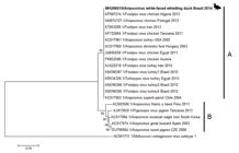
Evolutionary relationships among the sequences of Avipoxvirus detected in tumor-like lesions in white-faced whistling duck (Dendrocygna viduata) (MH286510) and other avian poxvirus sequences available in the GenBank database (NCBI). The sequences accession numbers, avian poxviruses, avian hosts, countries and years of viral detection are shown. Construction of Neighbour-joining tree was performed using Kimura 2-parameter with bootstrap values of 1000 replicates.
Discussion and Conclusion
This is the first report of Avipoxvirus detected in tumor-like lesions in white-faced whistling duck (Dendrocygna viduata). The site, gross and histological features revealed tumor-like lesions. Moreover, histopathology revealed microscopic evidence of poxvirus infection in some hyperplastic epithelial cells, which was represented by eosinophilic intracytoplasmic inclusion bodies in these cells. These inclusion bodies in the cytoplasm of infected cells consist of an aggregation of virus particles and makes histological examination one of the most effective tools for the diagnosis of fowlpox, since they can be detected in smears or tissue sections stained with Wright’s, Giemsa or other stains (Eaves & Flewett 1955Eaves G. & Flewett T. 1955. The structure of fowl-pox inclusions (Bollinger bodies). Epidemiol. Infect. 53(1):102-105. <https://dx.doi.org/10.1017/s0022172400000541> <PMid:14367806>
https://doi.org/10.1017/s002217240000054...
).
Atypical fowlpox lesions and dermal SCC begin and progress in the feather follicle epithelium (Langheinrich 1991Langheinrich K. 1991. Pathology of squamous cell carcinomas in broilers, p.58-62. In: Langheinrich K. (Eds), Proceedings of the Symposium on Avian Tumor Virus, Seattle, Washington., Fallavena et al. 1993Fallavena L.C., Rodrigues N.C., Scheufler W., Martins N.R., Braga A.C., Salle C.T. & Moraes H.L. 1993. Atypical fowlpox in broiler chickens in southern Brazil. Vet. Rec. 132(25):635. <https://dx.doi.org/10.1136/vr.132.25.635> <PMid:8394610>
https://doi.org/10.1136/vr.132.25.635...
), and this cell transformation, induced by viruses, is accompanied by the persistence of at least a part of the viral genome (Cann 1993Cann A.J. 1993. Infection, p.173-219. In: Cann A.J. (Eds), Principles of Molecular Virology. 6ª ed. Academic Press, London.). In cases of avian dermal SCC, the pathogenesis may be related to an error that may occur during the G1 or S phases of the cell cycle in the feather follicular epithelium, which could result in dysplastic changes able to trigger dermal SCC lesions (Langheinrich 1991Langheinrich K. 1991. Pathology of squamous cell carcinomas in broilers, p.58-62. In: Langheinrich K. (Eds), Proceedings of the Symposium on Avian Tumor Virus, Seattle, Washington.).
The PCR confirmed the presence of the 4b core protein gene (p4b) of Avipoxvirus in the tumor-like lesions. This gene is usually chosen for comparative genetic analysis and its amplification by PCR has been used as a molecular tool for the detection of avian poxviruses (Manarolla et al. 2010Manarolla G., Pisoni G., Sironi G. & Rampin T. 2010. Molecular biological characterization of avian poxvirus strains isolated from different avian species. Vet. Microbiol. 140(1/2):1-8. <https://dx.doi.org/10.1016/j.vetmic.2009.07.004>
https://doi.org/10.1016/j.vetmic.2009.07...
), being one of the most sensitive techniques for the etiologic diagnosis (Nayeri Fasaei et al. 2014Nayeri Fasaei B., Madadgar O., Ghalyanchi Langeroodi A. & Ghafari M. 2014. Molecular detection and phylogenetic analysis of Avipoxvirus strains isolated from different bird species. Iran. J. Vet. Res. 15(1):40-44. <https://dx.doi.org/10.22099/IJVR.2014.1980>
https://doi.org/10.22099/IJVR.2014.1980...
).
Some members of Poxviridae family have already been consistently associated with tumors in other animal species, such as rabbit fibroma virus (RFV) and Myxoma virus (MYXV), which have been associated with fibroma and myxoma. In nonpermissive cells, viral DNA is mostly integrated into the different sites of cell chromosomes, encoding binding proteins and inactivating cell growth, regulating proteins such as p53. This results in cell transformation due to the expression of proteins that control viral and cellular DNA synthesis (Şevik 2012Şevik M. 2012. Oncogenic viruses and mechanisms of oncogenesis. Turk. J. Vet. Anim. Sci. 36(4):323-329. <https://dx.doi.org/10.3906/vet-1104-2>
https://doi.org/10.3906/vet-1104-2...
). Although this is known to be the case for the previously mentioned Poxviridae family members, there is still a lack of studies regarding Avipoxvirus.
Sequencing of the Avipoxvirus DNA detected in tumor-like lesions was performed to study the genetic relationships among this strain and other avian poxviruses detected in lesions from diverse avian species. The phylogenetic analysis showed that our strain was genetically similar to other Avipoxvirus involved in cutaneous and/or diphtheritic forms of the disease in domestic and wild avian species, but not in tumor-like lesions. Molecular analysis also showed that the Avipoxvirus strain detected in the tumor-like lesions of this case was more genetically related to domestic bird strains than to wild bird strains.
This report provides the first molecular characterization of Avipoxvirus detected in tumor-like lesions in avian species. While previous reports have described simultaneous occurrences of SCC or tumor-like lesions and poxvirus detection in lesions in black-bellied whistling duck (Dendrocygna autumnalis) (Pereira et al. 2014Pereira W.L.A., Gabriel A.L.M., Monger S.G.B., Moraes L.A., Queiroz D.K.S. & Souza A.J.S. 2014. Lesões cutâneas tipo tumorais associadas à infeção por Avipoxvirus em uma marreca-cabocla (Dendrocygna autumnalis). Ciênc. Anim. Bras. 15(2):234-238. <https://dx.doi.org/10.1590/1809-6891v15i217202>
https://doi.org/10.1590/1809-6891v15i217...
) and in broilers in Brazil (Fallavena et al. 2002Fallavena L.C., Canal C.W., Salle C.T., Moraes H.L., Rocha S.L., Pereira R.A. & Silva A.B. 2002. Presence of avipoxvirus DNA in avian dermal squamous cell carcinoma. Avian Pathol. 31(3):241-246. <https://dx.doi.org/10.1080/03079450220136558> <PMid:12396347>
https://doi.org/10.1080/0307945022013655...
), and pink-backed pelican in Italy (Pesaro et al. 2009Pesaro S., Biancani B., Fabbrizi G. & Rossi G. 2009. Squamous cell carcinoma with presence of poxvirus-like inclusions in the foot of a pink-backed pelican (Pelecanus rufescens). Avian Pathol. 38(3):229-231. <https://dx.doi.org/10.1080/03079450902912176> <PMid:19468940>
https://doi.org/10.1080/0307945090291217...
), there is a lack of molecular characterization of these viruses. Poxvirus involved in the neoplasm of the white-faced whistling duck (D. viduata) exhibited high genetic similarity to other avian poxviruses detected mainly in domestic fowl, such as chickens and turkeys (Cluster A). Among these highly similar strains are Brazilian fowlpox virus detected in non-neoplastic skin lesions in commercial turkeys (Kunert-Filho et al. 2016Kunert-Filho H.C., Cibulski S.P., Finkler F., Grassotti T.T., Jaenisch F.R.F., Brito K.C.T., Carvalho D., Lovato M. & Brito B.G. 2016. First phylogenetic analysis of Avipoxvirus (APV) in Brazil. Pesq. Vet. Bras. 36(5):357-362. <https://dx.doi.org/10.1590/S0100-736X2016000500001>
https://doi.org/10.1590/S0100-736X201600...
). The phylogenetic analysis also showed major genetic differences between our strain and those obtained from non-neoplastic lesions in some pigeon species, eagle own, great bustard and Harris’s hawk.
There is a lack of information regarding diseases and their frequency in white-faced whistling ducks (D. viduata). A retrospective study over a period of 22 years (1974-1996), in Brazil, described only one case of clinical care for this species and the bird had an undifferentiated carcinoma in the coelomic cavity (Werner et al. 1998Werner P.R., Chiquito M. & Pachaly J.R. 1998. Estudo retrospectivo das neoplasias diagnosticadas em animais selvagens ou exóticos pelo Serviço de Patologia do Hospital Veterinário da Universidade Federal do Paraná entre 1974 e 1996. Arch. Vet. Sci. 3(1):39-44. <https://dx.doi.org/10.5380/avs.v3i1.3737>
https://doi.org/10.5380/avs.v3i1.3737...
).
Molecular characterization showed the genetic similarity of the Avipoxvirus detected in tumor-like lesions of this white-faced whistling duck (D. viduata) to other avian poxviruses detected mainly in domestic chicken and turkeys worldwide. As the information about the association between poxvirus and the development of neoplasm is scarce, our understanding is still limited. Therefore, experimental studies are required to elucidate the possible association between the Avipoxvirus and tumor-like lesions in avian species.
Acknowledgements
We would like to thank MSc. Bruna C. Ferreira for kindly provide the Avipoxvirus DNA for Polymerase Chain Reaction (PCR) positive control.
References
- Arai S., Arai C., Fujimaki M., Iwamoto Y., Kawarada M., Saito Y., Nomura Y. & Suzuki T. 1991. Cutaneous tumour-like lesions due to poxvirus infection in Chilean flamingos. J. Comp. Pathol. 104(4):439-441. <https://dx.doi.org/10.1016/S0021-9975(08)80154-5>
» https://doi.org/10.1016/S0021-9975(08)80154-5 - Back A., Soncini R.A., Ruthes O., Madureira Jr. S. & Flores R. 1995. An atypical fowl pox outbreak in broilers in southern Brazil. Avian Dis. 39(4):902-906. <https://dx.doi.org/10.2307/1592431>
» https://doi.org/10.2307/1592431 - Bolte A.L., Meurer J. & Kaleta E.F. 1999. Avian host spectrum of avipoxviruses. Avian Pathol. 28(5):415-432. <https://dx.doi.org/10.1080/03079459994434> <PMid:26911595>
» https://doi.org/10.1080/03079459994434 - Cann A.J. 1993. Infection, p.173-219. In: Cann A.J. (Eds), Principles of Molecular Virology. 6ª ed. Academic Press, London.
- Eaves G. & Flewett T. 1955. The structure of fowl-pox inclusions (Bollinger bodies). Epidemiol. Infect. 53(1):102-105. <https://dx.doi.org/10.1017/s0022172400000541> <PMid:14367806>
» https://doi.org/10.1017/s0022172400000541 - Fallavena L.C., Canal C.W., Salle C.T., Moraes H.L., Rocha S.L., Pereira R.A. & Silva A.B. 2002. Presence of avipoxvirus DNA in avian dermal squamous cell carcinoma. Avian Pathol. 31(3):241-246. <https://dx.doi.org/10.1080/03079450220136558> <PMid:12396347>
» https://doi.org/10.1080/03079450220136558 - Fallavena L.C., Rodrigues N.C., Moraes H.L., Salle C.T., Silva A.B., Nascimento V.P. & Rodrigues O. 1997. Squamous cell carcinoma-like and pox lesions occurring simultaneously in chorioallantoic membranes of chicken embryos inoculated with materials from squamous cell carcinoma and pox lesions in broiler chickens. Avian Dis. 41(2):469-471. <PMid:9201417>
- Fallavena L.C., Rodrigues N.C., Scheufler W., Martins N.R., Braga A.C., Salle C.T. & Moraes H.L. 1993. Atypical fowlpox in broiler chickens in southern Brazil. Vet. Rec. 132(25):635. <https://dx.doi.org/10.1136/vr.132.25.635> <PMid:8394610>
» https://doi.org/10.1136/vr.132.25.635 - Friend M. & Franson J.C. 1999. Avian pox, p.163-170. In: Friend M. & Franson J.C. (Eds), Field Manual of Wildlife Diseases: General Field Procedures and Diseases of Birds. Biological Resources Division, Madison.
- Gerlach H. 1994. Viruses, p.862-948. In Ritchie B.W., Harrison G.J. & Harrison L.R. (Eds), Avian Medicine: principles and application. Wingers publishing Lake Worth, Florida.
- Hafner S. & Goodwin M.A. 1997. Dermal squamous cell carcinoma, p.1044-1046. In: Calnek B.W., Barnes H.J., Beard C.W., McDougald L.R. & Saif Y.M. (Eds), Diseases of Poultry. 10th ed. Iowa State University Press, Iowa.
- Kimura M. 1980. A simple method for estimating evolutionary rates of base substitutions through comparative studies of nucleotide sequences. J. Mol. Evol. 16(2):111-120. <https://dx.doi.org/10.1007/BF01731581> <PMid:7463489>
» https://doi.org/10.1007/BF01731581 - Kunert-Filho H.C., Cibulski S.P., Finkler F., Grassotti T.T., Jaenisch F.R.F., Brito K.C.T., Carvalho D., Lovato M. & Brito B.G. 2016. First phylogenetic analysis of Avipoxvirus (APV) in Brazil. Pesq. Vet. Bras. 36(5):357-362. <https://dx.doi.org/10.1590/S0100-736X2016000500001>
» https://doi.org/10.1590/S0100-736X2016000500001 - Langheinrich K. 1991. Pathology of squamous cell carcinomas in broilers, p.58-62. In: Langheinrich K. (Eds), Proceedings of the Symposium on Avian Tumor Virus, Seattle, Washington.
- Lawson B., Lachish S., Colvile K.M., Durrant C., Peck K.M., Toms M.P., Sheldon B.C. & Cunnhingham A.A. 2012. Emergence of a novel avian pox disease in British tit species. PLoS One 7(11):e40176. <https://dx.doi.org/10.1371/journal.pone.0040176> <PMid:23185231>
» https://doi.org/10.1371/journal.pone.0040176 - Lee L.H. & Lee K.H. 1997. Application of the polymerase chain reaction for the diagnosis of fowl poxvirus infection. J. Virol. Methods 63(1/2):113-119. <https://dx.doi.org/10.1016/s0166-0934(96)02119-2> <PMid:9015281>
» https://doi.org/10.1016/s0166-0934(96)02119-2 - Luna L.G. 1968. Manual of Histologic Staining Methods of the Armed Forces Institute of Pathology. 3rd ed. American Registry of Pathology, New York. 258p.
- Manarolla G., Pisoni G., Sironi G. & Rampin T. 2010. Molecular biological characterization of avian poxvirus strains isolated from different avian species. Vet. Microbiol. 140(1/2):1-8. <https://dx.doi.org/10.1016/j.vetmic.2009.07.004>
» https://doi.org/10.1016/j.vetmic.2009.07.004 - McFerran J. & McNulty M. 1993. Poxviridae, p.1-15. In: McFerran J. & McNulty M. (Eds), Virus Infections of Birds. Elsevier Scientific Publishing, Oxford.
- Mete A., Borst G.H. & Dorrestein G.M. 2001. Atypical poxvirus lesions in two Galapagos doves (Nesopelia g. galapagoensis). Avian Pathol. 30(2):159-162. <https://dx.doi.org/10.1080/03079450120044560> <PMid:19184890>
» https://doi.org/10.1080/03079450120044560 - Morton J.K. & Dieterich R.A. 1979. Avian pox infection in an American green-winged teal (Anas crecca carolinensis) in Alaska. J. Wildl. Dis. 15(3):451-453. <https://dx.doi.org/10.7589/0090-3558-15.3.451>
» https://doi.org/10.7589/0090-3558-15.3.451 - Nayeri Fasaei B., Madadgar O., Ghalyanchi Langeroodi A. & Ghafari M. 2014. Molecular detection and phylogenetic analysis of Avipoxvirus strains isolated from different bird species. Iran. J. Vet. Res. 15(1):40-44. <https://dx.doi.org/10.22099/IJVR.2014.1980>
» https://doi.org/10.22099/IJVR.2014.1980 - Pereira W.L.A., Gabriel A.L.M., Monger S.G.B., Moraes L.A., Queiroz D.K.S. & Souza A.J.S. 2014. Lesões cutâneas tipo tumorais associadas à infeção por Avipoxvirus em uma marreca-cabocla (Dendrocygna autumnalis). Ciênc. Anim. Bras. 15(2):234-238. <https://dx.doi.org/10.1590/1809-6891v15i217202>
» https://doi.org/10.1590/1809-6891v15i217202 - Pesaro S., Biancani B., Fabbrizi G. & Rossi G. 2009. Squamous cell carcinoma with presence of poxvirus-like inclusions in the foot of a pink-backed pelican (Pelecanus rufescens). Avian Pathol. 38(3):229-231. <https://dx.doi.org/10.1080/03079450902912176> <PMid:19468940>
» https://doi.org/10.1080/03079450902912176 - Ritchie B.W. & Carter K. 1995. Poxviridae, p.285-311. In: Ritchie B.W. (Ed.), Avian Viruses: function and control. Winger’s Publishing, Lake Worth, Florida.
- Saitou N. & Nei M. 1987. The neighbor-joining method: a new method for reconstructing phylogenetic trees. Mol. Biol. Evol. 4(4):406-425. <https://dx.doi.org/10.1093/oxfordjournals.molbev.a040454> <PMid:3447015>
» https://doi.org/10.1093/oxfordjournals.molbev.a040454 - Schafer K. 1998. The cell cycle: a review. Vet. Pathol. 35(6):461-478. <https://dx.doi.org/10.1177/030098589803500601> <PMid:9823588>
» https://doi.org/10.1177/030098589803500601 - Şevik M. 2012. Oncogenic viruses and mechanisms of oncogenesis. Turk. J. Vet. Anim. Sci. 36(4):323-329. <https://dx.doi.org/10.3906/vet-1104-2>
» https://doi.org/10.3906/vet-1104-2 - Tamura K., Stecher G., Peterson D., Filipski A. & Kumar S. 2013. MEGA6: molecular evolutionary genetics analysis version 6.0. Mol. Biol. Evol. 30(12):2725-2729. <https://dx.doi.org/10.1093/molbev/mst197> <PMid:24132122>
» https://doi.org/10.1093/molbev/mst197 - Thompson J.D., Higgins D.G. & Gibson T.J. 1994. CLUSTAL W: improving the sensitivity of progressive multiple sequence alignment through sequence weighting, position-specific gap penalties and weight matrix choice. Nucleic Acids Res. 22(22):4673-4680. <https://dx.doi.org/10.1093/nar/22.22.4673> <PMid:7984417>
» https://doi.org/10.1093/nar/22.22.4673 - Tripathy D. & Reed W. 2008. Pox, p.291-307. In: Saif Y.M., Fadly A.M., Glisson J.R., McDougald L.R., Nolan L.K. & Swayne D.E. (Eds), Diseases of Poultry. 12nd ed. Iowa State Press, Iowa.
- Tsai S.S., Chang T.C., Yang S.F., Chi Y.C., Cher R.S., Chien M.S. & Itakura C. 1997. Unusual lesions associated with avian poxvirus infection in rosy-faced lovebirds (Agapornis roseicollis). Avian Pathol. 26(1):75-82. <https://dx.doi.org/10.1080/03079459708419195> <PMid:18483891>
» https://doi.org/10.1080/03079459708419195 - Van Riper C. III. & Forrester D. 2007. Avian pox, p.131-176. In: Van Riper C. III & Forrester D. (Eds), Infectious Diseases of Wild Birds. Blackwell Publishing, Iowa.
- Werner P.R., Chiquito M. & Pachaly J.R. 1998. Estudo retrospectivo das neoplasias diagnosticadas em animais selvagens ou exóticos pelo Serviço de Patologia do Hospital Veterinário da Universidade Federal do Paraná entre 1974 e 1996. Arch. Vet. Sci. 3(1):39-44. <https://dx.doi.org/10.5380/avs.v3i1.3737>
» https://doi.org/10.5380/avs.v3i1.3737
Publication Dates
-
Publication in this collection
14 Dec 2020 -
Date of issue
Oct 2020
History
-
Received
23 May 2020 -
Accepted
15 July 2020


