ABSTRACT:
This study describes, through a retrospective study, the epidemiological and clinical-pathological findings of compression in the central nervous system (CNS) of buffaloes. The study includes observations made in 15 animals from 1998 to 2021 by reviewing the clinical records of animals with compressive injuries of the CNS treated at the Veterinary Hospital of the Veterinary Medicine Institute of the Federal University of Pará. The animals treated with clinical signs compatible with CNS compressive lesions were subjected to general and specific clinical examinations of the nervous system. Blood samples were collected from four animals for complete blood counts, and cerebrospinal fluid (CSF) samples were collected from three animals for physical evaluation. Thirteen animals were necropsied. The age range of the affected animals ranged from four months to 11 years of age, with a greater frequency over age 12 months (80%, 13/15). The most affected vertebral segment was between T3 and L3 (60%, 9/15), followed by brain injury (20%, 3/15), the L4-S2 segment (13.3%, 2/15) and the C1-C5 segment (6.7%, 1/15). The clinical findings varied according to the location of the lesion. The necropsy findings revealed paraypophyseal abscess in the brainstem and vertebral body, subarachnoid hematoma, lymphoma and vertebral fractures. The performance of a thorough clinical examination of the CNS combined with the necropsy findings was important to characterize the clinical picture and to locate the cause and the affected CNS segments in the buffaloes studied. It is important to include CNS compressive lesions among the neurological diseases of buffaloes.
INDEX TERMS:
Central nervous system; buffalo; compressive lesions; neurological clinical signs
RESUMO:
Esse trabalho descreve, através de estudo retrospectivo, os achados epidemiológicos e clínico-patológicos de compressão no sistema nervoso central (SNC) de bubalinos. O estudo compreendeu as observações realizadas em 15 animais, durante os anos de 1998 a 2021, por meio da revisão dos arquivos de fichas clínicas de animais com lesões compressivas no SNC atendidos pelo Hospital Veterinário do Instituto de Medicina Veterinária da Universidade Federal do Pará. Os animais atendidos com sinais clínicos compatíveis com lesões compressivas no SNC foram submetidos a exames clínicos geral e específico do sistema nervoso. Foram coletadas amostras de sangue de quatro animais para realização de hemograma e amostras de líquido cefalorraquidiano (LCR) de três animais para avaliação física. Foram necropsiados 13 animais. A faixa etária dos animais acometidos variou de quatro meses a 11 anos de idade, com maior frequência na faixa acima de 12 meses (80%, 13/15). O segmento vertebral mais acometido foi entre T3-L3 (60%, 9/15), seguida por lesão no encéfalo (20%, 3/15), pelo segmento L4-S2 (13,3%, 2/15) e pelo segmento C1-C5 (6,7%, 1/15). Os achados clínicos variaram de acordo com a localização da lesão. Os achados de necropsia revelaram abscesso parahipofisário, no tronco encefálico e no corpo da vértebra, hematoma subaracnoidea, linfoma e fraturas vertebrais. A realização de um minucioso exame clínico do SNC associado aos achados de necropsia foram importantes para caracterizar o quadro clínico e localizar a causa e os seguimentos acometidos do SNC nos búfalos estudados. Torna-se importante incluir as lesões compressivas do SNC entre as enfermidades neurológicas dos bubalinos.
TERMOS DE INDEXAÇÃO:
Sistema nervoso central; bubalino; lesões compressivas; sinais clínicos neurológicos
Introduction
The diseases that affect the central nervous system (CNS) of ruminants constitute an important group of diseases. Among the infectious causes, rabies is considered the most important disease because it causes the most deaths of ruminants in Brazil, followed by botulinum toxins and toxic plants, causing extensive economic losses to Brazilian livestock (Riet-Correa et al. 1998Riet-Correa F., Schild A.L. & Fernandes C.G. 1998. Enfermidades do sistema nervoso dos ruminantes no sul do Rio Grande do sul. Ciência Rural 28(2):341-348. <https://dx.doi.org/10.1590/S0103-84781998000200028>
https://doi.org/10.1590/S0103-8478199800...
). However, other causes of CNS changes in production animals may be compression injuries caused by abscesses, vertebral column fractures and neoplasms (Barros et al. 2006Barros C.S.L., Driemeier D., Dutra I.S. & Lemos R.A.A. 2006. Doenças do Sistema Nervoso de Bovinos no Brasil. Vallée, Montes Claros. 207p.). CNS compressions have already been described by several authors (Riet-Correa et al. 2002Riet-Correa F., Riet-Correa G. & Schild A.L. 2002. Importância do exame clínico para o diagnóstico das enfermidades do sistema nervoso em ruminantes e eqüídeos. Pesq. Vet. Bras. 22(4):161-168. <https://dx.doi.org/10.1590/S0100-736X2002000400006>
https://doi.org/10.1590/S0100-736X200200...
, Borges et al. 2003Borges A.S., Silva D.P.G., Gonçalves R.C., Chiacchio S.B., Amorim R.M., Kuchembuck M.R.G., Vulcano L.C., Bandarra E.P. & Lopes R.S. 2003. Fraturas vertebrais em grandes animais: estudo retrospectivo de 39 casos (1987-2002). Arq. Bras. Med. Vet. Zootec. 55(2):127-132. <https://dx.doi.org/10.1590/S0102-09352003000200001>
https://doi.org/10.1590/S0102-0935200300...
, Marques et al. 2004Marques L.C., Cadioli F.A., Castro Netto A., Ávila L.G., Canola J.C. & Alessi A.C. 2004. Abscessos em coluna vertebral de bezerros e cordeiros: aspectos neurológicos. Rev. Educ. Contin. CRMV-SP 7(1/3):15-22. <https://dx.doi.org/10.36440/recmvz.v7i1/3.3233>
https://doi.org/10.36440/recmvz.v7i1/3.3...
, Rissi et al. 2010Rissi D.R., Pierezan F., Oliveira-Filho J.C., Lucena R.B., Carmo P.M.S. & Barros C.S.L. 2010. Abordagem diagnóstica das principais doenças do sistema nervoso de ruminantes e equinos no Brasil. Pesq. Vet. Bras. 30(11):958-967. <https://dx.doi.org/10.1590/S0100-736X2010001100010>
https://doi.org/10.1590/S0100-736X201000...
, Câmara et al. 2014Câmara A.C.L., Vale A.M., Batista J.S., Feijó F.M.C. & Soto-Blanco B. 2014. Suppurative intracranial processes in 15 domestic ruminants. Pesq. Vet. Bras. 34(5):421-426. <https://dx.doi.org/10.1590/S0100-736X2014000500006>
https://doi.org/10.1590/S0100-736X201400...
) in bovine, ovine and caprine species and equine, but there are few studies reporting compressive neurological disease and its epidemiological and clinical pathological aspects in buffaloes.
Some management conditions may predispose to the occurrence of vertebral fractures, such as slippery floors, many animals of different sizes or ages and animals with very rapid growth (Sherman 1987Sherman D.M. 1987. Localized diseases of the bovine brain and spinal cord. Vet. Clin. N. Am., Food Anim. Pract. 3(1):179-191. <https://dx.doi.org/10.1016/s0749-0720(15)31189-0> <PMid:3494493>
https://doi.org/10.1016/s0749-0720(15)31...
). According to Barros et al. (2006)Barros C.S.L., Driemeier D., Dutra I.S. & Lemos R.A.A. 2006. Doenças do Sistema Nervoso de Bovinos no Brasil. Vallée, Montes Claros. 207p., most abscesses that compress the spinal cord originate from hematogenous osteomyelitis of the vertebral body. Possible sources of infection in adult cattle include endocarditis and traumatic reticulitis, and in newborn calves, omphalophlebitis is the main source. The clinical signs vary, depending on the injured site, the degree of spinal compression and the involvement of the spinal anatomical tracts (Mackay & Van Metre 2015Mackay R.J. & Van Metre D.C. 2015. Diseases of the nervous system, p.917-1043. In: Smith B.P. (Ed.), Large Animal Internal Medicine. 5th ed. Elsevier, St. Louis.).
A complete neurological examination is based on anamnesis and evaluation of brain integrity, spinal cord and peripheral nerves. Complementary exams such as cerebrospinal fluid analysis, simple or contrasted radiography (myelography), electroencephalography, computed tomography and magnetic resonance imaging are also important to aid in the diagnosis of diseases that occur with CNS compressive injuries (Brewer 1987Brewer B.D. 1987. Examination of the bovine nervous system. Vet. Clin. N. Am., Food Anim. Pract. 3(1):13-24. <https://dx.doi.org/10.1016/S0749-0720(15)31176-2> <PMid:3494491>
https://doi.org/10.1016/S0749-0720(15)31...
, Braund 1994Braund K.G. 1994. Clinical Syndromes in Veterinary Neurology. 2nd ed. Mosby, St. Louis. 477p., Dirksen et al. 2005Dirksen G., Gründer H.D. & Stöber M. 2005. Medicina Interna y Cirurgía del Bovino. Vol. 1, 4th ed. Editora Inter-Medica, Buenos Aires, p.618-629., DeLahunta 2009DeLahunta A. 2009. Veterinary Neuroanatomy and Clinical Neurology. 3rd ed. Elsevier, St. Louis. 540p.).
In addition, the research and extension group in Clinical Medicine of Large Animals of the Federal University of Pará, Campus Castanhal, has been studying and diagnosing spinal fractures in animals with marked phosphorus deficiencies associated with neurological clinical signs in cattle and buffaloes (Peixoto et al. 2005Peixoto P.V., Malafaia P.A., Barbosa J.D. & Tokarnia C.H. 2005. Princípios de suplementação mineral em ruminantes. Pesq. Vet. Bras. 25(3):195-200. <https://dx.doi.org/10.1590/S0100-736X2005000300011>
https://doi.org/10.1590/S0100-736X200500...
, Barbosa Neto et al. 2007Barbosa Neto J.D., Oliveira C.M.C., Duarte M.D., Albernaz T.T., Oliveira Júnior C.A., Riet-Correa G. & Riet -Correa F. 2007. Phosphorus deficiency in buffaloes in the state of Pará, northern Brazil. Ital. J. Anim. Sci. 6(Supl.2):971-973. <https://dx.doi.org/10.4081/ijas.2007.s2.971>
https://doi.org/10.4081/ijas.2007.s2.971...
, 2014Barbosa J.D., Lima D.H.S., Belo Reis A.S., Pinheiro C.P., Souza M.G.S., Silva J.B., Salvarani F.M. & Oliveira C.M.C. 2014. Degenerative joint disease in cattle and buffaloes in the Amazon region: a retrospective study. Pesq. Vet. Bras. 34(9):845-850. <https://dx.doi.org/10.1590/S0100-736X2014000900007>
https://doi.org/10.1590/S0100-736X201400...
, 2016Barbosa F.B., Bomjardim H.A., Helayel M.J.S.A., Faial K.C.F., Oliveira C.M.C., Malafaia P., Brito M.F. & Barbosa J.D. 2016. Avaliação econômica de três tipos de suplementação mineral para bovinos de corte no Estado do Pará. Pesq. Vet. Bras. 36(7):600-604. <https://dx.doi.org/10.1590/S0100-736X2016000700007>
https://doi.org/10.1590/S0100-736X201600...
, 2021Barbosa J.D., Brito M.F., Oliveira C.M.C., Duarte M.D., Silveira N.S.S., Ferreira T.T.A., Silveira J.A.S., Bomjardim H.A., Barbosa C.C. & Malafaia P.A.M. 2021. Deficiências minerais em bovinos e bubalinos no Brasil: aspectos gerais, importância, diagnóstico, profilaxia e correção. Revta Bras. Buiatria 2(1):1-38. <https://dx.doi.org/10.4322/2763-955X.2021.010>
https://doi.org/10.4322/2763-955X.2021.0...
, Pinheiro et al. 2011Pinheiro C.P., Bomjardim H.A., Andrade S.J.T., Faial K.C.F., Oliveira C.M.C. & Barbosa J.D. 2011. Níveis de fósforo, cobre, cobalto e zinco em bubalinos (Bubalus bubalis) na Ilha de Marajó, Estado do Pará. Pesq. Vet. Bras. 31(3):193-198. <https://dx.doi.org/10.1590/S0100-736X2011000300002>
https://doi.org/10.1590/S0100-736X201100...
, Malafaia et al. 2014Malafaia P., Costa R.M., Brito M.F., Peixoto P.V., Barbosa J.D., Tokarnia C.H. & Döbereiner J. 2014. Equívocos arraigados no meio pecuário sobre deficiências e suplementação minerais em bovinos no Brasil. Pesq. Vet. Bras. 34(3):224-249. <https://dx.doi.org/10.1590/S0100-736X2014000300008>
https://doi.org/10.1590/S0100-736X201400...
, Bomjardim et al. 2015Bomjardim H.A., Oliveira C.M.C., Silveira J.A.S., Silva N.S., Duarte M.D., Faial K.C.F., Brito M.F. & Barbosa J.D. 2015. Deficiências minerais em vacas em lactação da bacia leiteira do município de Rondon do Pará, estado do Pará. Pesq. Vet. Bras. 35(5):409-416. <https://dx.doi.org/10.1590/S0100-736X2015000500004>
https://doi.org/10.1590/S0100-736X201500...
). Therefore, the objective of this study was to perform a retrospective study characterizing the clinical-pathological conditions caused by CNS compressive lesions in buffaloes.
Materials and Methods
Case selection and clinical evaluation. The retrospective study was conducted from 1998 to 2021 and enrolled 15 buffaloes (identified numerically from 1 to 15). The clinical records of the epidemiological data (age, race, sex and place of origin) and clinical pictures (clinical signs, clinical evolution, neurological lesion site, necropsy findings) of animals diagnosed with CNS compressive lesions treated at the Veterinary Hospital of the “Instituto de Medicina Veterinária” (Veterinary Medicine Institute - IMV) of the “Universidade Federal do Pará” (UFPA) were studied. As inclusion criteria in the study, all animals treated with neurological clinical signs compatible with CNS compression injury and subjected to general and specific clinical examinations of the nervous system were selected. The compressive injuries were characterized according to Dirksen et al. (2005)Dirksen G., Gründer H.D. & Stöber M. 2005. Medicina Interna y Cirurgía del Bovino. Vol. 1, 4th ed. Editora Inter-Medica, Buenos Aires, p.618-629.. All animals included in the study were negative for rabies, the main differential diagnosis was neurological disease; the animals from properties that did not perform mineral supplementation and located in phosphorus-deficient (P) regions. Exclusion criteria were as follows: animals with incomplete clinical records and those positive for rabies.
Complementary exams. All available complementary tests of the animals included in the study were reviewed. Therefore, hematological data from four animals (Buffaloes 2, 3, 7 and 15) related to the analysis of blood samples stored in 5ml tubes with anticoagulant (EDTA) were included in the study, and the blood counts were performed according to Jain (1993)Jain N.C. 1993. Essentials of Veterinary Hematology. Lea and Febiger, Philadelphia. 417p.. In addition, data from the cerebrospinal fluid (CSF) analysis collected from the cisterna magna of two animals with spinal cord injury (Buffaloes 6 and 9) and one with brainstem abscess (Buffalo 2) were included for physical evaluation of the CSF according to Dirksen et al. (2005)Dirksen G., Gründer H.D. & Stöber M. 2005. Medicina Interna y Cirurgía del Bovino. Vol. 1, 4th ed. Editora Inter-Medica, Buenos Aires, p.618-629.. In addition, the microbiological results obtained according to Rengifo et al. (2006)Rengifo S.A., Silva R.A., Pereira I.A., Souza M.M. & Botteon R.C.C.M. 2006. Isolamento de agentes microbianos a partir de amostras de sangue e umbigo de bezerros mestiços neonatos. Braz. J. Vet. Res. Anim. Sci. 43(4):442-447. <https://dx.doi.org/10.11606/issn.1678-4456.bjvras.2006.26458>
https://doi.org/10.11606/issn.1678-4456....
, the content of the abscess of Buffalo 3, and the results of the postmortem evaluation of 13 buffaloes with compressive lesions observed during the necropsy were reviewed. Only the histopathological result of Buffalo 11 had significant lesions and was included in the study.
Results
Epidemiological data and clinical signs
Fifteen buffaloes were treated, five animals from Marajó Island and the other 10 from properties located in the continental region of the state of Pará, properties located in regions deficient in phosphorus (P) and which had a history of problems with mineral supplementation for the animals. Regarding sex, 60% (9/15) of the buffaloes were males and 40% (6/15) were females; 86.7% (13/15) were Murrah, 6.7% (1/15) Mediterranean and 6.7% (1/15) were mixed Murrah/Mediterranean. The ages ranged from four months to 11 years. The most affected vertebral segment was T3-L3 60% (9/15), followed by brain injury 20% (3/15), by the L4-S2 segment 13.3% (2/15) and segment C1-C5 6.7% (1/15). The buffaloes with compression injury to the brain (Buffaloes 1-3) (Fig.1-6) showed sleepiness, decreased alertness, pressure of the head against objects, loss of vision, walking in a circular pattern, opisthotonus, and limb spasticity. The animal (Buffalo 4), with a spinal cord injury between C1 and C5, showed ataxia and postural change characterized by cervical scoliosis. In buffaloes with lesions in the T3-L3 segment, there were no neurological changes in the thoracic limbs, but they showed hyperreactive pain reflexes, ataxia, paresis and muscle weakness in the pelvic limbs (Fig.7-8). The buffaloes diagnosed with injury in the L4-S2 segment had unaltered pain reflexes in the cranial medullary segments and decreased pain in the caudal segments, ataxia and flaccid paresis of the pelvic limbs (Buffaloes 14 and 15). The epidemiological data and the individual diagnoses of each clinical case are described in Table 1.

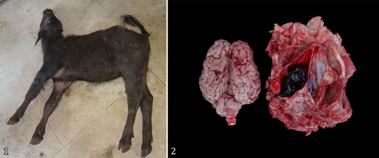
(1) Four-month-old Murrah Buffalo 1 with opisthotonus and limb spasticity. (2) Submeningian hematoma of Buffalo 1.

(3) Four-year-old Murrah Buffalo 3, with decreased alertness and head resting on the trough due to a parahypophyseal abscess. (4) Buffalo 3 pressing the head against the wall of the stall. This animal had a circular walking pattern, as demonstrated by the arrangement of feces on the floor of the stall.

(5) Necropsy findings of Buffalo 3, in which a parahypophyseal abscess is observed. (6) Aspiration puncture of the paraphypophyseal abscess of Buffalo 3.

(7) Buffalo 11, Murrah breed, 11 years old, exhibiting paresis and proprioceptive deficit of the pelvic limbs. (8) Transmeningean lymphoma (epi and subdural) at the height of the 8th thoracic vertebra, which compressed the spinal cord.
Clinical pathology, microbiology and anatomical histopathological findings
In the data of the four hemograms performed, leukocytosis by neutrophilia was observed. In the macroscopic examination of the CSF of Buffalo 2 with brainstem abscess, the CSF was turbid, with a flocculent deposit, which coagulated after a few seconds of collection. In Buffaloes 6 and 9, with vertebral fractures, the CSF showed no variations in the macroscopic aspect. In the microbiological examination, Streptococcus spp was isolated in the abscess content (Buffalo 3). The postmortem findings of the 13 buffaloes evaluated with CNS compression are shown in Table 2, as well as the findings of all complementary tests. In most of the animals studied, there were no significant histological lesions, except for Buffalo 11, which presented malignant neoplastic proliferation of round cells forming a dense mantle, which surrounded the nerve fibers adjacent to the meninges. The cells showed moderate pleomorphism, round to oval shape, large round to oval nucleus with dense and basophilic chromatin, sparse and eosinophilic cytoplasm and sometimes evident nucleolus.
Discussion
The data obtained in this manuscript are unprecedented and are discussed throughout this section compared with the corresponding lesions in cattle. Male animals were the most affected (60%, 9/15) when compared to females (40%, 6/15), which differs from the study by Borges et al. (2003)Borges A.S., Silva D.P.G., Gonçalves R.C., Chiacchio S.B., Amorim R.M., Kuchembuck M.R.G., Vulcano L.C., Bandarra E.P. & Lopes R.S. 2003. Fraturas vertebrais em grandes animais: estudo retrospectivo de 39 casos (1987-2002). Arq. Bras. Med. Vet. Zootec. 55(2):127-132. <https://dx.doi.org/10.1590/S0102-09352003000200001>
https://doi.org/10.1590/S0102-0935200300...
, who observed a predominance of lesions in females (64.1%, 25/39). In the present retrospective study, the great variation in the age of buffaloes treated with CNS compression was demonstrated; however, the largest number of affected animals were those over age 12 months (80%, 12/15); therefore, adult animals, again different from the data presented by Borges et al. (2003)Borges A.S., Silva D.P.G., Gonçalves R.C., Chiacchio S.B., Amorim R.M., Kuchembuck M.R.G., Vulcano L.C., Bandarra E.P. & Lopes R.S. 2003. Fraturas vertebrais em grandes animais: estudo retrospectivo de 39 casos (1987-2002). Arq. Bras. Med. Vet. Zootec. 55(2):127-132. <https://dx.doi.org/10.1590/S0102-09352003000200001>
https://doi.org/10.1590/S0102-0935200300...
, who reported a higher occurrence of compression (56.4%, 22/39) in animals up to 12 months of age. However, in both studies, the most frequent cause that led to the diagnosis of CNS injury with the presence of neurological signs was vertebral fractures, proving to be important data for routine clinical care.
The clinical signs varied according to the segment of the spinal cord affected and the intensity of the injury; however, in general, ataxia, paresis or paralysis of the limbs, inability to stand and walk, postural changes, and hyperesthesia were observed in the extremities, in addition to loss of skin sensitivity directly related to the location of the lesion. All these clinical signs described in buffaloes corroborate those already described in the literature by several authors in other animal species (Riet-Correa et al. 2002Riet-Correa F., Riet-Correa G. & Schild A.L. 2002. Importância do exame clínico para o diagnóstico das enfermidades do sistema nervoso em ruminantes e eqüídeos. Pesq. Vet. Bras. 22(4):161-168. <https://dx.doi.org/10.1590/S0100-736X2002000400006>
https://doi.org/10.1590/S0100-736X200200...
, Borges et al. 2003Borges A.S., Silva D.P.G., Gonçalves R.C., Chiacchio S.B., Amorim R.M., Kuchembuck M.R.G., Vulcano L.C., Bandarra E.P. & Lopes R.S. 2003. Fraturas vertebrais em grandes animais: estudo retrospectivo de 39 casos (1987-2002). Arq. Bras. Med. Vet. Zootec. 55(2):127-132. <https://dx.doi.org/10.1590/S0102-09352003000200001>
https://doi.org/10.1590/S0102-0935200300...
, Marques et al. 2004Marques L.C., Cadioli F.A., Castro Netto A., Ávila L.G., Canola J.C. & Alessi A.C. 2004. Abscessos em coluna vertebral de bezerros e cordeiros: aspectos neurológicos. Rev. Educ. Contin. CRMV-SP 7(1/3):15-22. <https://dx.doi.org/10.36440/recmvz.v7i1/3.3233>
https://doi.org/10.36440/recmvz.v7i1/3.3...
, Rissi et al. 2010Rissi D.R., Pierezan F., Oliveira-Filho J.C., Lucena R.B., Carmo P.M.S. & Barros C.S.L. 2010. Abordagem diagnóstica das principais doenças do sistema nervoso de ruminantes e equinos no Brasil. Pesq. Vet. Bras. 30(11):958-967. <https://dx.doi.org/10.1590/S0100-736X2010001100010>
https://doi.org/10.1590/S0100-736X201000...
, Câmara et al. 2014Câmara A.C.L., Vale A.M., Batista J.S., Feijó F.M.C. & Soto-Blanco B. 2014. Suppurative intracranial processes in 15 domestic ruminants. Pesq. Vet. Bras. 34(5):421-426. <https://dx.doi.org/10.1590/S0100-736X2014000500006>
https://doi.org/10.1590/S0100-736X201400...
), who reiterate the information that the clinical signs varied according to the location and intensity of the lesion in the CNS. It was observed that buffaloes with lesions in the brain showed decreased alertness, pressed their heads against objects (Fig.3-4), had loss of vision and walked in circular patterns, which according to Riet-Correa et al. (2002)Riet-Correa F., Riet-Correa G. & Schild A.L. 2002. Importância do exame clínico para o diagnóstico das enfermidades do sistema nervoso em ruminantes e eqüídeos. Pesq. Vet. Bras. 22(4):161-168. <https://dx.doi.org/10.1590/S0100-736X2002000400006>
https://doi.org/10.1590/S0100-736X200200...
and Borges et al. (2003)Borges A.S., Silva D.P.G., Gonçalves R.C., Chiacchio S.B., Amorim R.M., Kuchembuck M.R.G., Vulcano L.C., Bandarra E.P. & Lopes R.S. 2003. Fraturas vertebrais em grandes animais: estudo retrospectivo de 39 casos (1987-2002). Arq. Bras. Med. Vet. Zootec. 55(2):127-132. <https://dx.doi.org/10.1590/S0102-09352003000200001>
https://doi.org/10.1590/S0102-0935200300...
, are clinical indicators that occur due to the loss of functions in the nuclei and pathways that regulate these activities in the brain.
In seven of the necropsied buffaloes (53.8%, 7/13), the causes of CNS compressions were of noninfectious origin, mainly represented by vertebral body fractures, which consequently led to spinal cord compression. Based on the epidemiological data, clinical signs and anatomopathological findings obtained from the affected animals, it is strongly believed that all seven buffaloes had classical and compatible necropsy findings of phosphorus deficiency, which would explain the bone fragility and the predisposition to spontaneous fractures in different bones, including the bodies of the vertebrae observed in this study (Fig.9-10). The suspicion of mineral deficiency is also reinforced by previous studies that have proven the occurrence of phosphorus deficiency in the state of Pará (Peixoto et al. 2005Peixoto P.V., Malafaia P.A., Barbosa J.D. & Tokarnia C.H. 2005. Princípios de suplementação mineral em ruminantes. Pesq. Vet. Bras. 25(3):195-200. <https://dx.doi.org/10.1590/S0100-736X2005000300011>
https://doi.org/10.1590/S0100-736X200500...
, Barbosa Neto et al. 2007Barbosa Neto J.D., Oliveira C.M.C., Duarte M.D., Albernaz T.T., Oliveira Júnior C.A., Riet-Correa G. & Riet -Correa F. 2007. Phosphorus deficiency in buffaloes in the state of Pará, northern Brazil. Ital. J. Anim. Sci. 6(Supl.2):971-973. <https://dx.doi.org/10.4081/ijas.2007.s2.971>
https://doi.org/10.4081/ijas.2007.s2.971...
, 2014Barbosa J.D., Lima D.H.S., Belo Reis A.S., Pinheiro C.P., Souza M.G.S., Silva J.B., Salvarani F.M. & Oliveira C.M.C. 2014. Degenerative joint disease in cattle and buffaloes in the Amazon region: a retrospective study. Pesq. Vet. Bras. 34(9):845-850. <https://dx.doi.org/10.1590/S0100-736X2014000900007>
https://doi.org/10.1590/S0100-736X201400...
, 2016Barbosa F.B., Bomjardim H.A., Helayel M.J.S.A., Faial K.C.F., Oliveira C.M.C., Malafaia P., Brito M.F. & Barbosa J.D. 2016. Avaliação econômica de três tipos de suplementação mineral para bovinos de corte no Estado do Pará. Pesq. Vet. Bras. 36(7):600-604. <https://dx.doi.org/10.1590/S0100-736X2016000700007>
https://doi.org/10.1590/S0100-736X201600...
, 2021Barbosa J.D., Brito M.F., Oliveira C.M.C., Duarte M.D., Silveira N.S.S., Ferreira T.T.A., Silveira J.A.S., Bomjardim H.A., Barbosa C.C. & Malafaia P.A.M. 2021. Deficiências minerais em bovinos e bubalinos no Brasil: aspectos gerais, importância, diagnóstico, profilaxia e correção. Revta Bras. Buiatria 2(1):1-38. <https://dx.doi.org/10.4322/2763-955X.2021.010>
https://doi.org/10.4322/2763-955X.2021.0...
, Pinheiro et al. 2011Pinheiro C.P., Bomjardim H.A., Andrade S.J.T., Faial K.C.F., Oliveira C.M.C. & Barbosa J.D. 2011. Níveis de fósforo, cobre, cobalto e zinco em bubalinos (Bubalus bubalis) na Ilha de Marajó, Estado do Pará. Pesq. Vet. Bras. 31(3):193-198. <https://dx.doi.org/10.1590/S0100-736X2011000300002>
https://doi.org/10.1590/S0100-736X201100...
, Malafaia et al. al. 2014Malafaia P., Costa R.M., Brito M.F., Peixoto P.V., Barbosa J.D., Tokarnia C.H. & Döbereiner J. 2014. Equívocos arraigados no meio pecuário sobre deficiências e suplementação minerais em bovinos no Brasil. Pesq. Vet. Bras. 34(3):224-249. <https://dx.doi.org/10.1590/S0100-736X2014000300008>
https://doi.org/10.1590/S0100-736X201400...
, Bomjardim et al. 2015Bomjardim H.A., Oliveira C.M.C., Silveira J.A.S., Silva N.S., Duarte M.D., Faial K.C.F., Brito M.F. & Barbosa J.D. 2015. Deficiências minerais em vacas em lactação da bacia leiteira do município de Rondon do Pará, estado do Pará. Pesq. Vet. Bras. 35(5):409-416. <https://dx.doi.org/10.1590/S0100-736X2015000500004>
https://doi.org/10.1590/S0100-736X201500...
), especially on Marajó Island (Barbosa Neto et al. 2007Barbosa Neto J.D., Oliveira C.M.C., Duarte M.D., Albernaz T.T., Oliveira Júnior C.A., Riet-Correa G. & Riet -Correa F. 2007. Phosphorus deficiency in buffaloes in the state of Pará, northern Brazil. Ital. J. Anim. Sci. 6(Supl.2):971-973. <https://dx.doi.org/10.4081/ijas.2007.s2.971>
https://doi.org/10.4081/ijas.2007.s2.971...
, 2014Barbosa J.D., Lima D.H.S., Belo Reis A.S., Pinheiro C.P., Souza M.G.S., Silva J.B., Salvarani F.M. & Oliveira C.M.C. 2014. Degenerative joint disease in cattle and buffaloes in the Amazon region: a retrospective study. Pesq. Vet. Bras. 34(9):845-850. <https://dx.doi.org/10.1590/S0100-736X2014000900007>
https://doi.org/10.1590/S0100-736X201400...
, Pinheiro et al. 2011Pinheiro C.P., Bomjardim H.A., Andrade S.J.T., Faial K.C.F., Oliveira C.M.C. & Barbosa J.D. 2011. Níveis de fósforo, cobre, cobalto e zinco em bubalinos (Bubalus bubalis) na Ilha de Marajó, Estado do Pará. Pesq. Vet. Bras. 31(3):193-198. <https://dx.doi.org/10.1590/S0100-736X2011000300002>
https://doi.org/10.1590/S0100-736X201100...
), including farms where buffaloes in this study were treated.

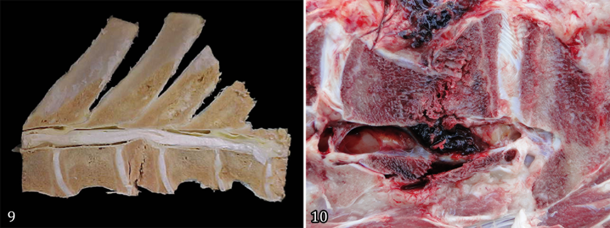
(9) Necropsy findings of Buffalo 8 showing fracture of the thoracic vertebra that compressed the spinal cord. (10) Recent fracture of the thoracic vertebra body with hematoma in the medullary canal and compression of the spinal cord of Buffalo 13.
The higher number of animals with lesions located between the T3-L3 vertebrae (60%, 9/15) reported in this study corroborates the observations made by Mackay & Van Metre (2015)Mackay R.J. & Van Metre D.C. 2015. Diseases of the nervous system, p.917-1043. In: Smith B.P. (Ed.), Large Animal Internal Medicine. 5th ed. Elsevier, St. Louis. in calves with abscesses in the thoracolumbar segment and by Borges et al. (2003)Borges A.S., Silva D.P.G., Gonçalves R.C., Chiacchio S.B., Amorim R.M., Kuchembuck M.R.G., Vulcano L.C., Bandarra E.P. & Lopes R.S. 2003. Fraturas vertebrais em grandes animais: estudo retrospectivo de 39 casos (1987-2002). Arq. Bras. Med. Vet. Zootec. 55(2):127-132. <https://dx.doi.org/10.1590/S0102-09352003000200001>
https://doi.org/10.1590/S0102-0935200300...
in cattle with vertebral fracture. This higher frequency of lesions in the regions between T3 and L3 can be explained by Hahn et al. (1999)Hahn C.N., Mayhew I.G. & Mackay R.J. 1999. Diseases of multiple or unknown sites, p.947-950. In: Colahan P.T., Merritt A.M., Moore J.N. & Mayhew I.G. (Eds), Equine Medicine and Surgery. 5th ed. Mosby, St. Louis., who describe the thoracic region as the one with greater dorsoventral movement, in addition to being the main site of body weight support, which confirms the predisposition of this anatomical region to the occurrence of CNS compressive injuries. Only one animal (Buffalo 4) showed clinical signs corresponding to the segment of cervical vertebrae between C1 and C5, as a result of cervical scoliosis, which, according to Damé et al. (2013)Damé M.C.F., Riet-Correa F. & Schild A.L. 2013. Doenças hereditárias e defeitos congênitos diagnosticados em búfalos (Bubalus bubalis) no Brasil. Pesq. Vet. Bras. 33(7):831-839. <https://dx.doi.org/10.1590/S0100-736X2013000700001>
https://doi.org/10.1590/S0100-736X201300...
, can be explained by the different congenital alterations in buffaloes, which may be associated with the degree of consanguinity that the species shows, leading to the emergence of the most different clinical signs, including those related to spinal cord compression.
In four buffaloes older than 12 months, the compressive lesions were possibly of infectious origin, represented by parahypophyseal abscess in Buffaloes 2 and 3 (Fig.5-6), abscess in the intervertebral disc of segment L4-S2 in Buffalo 15, and lymphoma in Buffalo 11. The presence of lymphoma in the spinal canal (Fig.7-8) may be associated with viral infection by bovine immunodeficiency virus, as reported by De Oliveira et al. (2016)De Oliveira C.H.S., Barbosa J.D., Damasceno K.A., Cassali G.D., Oliveira C.M.C., Leite R.C. & Reis J.K.P. 2016. Multicentric lymphoma in buffaloes in the Amazon region, Brazil. BMC Vet. Res. 12:238. <https://dx.doi.org/10.1186/s12917-016-0845-y>
https://doi.org/10.1186/s12917-016-0845-...
and Teixeira et al. (2018)Teixeira M.A.S., Machado F.M.C., Sarmento N.M.F.P., Oliveira Júnior C.A., Riet-Correa G., Cerqueira V.D., França T.N. & Bezerra Júnior P.S. 2018. Aspectos histopatológicos e imuno-histoquímicos da polisserosite em búfalos (Bubalus bubalis). Pesq. Vet. Bras. 38(1):59-64. <https://dx.doi.org/10.1590/1678-5150-PVB-5319>
https://doi.org/10.1590/1678-5150-PVB-53...
. It is inferred at this point that the cause of abscesses in the CNS in Buffaloes 2 and 3 may have occurred by extension of the adjacent suppurative lesions present at the base of the horn of these animals, as stated in their clinical records. It is believed that Streptococcus spp. isolated in the abscess content of Buffalo 3 may have reached the CNS by the hematogenous route because, according to Riet-Correa et al. (1998)Riet-Correa F., Schild A.L. & Fernandes C.G. 1998. Enfermidades do sistema nervoso dos ruminantes no sul do Rio Grande do sul. Ciência Rural 28(2):341-348. <https://dx.doi.org/10.1590/S0103-84781998000200028>
https://doi.org/10.1590/S0103-8478199800...
and Radostits et al. (2000)Radostits O.M., Gay C.C., Blood D.C. & Hinchcliff K.W. 2000. Veterinary Clinic: a treatise on the diseases of cattle, sheep, pigs, goats and horses. 9th ed. Guanabara Koogan, Rio de Janeiro, p.507-511., this is considered an important and frequent route for the access of infectious agents to the CNS. These authors also state that in general, there are four pathways for the arrival of an infectious agent in the CNS, which are hematogenous, by direct penetrating lesions, by extension of an adjacent suppurative lesion and by centripetal lesions via the peripheral nerve. In Buffalo 15 with an abscess in the L4-S2 segment, it was not possible to determine the origin of the infectious focus. In the case of buffaloes aged up to 12 months, the causes of CNS compressions were of noninfectious origin, represented by a submeningian hematoma (Buffalo 1), a fracture in the vertebral segment (Buffalo 6) and a congenital change in the cervical segment C1-C5 (Buffalo 4). The presence of hematoma (Fig.1-2) was not possible to determine the cause, but it is assumed that this type of injury is related to trauma, often caused by management failure.
The neutrophil leukocytosis found in four animals (Buffaloes 2, 3, 7 and 15) in this study was similar to those described by Marques et al. (2004)Marques L.C., Cadioli F.A., Castro Netto A., Ávila L.G., Canola J.C. & Alessi A.C. 2004. Abscessos em coluna vertebral de bezerros e cordeiros: aspectos neurológicos. Rev. Educ. Contin. CRMV-SP 7(1/3):15-22. <https://dx.doi.org/10.36440/recmvz.v7i1/3.3233>
https://doi.org/10.36440/recmvz.v7i1/3.3...
and Dirksen et al. (2005)Dirksen G., Gründer H.D. & Stöber M. 2005. Medicina Interna y Cirurgía del Bovino. Vol. 1, 4th ed. Editora Inter-Medica, Buenos Aires, p.618-629. in animals with CNS abscesses. However, this is a nonspecific finding, as it can be observed in several other diseases. The turbidity with flocculent deposits and the rapid coagulation of the CSF after a few seconds of collection observed in the macroscopic examination of Buffalo 2 coincide with the findings of Barros et al. (2006)Barros C.S.L., Driemeier D., Dutra I.S. & Lemos R.A.A. 2006. Doenças do Sistema Nervoso de Bovinos no Brasil. Vallée, Montes Claros. 207p., who mention that in inflammatory processes, such as spinal abscesses, meningitis and brain abscesses, the CSF may be turbid and viscous and coagulate upon exposure to air, as observed in the case studied. In Buffaloes 6 and 9, with vertebral fractures in the thoracolumbar region, the CSF showed no macroscopic change. It is possible that the collection site in the cisterna magna interfered with the result, since the lesion was in the thoracolumbar region, and according to Borges et al. (2003)Borges A.S., Silva D.P.G., Gonçalves R.C., Chiacchio S.B., Amorim R.M., Kuchembuck M.R.G., Vulcano L.C., Bandarra E.P. & Lopes R.S. 2003. Fraturas vertebrais em grandes animais: estudo retrospectivo de 39 casos (1987-2002). Arq. Bras. Med. Vet. Zootec. 55(2):127-132. <https://dx.doi.org/10.1590/S0102-09352003000200001>
https://doi.org/10.1590/S0102-0935200300...
, it is recommended that the collection be performed as close as possible to the lesion site in the CNS.
As limitations of this study, we report the difficulty of analyzing the clinical records, which were often incomplete, and the lack of complementary tests, such as blood count, cytology, microbiology and histopathology. This demonstrates the extreme importance of correct and complete clinical records for each animal in the clinical routine of veterinary hospitals or field visits. Data, such as those presented in this retrospective study, are important to maintain and share with the veterinary scientific and academic community to contribute to the continuous improvement of learning and clinical care.
Conclusions
The central nervous system (CNS) compressive lesions in the buffalo species occur in the routine of the medical clinic. The clinical signs are characterized mainly by ataxia, paresis or paralysis of the limbs, inability to stand and walk, postural changes, hyperesthesia in the extremities, and loss of skin sensitivity directly related to the location of the lesion.
Compression injuries in the CNS in buffaloes had various causes; however, the most frequent cause was fractures due to phosphorus deficiency, a recurrent problem in the Brazilian Amazon biome.
Acknowledgments
The authors thank the “Conselho Nacional de Desenvolvimento Científico e Tecnológico” (CNPq), “Fundação Amazônia de Amparo a Estudos e Pesquisas do Estado do Pará” (FAPESPA), “Coordenação de Aperfeiçoamento de Pessoal de Nível Superior” (CAPES) - Finance Code 001, “Programa de Pós-graduação em Reprodução Animal na Amazônia” (ReproAmazon), “Instituto de Medicina Veterinária” (IMV) and “Pró-Reitora de Pesquisa e Pós-Graduação da Universidade Federal do Pará” (PROPESP-UFPA) for funding the publication of this article by the “Programa de Apoio à Publicação Qualificada - edital PAPQ/2022”.
References
- Barbosa F.B., Bomjardim H.A., Helayel M.J.S.A., Faial K.C.F., Oliveira C.M.C., Malafaia P., Brito M.F. & Barbosa J.D. 2016. Avaliação econômica de três tipos de suplementação mineral para bovinos de corte no Estado do Pará. Pesq. Vet. Bras. 36(7):600-604. <https://dx.doi.org/10.1590/S0100-736X2016000700007>
» https://doi.org/10.1590/S0100-736X2016000700007 - Barbosa J.D., Brito M.F., Oliveira C.M.C., Duarte M.D., Silveira N.S.S., Ferreira T.T.A., Silveira J.A.S., Bomjardim H.A., Barbosa C.C. & Malafaia P.A.M. 2021. Deficiências minerais em bovinos e bubalinos no Brasil: aspectos gerais, importância, diagnóstico, profilaxia e correção. Revta Bras. Buiatria 2(1):1-38. <https://dx.doi.org/10.4322/2763-955X.2021.010>
» https://doi.org/10.4322/2763-955X.2021.010 - Barbosa J.D., Lima D.H.S., Belo Reis A.S., Pinheiro C.P., Souza M.G.S., Silva J.B., Salvarani F.M. & Oliveira C.M.C. 2014. Degenerative joint disease in cattle and buffaloes in the Amazon region: a retrospective study. Pesq. Vet. Bras. 34(9):845-850. <https://dx.doi.org/10.1590/S0100-736X2014000900007>
» https://doi.org/10.1590/S0100-736X2014000900007 - Barbosa Neto J.D., Oliveira C.M.C., Duarte M.D., Albernaz T.T., Oliveira Júnior C.A., Riet-Correa G. & Riet -Correa F. 2007. Phosphorus deficiency in buffaloes in the state of Pará, northern Brazil. Ital. J. Anim. Sci. 6(Supl.2):971-973. <https://dx.doi.org/10.4081/ijas.2007.s2.971>
» https://doi.org/10.4081/ijas.2007.s2.971 - Barros C.S.L., Driemeier D., Dutra I.S. & Lemos R.A.A. 2006. Doenças do Sistema Nervoso de Bovinos no Brasil. Vallée, Montes Claros. 207p.
- Bomjardim H.A., Oliveira C.M.C., Silveira J.A.S., Silva N.S., Duarte M.D., Faial K.C.F., Brito M.F. & Barbosa J.D. 2015. Deficiências minerais em vacas em lactação da bacia leiteira do município de Rondon do Pará, estado do Pará. Pesq. Vet. Bras. 35(5):409-416. <https://dx.doi.org/10.1590/S0100-736X2015000500004>
» https://doi.org/10.1590/S0100-736X2015000500004 - Borges A.S., Silva D.P.G., Gonçalves R.C., Chiacchio S.B., Amorim R.M., Kuchembuck M.R.G., Vulcano L.C., Bandarra E.P. & Lopes R.S. 2003. Fraturas vertebrais em grandes animais: estudo retrospectivo de 39 casos (1987-2002). Arq. Bras. Med. Vet. Zootec. 55(2):127-132. <https://dx.doi.org/10.1590/S0102-09352003000200001>
» https://doi.org/10.1590/S0102-09352003000200001 - Braund K.G. 1994. Clinical Syndromes in Veterinary Neurology. 2nd ed. Mosby, St. Louis. 477p.
- Brewer B.D. 1987. Examination of the bovine nervous system. Vet. Clin. N. Am., Food Anim. Pract. 3(1):13-24. <https://dx.doi.org/10.1016/S0749-0720(15)31176-2> <PMid:3494491>
» https://doi.org/10.1016/S0749-0720(15)31176-2 - Câmara A.C.L., Vale A.M., Batista J.S., Feijó F.M.C. & Soto-Blanco B. 2014. Suppurative intracranial processes in 15 domestic ruminants. Pesq. Vet. Bras. 34(5):421-426. <https://dx.doi.org/10.1590/S0100-736X2014000500006>
» https://doi.org/10.1590/S0100-736X2014000500006 - Damé M.C.F., Riet-Correa F. & Schild A.L. 2013. Doenças hereditárias e defeitos congênitos diagnosticados em búfalos (Bubalus bubalis) no Brasil. Pesq. Vet. Bras. 33(7):831-839. <https://dx.doi.org/10.1590/S0100-736X2013000700001>
» https://doi.org/10.1590/S0100-736X2013000700001 - De Oliveira C.H.S., Barbosa J.D., Damasceno K.A., Cassali G.D., Oliveira C.M.C., Leite R.C. & Reis J.K.P. 2016. Multicentric lymphoma in buffaloes in the Amazon region, Brazil. BMC Vet. Res. 12:238. <https://dx.doi.org/10.1186/s12917-016-0845-y>
» https://doi.org/10.1186/s12917-016-0845-y - DeLahunta A. 2009. Veterinary Neuroanatomy and Clinical Neurology. 3rd ed. Elsevier, St. Louis. 540p.
- Dirksen G., Gründer H.D. & Stöber M. 2005. Medicina Interna y Cirurgía del Bovino. Vol. 1, 4th ed. Editora Inter-Medica, Buenos Aires, p.618-629.
- Hahn C.N., Mayhew I.G. & Mackay R.J. 1999. Diseases of multiple or unknown sites, p.947-950. In: Colahan P.T., Merritt A.M., Moore J.N. & Mayhew I.G. (Eds), Equine Medicine and Surgery. 5th ed. Mosby, St. Louis.
- Jain N.C. 1993. Essentials of Veterinary Hematology. Lea and Febiger, Philadelphia. 417p.
- Mackay R.J. & Van Metre D.C. 2015. Diseases of the nervous system, p.917-1043. In: Smith B.P. (Ed.), Large Animal Internal Medicine. 5th ed. Elsevier, St. Louis.
- Malafaia P., Costa R.M., Brito M.F., Peixoto P.V., Barbosa J.D., Tokarnia C.H. & Döbereiner J. 2014. Equívocos arraigados no meio pecuário sobre deficiências e suplementação minerais em bovinos no Brasil. Pesq. Vet. Bras. 34(3):224-249. <https://dx.doi.org/10.1590/S0100-736X2014000300008>
» https://doi.org/10.1590/S0100-736X2014000300008 - Marques L.C., Cadioli F.A., Castro Netto A., Ávila L.G., Canola J.C. & Alessi A.C. 2004. Abscessos em coluna vertebral de bezerros e cordeiros: aspectos neurológicos. Rev. Educ. Contin. CRMV-SP 7(1/3):15-22. <https://dx.doi.org/10.36440/recmvz.v7i1/3.3233>
» https://doi.org/10.36440/recmvz.v7i1/3.3233 - Peixoto P.V., Malafaia P.A., Barbosa J.D. & Tokarnia C.H. 2005. Princípios de suplementação mineral em ruminantes. Pesq. Vet. Bras. 25(3):195-200. <https://dx.doi.org/10.1590/S0100-736X2005000300011>
» https://doi.org/10.1590/S0100-736X2005000300011 - Pinheiro C.P., Bomjardim H.A., Andrade S.J.T., Faial K.C.F., Oliveira C.M.C. & Barbosa J.D. 2011. Níveis de fósforo, cobre, cobalto e zinco em bubalinos (Bubalus bubalis) na Ilha de Marajó, Estado do Pará. Pesq. Vet. Bras. 31(3):193-198. <https://dx.doi.org/10.1590/S0100-736X2011000300002>
» https://doi.org/10.1590/S0100-736X2011000300002 - Radostits O.M., Gay C.C., Blood D.C. & Hinchcliff K.W. 2000. Veterinary Clinic: a treatise on the diseases of cattle, sheep, pigs, goats and horses. 9th ed. Guanabara Koogan, Rio de Janeiro, p.507-511.
- Rengifo S.A., Silva R.A., Pereira I.A., Souza M.M. & Botteon R.C.C.M. 2006. Isolamento de agentes microbianos a partir de amostras de sangue e umbigo de bezerros mestiços neonatos. Braz. J. Vet. Res. Anim. Sci. 43(4):442-447. <https://dx.doi.org/10.11606/issn.1678-4456.bjvras.2006.26458>
» https://doi.org/10.11606/issn.1678-4456.bjvras.2006.26458 - Riet-Correa F., Riet-Correa G. & Schild A.L. 2002. Importância do exame clínico para o diagnóstico das enfermidades do sistema nervoso em ruminantes e eqüídeos. Pesq. Vet. Bras. 22(4):161-168. <https://dx.doi.org/10.1590/S0100-736X2002000400006>
» https://doi.org/10.1590/S0100-736X2002000400006 - Riet-Correa F., Schild A.L. & Fernandes C.G. 1998. Enfermidades do sistema nervoso dos ruminantes no sul do Rio Grande do sul. Ciência Rural 28(2):341-348. <https://dx.doi.org/10.1590/S0103-84781998000200028>
» https://doi.org/10.1590/S0103-84781998000200028 - Rissi D.R., Pierezan F., Oliveira-Filho J.C., Lucena R.B., Carmo P.M.S. & Barros C.S.L. 2010. Abordagem diagnóstica das principais doenças do sistema nervoso de ruminantes e equinos no Brasil. Pesq. Vet. Bras. 30(11):958-967. <https://dx.doi.org/10.1590/S0100-736X2010001100010>
» https://doi.org/10.1590/S0100-736X2010001100010 - Sherman D.M. 1987. Localized diseases of the bovine brain and spinal cord. Vet. Clin. N. Am., Food Anim. Pract. 3(1):179-191. <https://dx.doi.org/10.1016/s0749-0720(15)31189-0> <PMid:3494493>
» https://doi.org/10.1016/s0749-0720(15)31189-0 - Teixeira M.A.S., Machado F.M.C., Sarmento N.M.F.P., Oliveira Júnior C.A., Riet-Correa G., Cerqueira V.D., França T.N. & Bezerra Júnior P.S. 2018. Aspectos histopatológicos e imuno-histoquímicos da polisserosite em búfalos (Bubalus bubalis). Pesq. Vet. Bras. 38(1):59-64. <https://dx.doi.org/10.1590/1678-5150-PVB-5319>
» https://doi.org/10.1590/1678-5150-PVB-5319
Publication Dates
-
Publication in this collection
29 Aug 2022 -
Date of issue
2022
History
-
Received
01 June 2022 -
Accepted
07 July 2022