Abstract
O objetivo do presente estudo foi determinar a dinâmica da produção de anticorpos anti-Babesia equi e B. caballi em éguas gestantes em condições naturais de campo em Minas Gerais e avaliar a eficiência da transferência passiva de anticorpos via colostro. Foram analisadas, pela imunofluorescência indireta, amostras de soros colhidas de 34 fêmeas aos 290, 305 e 320 dias de gestação, e no momento do parto, e de seus respectivos potros até 36 horas após o nascimento. Aos 290 dias de gestação, todas as fêmeas estavam positivas para B. equi e apenas duas estavam negativas para B. caballi. Todos os potros tornaram-se positivos para B. equi após ingestão de colostro, entretanto 42% permaneceram negativos para B. caballi 36 horas após o nascimento. Não se observou influência dos níveis de anticorpos durante a gestação na transferência passiva de anticorpos aos potros. A transferência passiva foi menos eficiente para B. caballi, sugerindo que os potros estão mais sujeitos a adquirirem infecção por esse agente.
Égua; Babesia equi; Babesia caballi; gestação; anticorpo
Babesia equi; Babesia caballi; pregnant mares; antibodies
Égua; Babesia equi; Babesia caballi; gestação; anticorpo
COMMUNICATION
(Comunicação)
Serological diagnosis of Babesia equi and B. caballi in pregnant mares
(Diagnóstico sorológico de Babesia equi e Babesia caballi em éguas gestantes)
L.M.F. Passos1, M.F.B. Ribeiro2, P.I. Anderegg1, R. Böse3
1
Caixa Postal 567
30123-970 - Belo Horizonte, Minas Gerais
2Instituto de Ciências Biológicas, UFMG
3Labor Dr. Böse Hanover Gmbh, Germany Recebido para publicação em 30 de abril de 1999.
E-mail: lygia@dedalus.lcc.ufmg.br
11kit. Labor Dr. Böse Hanover
Babesia equi and B. caballi are tick-transmitted hemoparasites that cause the equine babesiosis. B. caballi is transmitted by Anocentor nitens (Roby & Anthony, 1963) and, in Brazil, it is known that the Boophilus microplus tick can transmit B. equi (Guimarães et al., 1998 a,b; Heuchert et al., 1999).
During the clinical infection, the main symptoms of equine babesiosis are fever, petequial hemorrhages and high mortality. During the chronic phase, there is edema in the abdomen and extremities, apaty, anorexia and weight loss.
Maternal antibodies, against both B. equi and B. caballi, decline steadily to extinction by about four months from birth (Donnelly et al., 1982); however in endemic areas it is expected that primary infection takes place before they decrease. It has been demonstrated that under these conditions foals acquire the infection shortly after birth with the majority showing patent parasitemia before 42 days of age (Ribeiro et al., 1995). Regarding B. equi, transplacental transmission takes place during the first four months of gestation and appears to be a rule rather than an exception, as recently demonstrated by a DNA probe (Lewis et al., 1999).
Nowadays, one great importance of equine babesiosis is related to the introduction of animals from endemic areas (chronic carriers) into free areas, limiting the international trade of animals.
Although the equine babesioses are endemic, causing high economic losses in several countries in Latin America, little is yet known on the epidemiology of these diseases (Pfeifer Barbosa et al., 1995; Ribeiro et al., 1995; Heuchert et al., 1999).
The objective of this study was to determine the dynamic of antibody production against B. equi and B.caballi in pregnant mares living under natural field conditions of an endemic area and to evaluate the efficiency of the passive transfer of antibodies via colostrum.
The work was carried out in a farm located in the State of Minas Gerais, where serum samples were taken from 34 mares at 290, 305, 320 days of pregnancy and at the moment of delivery. Samples of sera were also taken from the foals up to 36 hours after birth.
Serial double dilutions of serum samples were analyzed by the indirect fluorescent antibody test (IFAT), according to Tenter & Friedhoff (1986), with modifications. The antigens had been prepared from cell cultures of each species (Böse & Daemen, 1992) and were part of a commercial
The degree of fluorescence was scored as 3+, 2+, 1+, trace and negative. The highest dilution showing a 1+ fluorescence was recorded as the IFAT titer. A titer of ³ 1:40 was considered positive for B. equi and ³ 1:80 for B. caballi. Data were analysed by multiple comparison by the Bonferroni test.
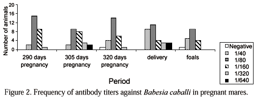
All animals were positive to B. equi at 290 days of pregnancy and the highest titters observed were 1:640 (Fig. 1). Two animals were negative to B.caballi and the highest titters observed were 1:320 (Fig. 2).
Antibody titters remained relatively high until the end of pregnancy, which had duration of 330 days. Nevertheless, antibody titters to both species decreased considerably at the moment of delivery (Fig. 3). This decrease was significant (P<0.01) only to antibody titters against B. equi.
All foals became positive to B. equi after ingestion of colostrum, with higher frequency of serological titers between 1:80 and 1:160 (Fig. 1).
From a total of 31 foals examined, 13 (42%) were negative to B. caballi at 36 hours after birth (Fig. 2).
The complement fixation test (CF) still remains the official test to detect positive animals, however it is less sensitive than the IFAT (Tenter & Friedhoff, 1986). Therefore the CF is unsuitable for epidemiological studies.
As Brazil is considered an endemic area, the high number of positive mares were not surprising and indicate that transmission is very efficient with probably abundant vectors. Under these conditions outbreaks of clinical disease in adults are not expected.
It was concluded that in endemic conditions there is no influence of levels of antibodies in pregnant mares on the passive transfer of antibodies. In addition, the passive transfer of antibodies against B. caballi is less efficient than that against B. equi, therefore young animals are at higher risk of acquiring this infection.
Keywords: Babesia equi, Babesia caballi, pregnant mares, antibodies
RESUMO
O objetivo do presente estudo foi determinar a dinâmica da produção de anticorpos anti-Babesia equi e B. caballi em éguas gestantes em condições naturais de campo em Minas Gerais e avaliar a eficiência da transferência passiva de anticorpos via colostro. Foram analisadas, pela imunofluorescência indireta, amostras de soros colhidas de 34 fêmeas aos 290, 305 e 320 dias de gestação, e no momento do parto, e de seus respectivos potros até 36 horas após o nascimento. Aos 290 dias de gestação, todas as fêmeas estavam positivas para B. equi e apenas duas estavam negativas para B. caballi. Todos os potros tornaram-se positivos para B. equi após ingestão de colostro, entretanto 42% permaneceram negativos para B. caballi 36 horas após o nascimento. Não se observou influência dos níveis de anticorpos durante a gestação na transferência passiva de anticorpos aos potros. A transferência passiva foi menos eficiente para B. caballi, sugerindo que os potros estão mais sujeitos a adquirirem infecção por esse agente.
Palavras chave: Égua, Babesia equi, Babesia caballi, gestação, anticorpo
ACKNOWLEDGMENTS
The authors thank Mr. A. F. Silva Neto for providing the serum samples and Ms. L. Marangunich for the statistical assistance.
REFERENCES
- BÖSE, R., DAEMEN, K. Demonstration of the humoral immune response of horses to Babesia caballi by Western blotting. Int. J. Parasitol, v.22, p.627-630, 1992.
- DONNELLY, J., PHIPPS, L.P., WATKINS, K.L. Evidence of maternal antibodies to Babesia equi and Babesia caballi in foals of seropositive mares. Equine Vet. J., v.14, p.126-128, 1982.
- GUIMARÃES, A.M., LIMA, J.D., RIBEIRO, M.F.B. et al. Ultrastructure of sporogony in Babesia equi in salivary glands of adult female Boophilus microplus ticks. Parasitol. Res, v.84, p.69-74, 1998a.
- GUIMARÃES, A.M., LIMA, J.D., RIBEIRO, M.F.B. Sporogony and experimental infection of Babesia equi by Boophilus microplus ticks. Parasitol. Res., v.84, p.323-327, 1998b.
- HEUCHERT, C.M.S., GIULLI JR., V., ATHAIDE, D.F. et al. Seroepidemiologic studies on Babesia equi and Babesia caballi infections in Brazil. Vet. Parasitol., v.85, p.1-11, 1999.
- LEWIS, B., PENZHORN, B., ALLSOPP, P. Transplacental transmission of Babesia (Theileria?) equi in horses: the rule rather than exception? Fifth Biennial Conference STVM, Proceedings..., Key West, p.113, 1999.
- PFEIFER BARBOSA, I., BÖSE, R., PEYMANN, B. et al. Epidemiological aspects of equine babesioses in a herd of horses in Brazil. Vet. Parasitol, v.58, p.1-8, 1995.
- RIBEIRO, M.F.B., SAITO, J.F., PIMENTEL, P.V. Babesiose equina. I. Primo-infecção de potros em área endêmica. Arq. Bras. Med. Vet. Zootec, v.47, p.641-647, 1995.
- ROBY, T.O., ANTHONY, D.W. Transmission of equine piroplasmosis by the tropical tick, Dermacentor nitens (Newman). J. Am. Vet. Med. Assoc., v.142, p.768-769, 1963.
- TENTER, A.M., FRIEDHOFF, K.F. Serodiagnosis of experimental and natural Babesia equi and B. caballi infections. Vet. Parasitol., v.20, p.49-61, 1986
Publication Dates
-
Publication in this collection
05 Apr 2001 -
Date of issue
Dec 1999
History
-
Received
30 Apr 1999