Abstracts
The use of ethyl-cyanoacrylate and octyl-cyanoacrylate were clinically and histopathologically compared on the corneas of 36 rabbits after lamellar keratectomy (standardized diameter and depth). The animals were distributed into two groups, one for each type of adhesive. From each group, six subgroups were histopathologically evaluated on the 3rd, 7th, 14th, 21st, 30th, and 60th day post-operative. General (daily) and ophthalmic (days 0, 1, 3, 5, 7, 14, 21, 30, 44, and 60) evaluations clinically indicated that there were significant differences for the variables water intake, attitude, blepharitis, corneal edema, and fluorescein test. The adhesive permanence time for octyl-cyanoacrylate (17.22 days) was greater than that for ethyl-cyanoacrylate (7.66 days). With respect to the histopathological evaluation, corneal epithelization and collagen organization occurred without severe complications. However, treatment with ethyl-cyanoacrylate led to a moderate inflammatory reaction in the initial phases. With octyl-cyanoacrylate, re-epithelization and collagen organization proceeded more slowly with a discrete inflammatory reaction in the initial phases. From clinical and histopathologic points of view, octyl-cyanoacrylate showed advantages over ethyl-cyanoacrylate, whereas wound healing was achieved in both groups without major complications.
rabbit; cornea; cyanoacrylate
Comparou-se o uso do etil-cianoacrilato e do octil-cianoacrilato em córneas de 36 coelhos após ceratectomia lamelar (diâmetro e profundidade padronizados). Os animais foram distribuídos em dois grupos, segundo o tipo de adesivo, e redistribuídos em seis subgrupos com três animais cada, para as avaliações histológicas aos 3, 7, 14, 21, 30 e 60 dias de pós-operatório. As avaliações clínicas gerais (diárias) e as oftálmicas (dias 0, 1, 3, 5, 7, 14, 21, 30, 44 e 60), indicaram diferença entre os dois grupos, quanto ao consumo de água, atitude, blefarite, edema da córnea e teste da fluoresceína. O Tempo de permanência, sobre o leito corneal, do adesivo octil-cianoacrilato (17,22 dias), foi maior que o do etil-cianoacrulato (7,66 dias). A histopatologia, para ambos os grupos, mostrou que a re-epitelização e a organização do colágeno ocorreram sem graves intercorrências. O grupo tratado com o etil-cianoacrilato apresentou, nas fases iniciais, reação inflamatória mais evidente que o tratado com octil-cianoacrilato. Neste, a re-epitelização e a organização do colágeno ocorreram mais lentamente e com reação inflamatória discreta. Sob os pontos de vista clínico e de avaliação histológica simples, os resultados mostraram vantagens do octil-cianoacrilato, entretanto, a cicatrização da córnea ocorreu em ambos os grupos.
coelho; córnea; cianoacrilato
VETERINARY MEDICINE
Effects of ethyl-cyanoacrylate and octyl-cyanoacrylate on experimental corneal lesions in rabbits
Efeitos do etil-cianoacrilato ou do octil-cianoacrilato sobre lesões corneais experimentais em coelhos
V.T. BarbosaI; R. ThiesenI; E.G. SoaresII; M.R.F. MachadoI; J.L. LausI,* * Autor para correspondência ( corresponding author) E-mail: jllaus@fcav.unesp.br
IFaculdade de Ciências Agrárias e Veterinárias - UNESP Via de Acesso Prof. Paulo Donato Castellane, s/n 14884-900 Jaboticabal, SP
IIFaculdade de Medicina de Ribeirão Preto - USP Ribeirão Preto, SP
ABSTRACT
The use of ethyl-cyanoacrylate and octyl-cyanoacrylate were clinically and histopathologically compared on the corneas of 36 rabbits after lamellar keratectomy (standardized diameter and depth). The animals were distributed into two groups, one for each type of adhesive. From each group, six subgroups were histopathologically evaluated on the 3rd, 7th, 14th, 21st, 30th, and 60th day post-operative. General (daily) and ophthalmic (days 0, 1, 3, 5, 7, 14, 21, 30, 44, and 60) evaluations clinically indicated that there were significant differences for the variables water intake, attitude, blepharitis, corneal edema, and fluorescein test. The adhesive permanence time for octyl-cyanoacrylate (17.22 days) was greater than that for ethyl-cyanoacrylate (7.66 days). With respect to the histopathological evaluation, corneal epithelization and collagen organization occurred without severe complications. However, treatment with ethyl-cyanoacrylate led to a moderate inflammatory reaction in the initial phases. With octyl-cyanoacrylate, re-epithelization and collagen organization proceeded more slowly with a discrete inflammatory reaction in the initial phases. From clinical and histopathologic points of view, octyl-cyanoacrylate showed advantages over ethyl-cyanoacrylate, whereas wound healing was achieved in both groups without major complications.
Keywords: rabbit, cornea, cyanoacrylate
RESUMO
Comparou-se o uso do etil-cianoacrilato e do octil-cianoacrilato em córneas de 36 coelhos após ceratectomia lamelar (diâmetro e profundidade padronizados). Os animais foram distribuídos em dois grupos, segundo o tipo de adesivo, e redistribuídos em seis subgrupos com três animais cada, para as avaliações histológicas aos 3, 7, 14, 21, 30 e 60 dias de pós-operatório. As avaliações clínicas gerais (diárias) e as oftálmicas (dias 0, 1, 3, 5, 7, 14, 21, 30, 44 e 60), indicaram diferença entre os dois grupos, quanto ao consumo de água, atitude, blefarite, edema da córnea e teste da fluoresceína. O Tempo de permanência, sobre o leito corneal, do adesivo octil-cianoacrilato (17,22 dias), foi maior que o do etil-cianoacrulato (7,66 dias). A histopatologia, para ambos os grupos, mostrou que a re-epitelização e a organização do colágeno ocorreram sem graves intercorrências. O grupo tratado com o etil-cianoacrilato apresentou, nas fases iniciais, reação inflamatória mais evidente que o tratado com octil-cianoacrilato. Neste, a re-epitelização e a organização do colágeno ocorreram mais lentamente e com reação inflamatória discreta. Sob os pontos de vista clínico e de avaliação histológica simples, os resultados mostraram vantagens do octil-cianoacrilato, entretanto, a cicatrização da córnea ocorreu em ambos os grupos.
Palavras-chave: coelho, córnea, cianoacrilato
INTRODUCTION
The cornea, because of its functions, is one of the group of structures most studied with regard to the vision apparatus (Samuelson, 1999). Deep ulcerations and traumatic perforations are among the illnesses of greatest morbidity that affects it.
Because of the need for sophisticated equipment, highly capable professionals, and training in micro-surgery, the use of classical techniques in keratoplasty is limited to specialized centers. Therefore, the study and development of new procedures that are simple, effective, and easy to perform are daily challenges.
Adhesive tissues, tested since the 1960s, are defined as polymerizeable substances with the capacity to keep tissues together or to cause a barrier against extravasation. These are materials that have undergone development and currently offer biocompatibility and efficacy (Reece et al., 2001). Such desirable characteristics are notably: provide local action for the period necessary for their effective function; degrade without the production of fragments; adhere to surfaces at body temperature; create a strong, flexible bridge between tissues; tolerate moisture; and have the capacity to spread out (Donkerwolke et al., 1998; Reece et al., 2001).
Cyanoacrylates belong to a group of materials derived from cyanoacrylic acid. Their liquid monomers solidify by polymerization after contact with a weak base released by a little heat (Watte et al., 2004). It is known that the biocompatibility of cyanoacrylates is strongly related to the number of carbons present in its side chain, and that the longer it is, the less tissue reaction will be expected (Woodward et al., 1965; Pani et al., 1968; Dellevigne et al., 1971; Vote and Elder, 2000).
Greater tissue reaction after subcutaneous implantation of short-chain cyanoacrylates was due to the release of toxic substances resulting from their degradation, notably cyanoacetate and formaldehyde. Difference related to tissue vascularization, variable among tissues, also equally contributes to the occurrence of such events (Trott, 1997).
Infiltration of eosinophils and neutrophils after the application of cyanoacrylates has been reported in different tissues like muscle (Woodward et al., 1965), subcutaneous (Pani, et al., 1968), and equine intestine (Duarte et al., 2007). The occurrence of severe infiltrate inflammation was also reported by Laus et al. (1993) when using a gel adhesive in healthy corneas of dogs.
In human ophthalmology, there are reports describing the application of cyanoacrylates in practice, with satisfactory results (Boruchoff et al., 1969; Hirst et al., 1982; Taravella and Chang, 2001; Setlik et al., 2005). However, in veterinary ophthalmology, the lack of technical knowledge of these products, the little experience with regard to their manipulation, and the costs involved have limited their utilization.
The aim of this study was to compare the clinical and histopathological results between ethyl-cyanoacrylate and octyl-cyanoacrylate when applied in lamellar keratectomy in rabbits.
MATERIAL AND METHODS
Thirty-six adult male New Zealand rabbits were used. They were 120-day-old and averaged 3kg b.w. The study rigorously followed the bioethical guidelines of the Association for Research in Vision and Ophthalmology. After a clinical and ophthalmic examination based on Shirmer's lacrimal test,1 1 Schirmer's test - Ophthalmos, Brazil. slit-lamp biomicroscopy2 2 Slit Lamp SL-14 - Kowa Company, USA. , and the fluorescein test3 3 Fluorescein strips - Ophthalmos, Brazil. , the animals were kept in individual cages, in a ventilated environment that was clean and dry, where they were fed on commercial ration4 4 Nutricoelho - Purina, Brazil. and water ad libitum.
The adhesives were evaluated in the right eye of the animals, separated into the ethyl-cyanoacrylate5 5 Super Bonder - Loctite, 3M, Brazil. and octyl-cyanoacrylate6 6 Dermabond - Johnson & Johnson, USA. groups. The rabbits from both groups were evenly matched and randomly distributed into six subgroups of three animals, for different intervals for evaluation of the corneas, after the application of the adhesives.
Anesthesia was induced with the use of a combination of ketamine7 7 Cetamin - Syntec, Brazil. and midazolam8 8 Dormire - Cristália, Brazil. , at a dose of 40 and 0.2mg/kg, respectively, intramuscularly administered. It was maintained by inhalational halothane9 9 Tanohalo - Cristália, Brazil. , supplied with face mask, in a semi-closed circuit. Immediately before lamellar keratectomy, two drops of tetracaine anesthetic with 0.1% phenylephrine were instilled10 10 Colírio Anestésico - Allergan, Brazil. .
The surgical procedures were carried out using a surgical microscope11 11 [1]M-9 - DF Vasconcelos, Brazil. , at 10 times magnification. After routine antiseptic measures, protection of the operating field, blepharostasis, and fixation of the eyeball; a lamellar button was created by 3mm in diameter trepanning12 12 Perforator for cornea with deepness measurer - Steel Inox, Brazil. , composed of epithelium and about half the thickness of the stroma in the central region of the cornea. It was excised with an angled scalpel13 13 CM Phaco Slit Blade Angled 5.2mm; 45 Deg.; Bevel up - Eagle, USA. and in its place, one of the two adhesives was deposited using a syringe14 14 1mL/cc insuline U-100 - Injex, Brazil. with a hypodermic needle15 15 0.70x25 22 G1 - BD, Brazil. . The area was then covered with a 3mm diameter cellulose sterile film16 16 Bionext - Bionext Produtos Biotecnológicos Ltda., Brazil. , for the purpose of creating a smooth surface over the lesion.
In the postoperative period, 0.3% tobramycin17 17 Tobramycin 0.3%, ophtalmic solution - Cristália, Brazil. was instilled every 6h for 10 days and systemic analgesia was subcutaneously administered with buprenorphine18 18 Tengesic - Schering Plough, Brazil. , at a dose of 0.02mg/kg, at intervals of 12 hours, for five consecutive days. The animals were fitted with an Elisabethan collar.
In the postoperative period, ophthalmic evaluations were performed on days 1, 3, 5, 7, 10, 14, 21, 30, 45, and 60, at night, and consisted of Shirmer's lacrimal test, determination of the degree of blepharospasm, ocular discharge, conjunctival hyperemia and chemosis, and fluorescein test. Parameters related to corneal conditions (edema, vascularization, and pigmentation) and to the anterior uvea were evaluated. In addition, in the first 10 days of the postoperative period, on a daily basis and in the morning, the animals were observed for behavior, and food and water consumption. Retention of the adhesives on the cornea was daily evaluated in the morning. Scores were used for data compilation and statistical study with regard to each evaluated event (Table 1).
At the end of the each histopathological study period, the animals were sacrificed with letal dose of barbiturate19 19 Thiopentax - Cristália, Brazil. (100mg/kg) intravenously applied. Subconjunctival enucleation was performed and the eyes were kept in modified Karnovsky fixative for histological examination. The used stains were hematoxylin-eosin and Masson's trichrome. The preparations were analyzed and the most important events documented by photomicroscopy20 20 DM 5000B - Leica, Germany. .
The experimental design was based on the factorial arrangement of two treatments x six periods with three repetitions (36 animals). The data for the different studied parameters were examined by analysis of variance and the means were compared by a t-test, at 5% level of significance, utilizing generalized linear models and chi-squared test on Statistical Analysis System.
RESULTS
Table 2 presents the means determined for the variables behavior, and food and water consumption for the two treatments. Animals that received octyl-cyanoacrylate were more alert and consumed more water (P<0.05).
The lacrimal production showed a mean baseline level (day 0) of 6.2±0.6mm/min. There was no significant difference between the treatments. Ethyl-cyanoacrylate showed a mean of 8.55mm/min and octyl-cyanoacrylate a mean of 9.04mm/min, but these values did significantly differ from the baseline value.
In the postoperative period, no significant differences were found between the two treatments with regard to extent of blepharospasm, ocular discharge, conjunctival hyperemia, and chemosis. About the clinical postoperative evaluation, there was a significant increase on the first day for all the variables, with a tendency for their normalization in the course of the postoperative period and return to baseline values by day 45. Ocular discharge, predominantly of the mucoid and/or seromucoid type, was observed in all the animals, with the greatest intensity on days one and three postoperative.
Corneal edema was peri-lesional and discrete. This was seen on the first day postoperative in 61% of the rabbits that received ethyl-cyanoacrylate. At three days, 11% of the animals of this group still manifested edema. In those that received octyl-cyanoacrylate, corneal edema was found in only 16% of the animals in the first day postoperative. At the fifth day postoperative, no more edema was observed in animal from both groups.
Uveitis occurred in animals from both groups and it was considered discrete when there was observed just iris hyperemia. When this signal was accomplished with edema and miosis, the uveitis was considered moderate. There was no significant difference in mean frequency of anterior uveitis between treatments with octyl-cyanoacrylate (m = 0.75) and ethyl-cyanoacrylate (m = 0.76).
The fluorescein test was positive in 61% of the animals of the ethyl-cyanoacrylate group on day one, 3% on day three, 2% on days five and seven, and 11% on day 10. With regard to octyl-cyanoacrylate, 33% of the animals showed a positive result on the first day postoperative and 5% on the second. There was a significant difference in the mean frequency of positive fluorescein test between the octyl-cyanoacrylate (m = 0.72) and ethyl-cyanoacrylate groups (m = 0.79) (P<0.05).
The estimated means (for all the periods) for retention time of the adhesive on the cornea were 17.22 days for the octyl-cyanoacrylate group and 7.66 days for the ethyl-cyanoacrylate group. There was a significant difference between the two treatments (P<0.05). The observed means (simple means for each time. each period) are presented in Table 3.

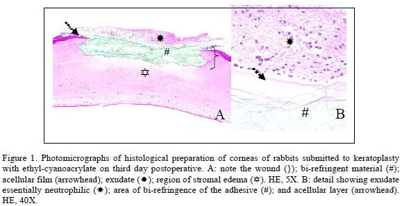
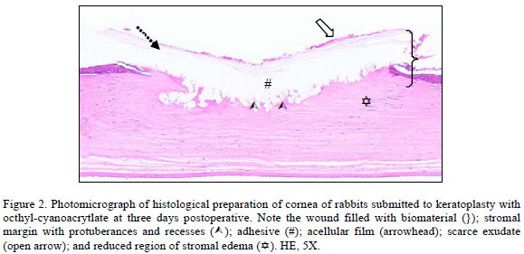
In corneas evaluated at three days postoperative, the treatment with ethyl-cyanoacrylate showed a fracture of the adhesive corresponding to a third of the corneal thickness, which was filled with fragmented be-refringent material, covered by an acellular layer on which there was a neutrophilic exudate; the corneal stroma on the adhesive was shown to be edemous (Fig. 1). Treatment with octyl-cyanoacrylate showed a fractured space, which was filled by adhesive, appeared transparent, with protuberances and recesses at the stromal edges, suggesting possible penetration of the biomaterial. Scarce exudate rested on an acellular layer that covered the biomaterial. A reduced area of stromal edema was demonstrated on the adhesive (Fig. 2).
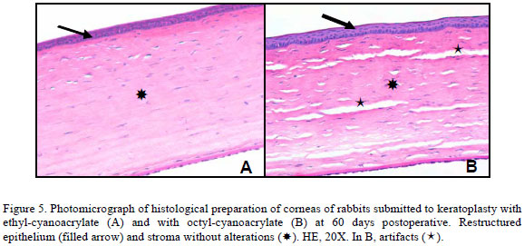
At seve days postoperative, animals from both groups exhibited hyperplasia and hypertrophy of the germinative cells of the epithelium on the edges of the lesion, which had migrated over the lesioned stroma. In the animals from ethyl-cyanoacrylate group, there was a mixed inflammatory infiltrate represented by polymorphonuclear (neutrophils) and phagocytic mononuclear cells. At 14 days postoperative, the corneas of the animals from ethyl-cyanoacrylate group showed complete re-epithelization, while the stroma, still disorganized, was permeated by various keratocytes and the neo-epithelium still displayed cellular hyperplasia (Fig. 3). Attenuation of hyperplasia and organization of collagen fibers were gradually seen in subsequent evaluations (21 and 30 days postoperative). In the animals from octyl-cyanoacrylate group, at 14, 21, and 30 days, there was a slow migration of hyperplastic epithelial cells and moderate number of inflammatory cells (neutrophils), intact and in a state of degeneration, on the detached portions of the adhesive. The stroma, despite remaining relatively organized, showed a discrete concentration of keratocytes in the portions immediately subjacent to the adhesive (Fig. 4). By the 60th day postoperative, the animals from both groups showed complete re-epithelization and organized stroma (Fig. 5).
DISCUSSION
The evaluations relative to food and water consumption and behavior were devised for the purpose of identifying some indication of ocular pain, since the cornea has nociceptive innervation on its surface. According to Johnston (2005), the ingestion of food and water, and consequently body weight, tend to decrease notably in cases of pain. Regarding water consumption and behavior, it can suggest that octyl-cyanoacrylate induced less discomfort.
The mean baseline value for the Shirmer lacrimal test was 6.20.6 mm/min. The result is similar to that reported by Abrams et al. (1990), who evaluated 142 healthy rabbits and obtained a mean of 5.32.96. The increase observed in both treatments which did not significantly differ, is believed to be due to conjunctival and corneal irritation after the surgery procedure.
About blepharospasm, ocular discharge, hyperemia, and chemosis, the findings were similar to those of Ollivier et al. (2001), who tested butyl-cyanoacrylate in rabbit corneas and showed a significant increase on the first day postoperative, with gradual decline to levels not significantly different than baseline levels. According to these authors, the tendency toward normalization is due to the fact that the surface over the adhesive becomes less irregular with time, causing less discomfort. Using butyl-cyanoacrylate in dogs, Bromberg (2002) and Watte et al. (2004) observed discrete blepharospasm which disappeared over a course of two to three days. In human patients, protection with a contact lens reduced the occurrence of such symptoms (Taravella and Chang, 2001; Feldberg et al., 2003; Setlik, 2005).
Peri-lesional corneal edema was observed on the first day postoperative in 61% of the animals treated with ethyl-cyanoacrylate and in 10% of those who received octyl-cyanoacrylate. These observations suggest that octyl-cyanoacrylate produced a better impermeabilization of the cornea in the lesioned areas. Mota et al. (2004) showed less occurrence of edema in lesions treated with butyl-cyanoacrylate compared to the sutured cornea. According to the authors, the adhesive provided better apposition between the edges of the lesion, impermeabilizing it and aiding in stromal re-organization.
Feldberg et al. (2003) studied ethyl-cyanoacrylate in 22 human patients, to whom a second application was needed in 50% of the cases. This group found a significant difference between treatments in the fluorescein test. Such a finding, together with corneal edema, indicated that octyl-cyanoacrylate provided better plasticity and adherence to the tissue receptor.
The uveitis identified in both groups, did not show significant differences and was a reflex response to corneal irritation. Gasset et al. (1970) demonstrated minor iritis after intracorneal application of butyl-cyanoacrylate in rabbits. In humans, the utilization of therapeutic contact lenses in the postoperative period has been shown to minimize mechanical irritation and consequently reflex uveitis.
With respect to the mechanical function of adhesives as a temporary tectonic procedure, there was a difference between the estimated mean (which involved all the periods) and the observed mean (simple mean for each individual period). For the treatment with ethyl-cyanoacrylate, the estimated mean retention time was 7.66 days. When considering only the period of 60 days, the mean was 11.6 days. This finding is in contrast to that of Feldberg al. (2003), who found a variation of three days to five months in humans. The difference among species, size and depth of the lesions, as well as the utilization of therapeutic contact lenses which limit friction by the eyelids on the adhesive could have accounted for the differences. With regard to octyl-cyanoacrylate, the estimated mean was 17.22 days and that observed at 60 days was 32 days. Bromberg (2002) reported a mean of 247 days in a study on the use of butyl-cyanoacrylate in the treatment of refractory ulcers in dogs. Watte et al. (2004), in conditions of clinical application in dogs, found values varying form one week to six months. Mota et al. (2004) reported no detachment of butyl-cyanoacrylate for up to 30 days of observation in rabbits.
To make use of a corneal adhesive, it is necessary that it remain until tissue repair is complete. The fact that octyl-cyanoacrylate remains on the corneal surface 2.5 times longer than ethyl-cyanoacrylate, together with the significant difference in fluorescein test, demonstrates the advantages of the octyl-cyanoacrylate.
With regard to histopathology, the presence of a considerable amount of exudate, predominantly neutrophilic, on the acellular film on the third day postoperative in the animals treated with ethyl-cyanoacrylate, resulted in an acute inflammatory response of the conjunctiva, as a reflex of corneal irritation. In the group that received octyl-cyanoacrylate, a very slightly amount of neutrophilic exudate was found. Inflammatory infiltrate is a common report when cyanoacrylates (Woodward et al., 1965; Pani, et al., 1968; Duarte et al., 2007) or other types of adhesives were used in tissues (Laus et al., 1993).
Differences were found with respect to the appearance and bi-refringence of the adhesives by light microscopy. Ethyl-cyanoacrylate showed little advantage due to elevated bi-refringence and blotchy appearance. According to Reece et al. (2001), the adhesive should degrade without leaving fragments. Octyl-cyanoacrylate expressed minimal bi-refringence and showed no fragments. It was found to be interwoven with and fixed to the corneal stroma, with protuberance and recesses. Donkerwolke et al. (1998) reported the importance of a strong and flexible bridge to allow tissue migration.
Hyperplasia and hypertrophy of the epithelial cell layers, in migration, separated the adhesive from the corneal stroma in both treatments by the seventh day postoperative. Similar results were described in rabbits by Gasset et al. (1970) and Mota et al. (2004), after the corneal application of cyanoacrylates. For the same period, inflammatory infiltrate containing neutrophils and some macrophages in the subjacent stromal portion was found in the animals that received ethyl-cyanoacrylate.
Two weeks after the application of the adhesives, hypertrophy and cellular hyperplasia were present in animals from both groups. In the treatment with ethyl-cyanoacrylate, re-epithelization of the lesion was evident. The subjacent stroma showed intense fibroplasia and substantial disorganization. In animals from octyl-cyanoacrylate group, hyperplasia and hypertrophy were observed with epithelial migration of considerably less intensity. There was organization of the stromal collagen fibers which were found to be permeated by new keratocytes. These findings are similar to those reported by Vote and Elder (2000) describing the slow degradation of octyl-cyanoacrylate.
Histopathological changes at 21 and 30 days postoperative for ethyl-cyanoacrylate included stromal organization with a gradual decrease in intensity of epithelial hyperplasia and hypertrophy, and keratocytes infiltrating the stroma undergoing repair. In the group of animals that received octyl-cyanoacrylate, at 21 and 30 days postoperative, the observation of inflammatory cells, intact and undergoing degeneration at the interface between adhesive and tissue, already noted, and of the re-structured epithelium, emphasizes the necessity of monitoring individuals who are treated with slow degrading cyanoacrylates, in light of the possibility of development of infections. Such reservations were also noted by Gasset et al. (1970), Vote and Elder (2000), and Bromberg (2002).
At 60 days postoperative, corneal re-structuring was seen in both groups, without differences with respect to cellular pattern. Gasset et al. (1970) and Ollivier et al. (2001) reported similar findings in experimental studies of cyanoacrylates.
CONCLUSION
It is concluded that octyl-cyanoacrylate is more advantageous than ethyl-cyanoacrylate in the repair of lesioned corneas, whereas both adhesives showed final satisfactory results when performed with the objective of an emergency tectonic coverage for this clinical situation.
ACKNOWLEDGMENTS
The authors thank the Conselho Nacional de Desenvolvimento Cientifico and Tecnologico (CNPq) for the financial support and collaborators Marco Augusto Giannoccaro da Silva, Bianca da Costa Martins, Alexandre Pinto Ribeiro, Ana Leticia de Souza, and Vera Cippoli Musci for their help during the course of this work. The authors also grateful to Prof. Dr. Antônio Augusto Coppi Maciel Ribeiro and Dr. Pedro Franklin Barbosa for the valuable collaboration. Dr. Albert Leyva assisted with the translation of the manuscript.
Recebido em 10 de dezembro de 2008
Aceito em 31 de agosto de 2009
- ABRAMS, K.L.; BROOKS, D.E.; FUNK, R.S. et al Evaluation of the Shirmer tear test in clinically normal rabbits. Am. J. Vet. Res., v.51, p.1912-1913, 1990.
- BORUCHOFF, S.A.; REFOJO, M.; SLANSKY, H.H. et al. Clinical applications of adhesives in corneal surgery. Trans. Am. Acad. Ophthalmol. Otolaryngol, v.73, p.499-505, 1969.
- BROMBERG, N.M. Cyanoacrylate tissue adhesive for treatment of refractory corneal ulceration. Vet. Ophthalmol., v.5, p.55-60, 2002.
- DELLEVIGNE, W.; WOLFERTH, C.C.; JONES, N. et al. Cyanoacrylate monomers as an adhesive. Arch. Surg, v.102, p.493-495, 1971.
- DONKERWOLCKE, M.; BURNY, F.; MUSTER, D. Tissues and bone adhesives historical aspects. Biomaterials, v.19, p.1461-1466, 1998.
- DUARTE, C.A.; CATTELAN, J.W.; LUCAS, F.A. et al. Aspectos morfométricos da cicatrizaçăo do cólon descendente de eqüinos submetidos a enterorrafias aposicionais com poliglactina 910 e com cianoacrilato. Arq. Bras. Med. Vet. Zootec, v.59, p.49-55, 2007.
- FELBERG, S.; LAKE, J.C.; LIMA, F.A. et al. Adesivo de cianoacrilato no tratamento de afinamentos e perfuraçőes corneais: técnica e resultados. Arq. Bras. Oftalmol, v.66, p.345-349, 2003.
- GASSET, A.R.; HOOD, C.I.; ELLISON, E.D. et al. Ocular tolerance to cyanoacrylate monomer tissue adhesive analogues. Invest. Ophthalmol, v.9, p.3-11, 1970.
- HIRST, L.W.; SMIDDY, W.E.; STARK, W.J. Corneal perforations: changing methods of treatment, 1960-1980. Ophthalmology, v.89, p.630-635, 1982.
- JOHNSTON, M.S. Clinical approaches to analgesia in ferrets and rabbits. Top. Med. Surg, v.14, p.229-235, 2005.
- LAUS, J.L.; ROSSI, M.A.S.; SOUZA, M.S.B. et al. Avaliaçăo dos efeitos de um novo adesivo para fins biológicos (Colagel) na ceratoplastia experimental em căes. Braz. J. Vet. Res. Anim. Sci, v.30, suppl., p.183-193, 1993.
- MOTA, F.C.D.; EURIDES, D.; FREITAS, P.M.C. et al. Use of the n-butyl cyanoacrylate adhesive and the polyglactine thread suture for corneal rhaphy in rabbit (Oryctolagus cunicullus). J. Vet. Sci, v.5, p.267-270, 2004.
- OLLIVIER, F.; DELVERDIER, M.; REGNIER, A. Tolerante of the rabbit cornea to an n-butyl-ester cyanoacrylate adhesive (Vetbond). Vet. Ophthalmol, v.4, p.261-266, 2001.
- PANI, K.C.; GLADIEUX, G.; BRANDES, G. et al. The degradation of N-butil-alpha-cianoacrolato tissue adhesive. Surgery, v.63, p.481-489, 1968.
- REECE, T.B.; MAXEY, T.S.; KRON, I.L. A prospectus on tissue adhesives. Am. J. Surg., v.182, p.40-44, 2001.
- SAMUELSON, D.A. Ophthalmic Anatomy. In: GELATT, K.N. Veterinary ophthalmology, 3.ed. Baltimore: Lippincott Williams & Wilkins, 1999. cap.2, p.31-150.
- SETLIK, D.E.; SELDOMRIDGE, D.L.; ADELMAN, R.A. et al. The effectiveness of isobutyl cyanoacrylate tissue adhesive for the treatment of corneal perforations. Am. J. Ophtahlmol., v.140, p.920-921, 2005.
- TARAVELLA, M.J.; CHANG, C.D. 2-octilcyanoacrylate medical adhesive in treatment of a corneal perforation. Cornea, v.20, p.220-221, 2001.
- TROTT, A.T. Cyanoacrylate tissue adhesives an advance in wound care. J. Am. Med. Assoc., v.277, p.1559-1560, 1997.
- VOTE, B.J.T.; ELDER, M.J. Cyanoacrylate glue for corneal perforations: a description of a surgical technique and a review of the literature. Clin. Exp. Ophthalmol, v.28, p.437-442, 2000.
- WATTÉ, C.M.; ELKS, R.; MOORE, D.L. et al. Clinical experience with butyl-2-cyanoacrylate adhesive in the management of canine and feline corneal disease. Vet. Ophthalmol, v.7, p.319-326, 2004.
- WOODWARD, S.C.; HERRMAN, J.B.; CAMERON, J.L. et al. Histotoxicity of cyanoacrylate tissue adhesive in the rat. Ann. Surg, v.162, p.113-122, 1965.
Publication Dates
-
Publication in this collection
23 Nov 2009 -
Date of issue
Oct 2009