Abstracts
Morphological investigations were conducted on four bodies of dogs who died due to severe clinical symptoms following a massive invasion of cardiac and pulmonary Dirofilaria. The subjects were monitored clinically and diagnosed serologically positive for the Heartworm disease. The necropsy examination of the cardiovascular system (right ventricle and pulmonary artery) revealed the presence of 25 adult parasites in one dog with length ranging between 8 and 33cm. Macroscopically, lesions consistently observed were represented by the right ventricular dilatation and the diffuse wall thickening of the pulmonary artery. Parasitic invasion secondary lesions were present in the lungs, liver and kidneys (cardiac and vascular lesions). The histological examination mainly revealed myocardial injury, vascular (dystrophic), pulmonary (circulatory and inflammatory), hepatic (degenerative) and renal (degenerative and inflammatory) damage.
dog; injury; Dirofilaria immitis
Investigações morfológicas foram realizadas em quatro cadáveres de cães, mortos em razão de graves sintomas clínicos atribuídos à invasão massiva cardíaca e pulmonar por Dirofilaria immitis. Os indivíduos foram acompanhados sob o ponto de vista clínico, sendo diagnosticados serologicamente positivos para dirofilariose. À necropsia do sistema cardiovascular (ventrículo direito e artéria pulmonar,) revelou-se em um indivíduo a presença de 25 parasitas adultos, com comprimento entre 8-33 centímetros. Macroscopicamente, as lesões observadas foram a dilatação do ventrículo direito e o espessamento difuso da parede da artéria pulmonar. Lesões secundárias à invasão parasitária estavam presentes nos pulmões, no fígado e nos rins (lesões cardíacas e vasculares). O exame histológico revelou, principalmente, lesões do miocárdio, lesões vasculares (distróficos) e pulmonares (circulatórias e inflamatórias), mas também no fígado (degenerativas) e nos rins (degenerativas e inflamatórias).
cão; lesão; Dirofilaria immitis
VETERINARY MEDICINE
Vascular, hepatic and renal lesions by Dirofilaria immitis invasion in dogs
Lesões vasculares, hepáticas e renais na invasão por Dirofilaria immitis no cão
S.A. Pasca; D. Acatrinei; O.Z. Oprean; M. Lazar
Faculty of Veterinary Medicine - "Ion Ionescu de la Brad" University - Iasi, 8, M. Sadoveanu Alley, 700490, Romania
ABSTRACT
Morphological investigations were conducted on four bodies of dogs who died due to severe clinical symptoms following a massive invasion of cardiac and pulmonary Dirofilaria. The subjects were monitored clinically and diagnosed serologically positive for the Heartworm disease. The necropsy examination of the cardiovascular system (right ventricle and pulmonary artery) revealed the presence of 25 adult parasites in one dog with length ranging between 8 and 33cm. Macroscopically, lesions consistently observed were represented by the right ventricular dilatation and the diffuse wall thickening of the pulmonary artery. Parasitic invasion secondary lesions were present in the lungs, liver and kidneys (cardiac and vascular lesions). The histological examination mainly revealed myocardial injury, vascular (dystrophic), pulmonary (circulatory and inflammatory), hepatic (degenerative) and renal (degenerative and inflammatory) damage.
Keywords: dog, injury, Dirofilaria immitis
RESUMO
Investigações morfológicas foram realizadas em quatro cadáveres de cães, mortos em razão de graves sintomas clínicos atribuídos à invasão massiva cardíaca e pulmonar por Dirofilaria immitis. Os indivíduos foram acompanhados sob o ponto de vista clínico, sendo diagnosticados serologicamente positivos para dirofilariose. À necropsia do sistema cardiovascular (ventrículo direito e artéria pulmonar,) revelou-se em um indivíduo a presença de 25 parasitas adultos, com comprimento entre 8-33 centímetros. Macroscopicamente, as lesões observadas foram a dilatação do ventrículo direito e o espessamento difuso da parede da artéria pulmonar. Lesões secundárias à invasão parasitária estavam presentes nos pulmões, no fígado e nos rins (lesões cardíacas e vasculares). O exame histológico revelou, principalmente, lesões do miocárdio, lesões vasculares (distróficos) e pulmonares (circulatórias e inflamatórias), mas também no fígado (degenerativas) e nos rins (degenerativas e inflamatórias).
Palavras-chave: cão, lesão, Dirofilaria immitis
INTRODUCTION
The lesions caused by D. immitis can be found at cardiovascular (Kaiser et al., 2004; Mehlhorn 2008), pulmonary, hepatic (McGavin et al., 2007) and renal (Paes-de-Almeida et al., 2003) levels, and generally consist of right heart dilation, profilerative endarteritis, strokes, myocardial collagen matrix thromboses (Wang et al., 2005) degenerative liver damage and kidney ultrastructure alterations. One of the most serious complications is the Caval syndrome. At its origin, there are the pulmonary artery localizations, due to the retrograde movements of parasites that lesion the valvular apparatus and lead to tricuspid retching, and the stronger they are, the higher the pulmonary pressure is. The result of these disorders is heart failure, in which case the prognosis is extremely reserved and the antiparasitic and symptomatic treatment has uncertain results. The simultaneous death of all helminths leads to the pulmonary artery ramifications embolism and the death of the host.
MATERIAL AND METHODS
The investigation period was October 2010 to January 2011, on four bodies of dogs that have died from severe clinical signs due to Dirofilaria immitis infestation. The dogs were closely monitored, knowing that they are seropositive for infestation with Dirofilaria immitis. The attention was mainly focused on vascular damages caused by the parasite.
Necropsy and histopathological examinations were thus performed. After taking the samples, the pieces were fixed in 10% formaldehyde solution, inserted in paraffin and sectioned in 5µm thickness. We used haematoxylin-eosin-methylene blue stain (Masson Tricromic), Periodic Acid-Schiff Fuxin stain (PAS) and Alcian blue stain.
RESULTS AND DISCUSSION
Lesions were noticed in all organs. They were caused by circulatory disorders produced by severe cardiac and vascular parasitism and by the toxic action of the parasites.
The heart showed a right ventricular dilatation, induced by the presence of adult parasites in the ventricular cavity, also the ventricular wall appears slightly soft and extremely thin. The clipping of the pulmonary artery and of the lobar branches revealed the presence of a large number of Dirofilaria (approx. 6-7 worms) (Fig. 1 and 2).
After opening the right ventricle by an incision in its wall, parallel to the paraconal trench, a bundle of four dirofilaria was found inside, included in a thrombus occupying almost the entire ventricular cavity (Kaiser et al., 2004; Mehlhorn 2008) (Fig. 3). In all examined cadavers, hepatic stasis was present even in the chronic form. The liver was enlarged in volume, blackish red, endured and with an obvious lobular design (Fig. 4). The kidneys were slightly increased in volume, with brown-greyish coloring, sprinkled with small white-greyish outbreaks, with a diameter of 1mm. (Fig. 5).
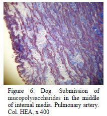
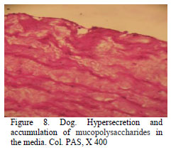
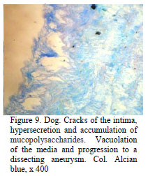
On the histological examination, the largest changes were noticed in the pulmonary artery and lungs. In the media of the pulmonary artery, the presence of acid mucopolysaccharides deposits between the smooth muscle fibres, with fibrillar aspect, weakly basophile in tricromic Masson stainin, PAS-positive and Alcian-positive was observed. The mucopolysaccharides accumulation led to the dissociation of the media and the fragmentation of the smooth muscle fibres. The subendotelial accumulation of mucopolysaccharides sometimes caused the appearance of the dissecting aneurysm with endothelial cleavage and the internal side of the media (Fig. 6, 7, 8, and 9 ).
The external half of the media presented the hyalinisation of muscle fibers and collagenisation (Fig. 10). In the pulmonary artery branches, we could observe the histological endothelial erosion and villous proliferations (like pseudoviles) as a result of the mechanical action of the luminal parasites and the subendotelial migration of miofibroblasts (Fig.11, 12 and 13 ). Pulmonary arterial constriction causes increased flow velocity, especially with exertion, and resultant shear stresses further damage to the endothelium. Previously mentioned parietal lesions imply a high risk of thrombosis and spontaneous hemorrhages.
In the lung, the lesions were more diverse, being a product of the heart failure and the location of intrapulmonary microfilaria. At the intrapulmonary bronchi, we observed the presence of an intraluminal hemorrhagic infiltration caused by the parasitic migrations, the proliferation of the ciliated pseudostratified epithelium and conjunctive peribronchial hyperplasia (McGavin et al., 2007) (Fig. 14). Subpleural pulmonary emphysema and pleural fibrosis were also observed in certain portions of the surface (Fig. 15).
On the lung tissue signs of congestion and subacute-chronic interstitial pneumonia followed by interstitial collagenisations and the sclerosis of the interstitial blood vessels (Fig. 16) was observed.
Microfilaria were also observed in the lung alveoli, interstitial and the septum capillaries (Fig. 17 and 18).
The livers in the four bodies constantly presented dystrophic changes probably due to parasite toxins released into circulation consisting in hydropic hepatoses with hiperhydrated hepatocytes, with picnotic nuclei centrally disposed (McGavin et al., 2007). The chronic venous stasis, the lymphatic stasis, and the perivenous edemas were observed in all cases examined, as a consequence of the right heart failure (Fig. 19, 20 and 21).
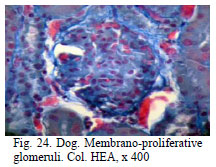
In the kidneys, signs of congestion and granular dystrophy were noted in the uriniferous tubules, in the cortical. The glomerular lesions consisted of membranous and membrano-proliferative glomeruli (Fig. 22, 23 and 24). Glomerular lesions are due to storage and to physical damage of glomerules by viable microphilariae (Paes-de-Almeida et al., 2003). In the renal medulla, the lesions consisted of infiltration with lymphocytes and plasmocytes on the interstitial cells (Fig. 25).
Dirofilaria immitis - associated inflammatory mediators that induce immune responses in the lungs and kidneys (immune complex glomerulonephritis) cause vasoconstriction and bronchoconstriction. Release of plasma and inflammatory mediators from small vessels and capillaries causes parenchymal lung inflammation. The endothelial damage, vasoconstriction, increased flow velocity, and local ischemia is a vicious cycle. Inflammation with ischemia can result in irreversible interstitial fibrosis.
Arterial thrombosis is caused by both blood clots and worms lodged within narrow lumen arterioles.
CONCLUSIONS
The examination of the 4 cases revealed the following conclusions: 1. The acute and chronic congestive lesions of the organs were due to the right heart failure following to the parasitism by Dirofilaria immitis; 2. The parietal arterial lesions were represented by secondary erosions of the mechanical action of parasites, the miofibroblasts hyperplasia and their subendotelial migration, the deposit of mucopolysaccharides mainly in the internal half of the media, with the fragmentation of leukocytes and the emergence of dissected aneurysms; 3. Lung injury represented consistently by microthrombosis, hemorrhagies, emphysema, interstitial pneumonia, interstitial, vascular and pleural sclerosis. Interstitial lung cellular hyperplasia causes a significant barrier to oxygenation. Most significantly due to restricted pulmonary vascular capacity and subsequent pathology. 4. The liver manifested hydrolytic dystrophy due to parasite toxicity, acute and chronic congestion and vascular sclerosis; 5. The kidneys have revealed the presence of membranous and membrano-proliferative glomeruli produced by immune complex deposits caused by systemic parasitism and tubular epithelial dystrophic processes.
ACKNOWLEDGMENTS
The authors thank UEFISCDI Romania for the financial support (Research Program TE 112/2010).
Recebido em 12 de outubro de 2011
Aceito em 16 de abril de 2012
E-mail: passorin@yahoo.com
- KAISER, L.; WILLIAMS, J.F. Dirofilaria immitis: worm burden and pulmonary artery proliferation in dogs from Michigan (United States). Vet. Parasitol, v.124, p.125-129, 2004.
- MEHLHORN, H. (ED) Encyclopedia of Parasitology 3rd Edition. New York: SPRINGER, 2008, p.200.
- McGAVIN, D.M.; ZACHARY, J.F. Pathologic Basis of Veterinary Disease 4nd, ed. St. Louis: Mosby Inc., 2007, p.582-582.
- PAES-DE-ALMEIDA, E.C.; FERREIRA, A.M.R.; LABARTHE, N.V. et al. Kidney ultrastructural lesions in dogs experimentally infected with Dirofilaria immitis (Leidy, 1856). Vet. Parasitol, v.113, p.157-168, 2003
- WANG, J.S.; TUNG, K.C.; HUANG, C.C.; LAI, C.H. Alteration of extracellular collagen matrix in the myocardium of canines infected with Dirofilaria immitis. Vet. Parasitol., v.131, p.261-265, 2005.


Publication Dates
-
Publication in this collection
31 Aug 2012 -
Date of issue
Aug 2012
History
-
Received
12 Oct 2011 -
Accepted
16 Apr 2012