Visceral leishmaniasis (VL) is a cosmopolitan parasitic zoonosis that can promote myocarditis and heart rate changes in canine and human hosts. Thus, histopathological aspects of the myocardium and clinical, hematological, biochemical, radiological and electrocardiographic data were evaluated in a group of 36 dogs naturally infected with VL (VLG) and compared to data from 15 non-infected dogs (CG=Control Group). A prevalence of asymptomatic dogs was present in the CG (100%) and polysymptomatic dogs in the VLG (66%). In addition, two dogs in the VLG demonstrated systolic murmurs in the mitral valve region: one with a II/VI intensity and the other with a III/VI intensity. The mean values of RBC, hemoglobin and hematocrit were lower in dogs in VLG and were associated with higher values of total protein, total leukocytes, neutrophils, creatine kinase overall (CK) and the CK-MB fraction (CK-MB). The absence of radiographic changes was accompanied by a predominance of respiratory sinus arrhythmia associated with episodes of migratory pacemaker and sinus arrest in dogs in VLG (75%), sinus rhythm in dogs in CG (60%) and decreased P wave amplitude in VLG electrocardiography. Mononuclear cell infiltration was detected in the myocardium of 77,8% of dogs in GVL and classified primarily as mild multifocal lymphohistioplasmacytic. Amastigotes were detected in only one dog, which did not allow the association between myocarditis and parasitism, although the myocardial lesions that were found constitute irrefutable evidence of myocarditis in the VLG dogs, accompanied by lenient electrocardiographic changes compared to CG.
Electrocardiography; Leishmania sp.; visceral leishmaniasis; dogs
RESUMO
A leishmaniose visceral (LV) é uma zoonose parasitária cosmopolita capaz de promover miocardite e alterações no ritmo cardíaco em cães e seres humanos. Dessa forma, os aspectos clínicos, hematimétricos, bioquímicos, radiográficos, eletrocardiográficos e histopatológicos do miocárdio foram avaliados em 36 cães naturalmente infectados com LV (GLV) e comparados a 15 cães não infectados (GC). Houve predomínio de cães assintomáticos no GC (100%) e polissintomáticos no GLV (66%). Dois cães do GLV apresentaram sopro sistólico de intensidade II/VI e III/VI, em região de foco mitral. Os valores médios de hemácia, hemoglobina e hematócrito foram inferiores nos cães do GLV, associados a maiores valores de proteína total, leucócitos totais, neutrófilos, creatinina quinase total (CK) e fração MB (CK-MB). Ausência de alterações radiográficas foi acompanhada de predomínio de arritmia sinusal respiratória associada a episódios de marcapasso migratório e sinus arrest nos cães do GLV (75%), ritmo sinusal nos cães do GC (60%) e diminuição da amplitude da onda P no GLV à eletrocardiografia. Infiltrado inflamatório mononuclear foi detectado no miocárdio de 77,8% dos cães do GLV, classificados, em sua maioria, como linfoistioplasmocitário multifocal leve. A forma amastigota foi detectada em apenas um cão, não permitindo a associação entre a miocardite e a parasitose, ainda que as lesões miocárdicas encontradas constituam prova irrefutável da miocardite nos cães do GLV, acompanhadas por alterações eletrocardiográficas brandas em comparação ao GC.
eletrocardiografia; Leishmania sp.; leishmaniose visceral; caninos
INTRODUCTION
Visceral leishmaniasis is a zoonotic disease caused by the flagellate protozoan parasite Leishmania sp. In Brazil, the disease is considered to be endemic in 21 states in the Federation (Ministry of Health, 2006), which collectively cover much of the national territory. VL is transmitted by the bite of insects called Phlebotominae, which belong to the genus Lutzomyia sp. and are considered the intermediate hosts (Monteiro et al., 2005MONTEIRO, E.M.; SILVA, J.C.F.; COSTA, R.T. et al Leishmaniose visceral: estudo de flebotomíneos e infecção canina em Montes Claros, Minas Gerais., Rev. Soc. Bras. Med. Trop. v.38, p.147-152, 2005.). This parasite is suitable for many definitive hosts, but only canines are considered natural hosts and reservoirs of the disease (Xavier et al., 2006XAVIER, S.C.; CHIARELLI, I.M.; LIMA, W.G. et al Canine visceral leishmaniasis: a remarkable histopathological picture of one asymptomatic animal reported from Belo Horizonte, Minas Gerais, Brasil. Arq. Bras. Med. Vet. Zootec. v.58, p.994-1000, 2006.).
In dogs, VL has nonspecific and variable clinical signs, which results in asymptomatic or mildly polysymptomatic animals, because one or more organ systems may be affected (Mancianti et al., 1988MANCIANTI, F.; GRAMICCIA, M.; GRADONI, L.; PIERI, S. Studies on canine leishmaniasis control. 1. Evolution of infection of different clinical forms of canine leishmaniasis following antimonial treatment. Trans. R. Soc. Trop. Med. Hyg. v.82, p.566-567, 1988.). Myocardial injury characterized by lymphocytic infiltration, necrosis and the presence of the amastigote form of the parasite in the myocardium were recently reported in dogs with VL, but were not associated with clinically relevant changes (Rosa et al., 2013ROSA, F.A.; LEITE, J.H.A.C.; BRAGA, E.T. et al Cardiac lesions in 30 dogs naturally infected with Leishmania infantum chagasi. Vet. Patholv.51, p.503-606; 2013.). Reports of dogs with VL that show mononuclear myocarditis, necrotizing vasculitis and pericarditis (Silvaet al., 2009SILVA, B.C.; RACHID, M.A.; VIEIRA, F.G. et al Chronic pericarditis in a naturally Leishmania chagasi infected dog. Braz. J.. Vet. Pathol v.2, p.107-10, 2009.; Alveset al., 2010ALVES, G.B.B.; PINHO, F.A.; SILVA, S.M. et al Cardiac and pulmonary alterations in symptomatic and asymptomatic dogs infected naturally withLeishmania (Leishmania) chagasi Braz. J. Med. Biol. Res, v.43, p.310-315, 2010.), and increased activity of the serum biomarkers CK, CK-MB (Medeiros et al., 2011MEDEIROS, A.A.; SOARES, N.P.; MUNDIM, A.V. et al Níveis de CK e CK-MB em cães naturalmente infectados por Leishmania. In: CONGRESSO BRASILEIRO DE MEDICINA VETERINÁRIA, 38., 2011, Florianópolis, Anais... Florianópolis: SBMV/Somevesc, 2011.. (Resumo)) and serum troponin I (cTnI) (Silvestrini et al, 2012SILVESTRINI, P.; PIVIANI, M.; ALBEROLA, J. et al Serum cardiac troponin I concentrations in dogs with leishmaniasis: correlation with age and clinicopathologic abnormalities. Vet. Clin. Pathol. v.41, p.568-574, 2012.) reinforce the hypothesis of cardiac injury associated with this parasite, but do not clarify the hypothesis of possible clinical consequences related to myocarditis.
However, the electrocardiographic assessment of 105 dogs with VL demonstrated the occurrence of sinus arrest (14.3%), premature atrial complexes (4.8%) and right bundle branch block (4.8%) (Sousa et al., 2013SOUSA, M.G.; CARARETO R.; SILVA J.G.; OLIVEIRA J. Assesment of the electrocardiogram in dogs with visceral leishmaniasis. Pesqui. Vet. Bras. v.33, p.643-647, 2013.), which suggests the possible influence of myocarditis on these parameters. Likewise, the suppression of R wave millivoltage associated with atrophy and the degeneration of cardiomyocytes was reported in a dog with VL (López-Peña et al., 2009LÓPEZ-PEÑA, M.; ALEMAÑ, N.; MUÑOZ, F. et al Visceral leishmaniasis with cardiac involvement in a dog: a case report. Acta Vet. Scand v.51, p.1-3, 2009.), as was first degree atrioventricular block and ST segment depression accompanied by myocarditis and necrotizing polyarteritis of the right atrium (Torrent et al., 2005TORRENT, E.; LEIVA, M.; SEGALÉS, J. et al Myocarditis and generalised vasculitis associated with leishmaniosis in a dog. J. Small Anim. Pract. v.46, p.549-552, 2005.).
Therefore, to verify the hypothesis of myocardial injury associated with clinical cardiovascular changes in dogs with VL, the aim of the present prospective study was to compare the general physical, hematological, serum biochemical (CK, MB-CK and cTnI), radiographic, electrocardiographic and myocardium-related histopathological features in dogs naturally infected by Leishmania sp.
MATERIALS AND METHODS
Fifty-one dogs assessed in a Veterinary Hospital of Pampa Federal University (Brazil) were included in the study, as was previously approved by the Ethics Committee on Institutional Animal Use (protocol 016/2012). All of the animals were tested for VL by indirect immunofluorescence assay (IFA) and enzyme linked immunosorbent assay (ELISA), and considered to be seropositive if the IFA showed a titration greater than or equal to 1:40 and if the ELISA test showed a positive reaction. The control group (CG) consisted of 15 clinically healthy dogs (5 males and 10 females) that tested seronegative for VL. The dogs in the CG ranged in age from 1 to 8 years old and had a mean body weight of 18.31±10.07kg. The Visceral Leishmaniasis group (VLG) was composed of 36 dogs that tested seropositive for VL (by natural infection). The 36 dogs in the VLG included 22 males and 14 females between 1 and 7 years old with a mean body weight of 15.25±8,047kg.
Each dog in this study was subjected to general physical examination, complete blood counts, serum biochemistry (CK, MB-CK and cTnI), thoracic radiography and electrocardiography. However, only the VLG dogs were subjected to post mortem histopathological examination of the myocardium because the CG dogs were not euthanized. On physical examination, the animals were classified into asymptomatic, mildly symptomatic and polysymptomatic (Mancianti et al., 1988MANCIANTI, F.; GRAMICCIA, M.; GRADONI, L.; PIERI, S. Studies on canine leishmaniasis control. 1. Evolution of infection of different clinical forms of canine leishmaniasis following antimonial treatment. Trans. R. Soc. Trop. Med. Hyg. v.82, p.566-567, 1988.).
Blood samples were collected by jugular venipuncture and suitably stored in vials containing tetraacetic acid for the assessment of hematological parameters according to Rebar et.al. (2003REBAR, A. H.; MAC WILLIANS, P.S.; FELDMAN, B.F. et al (Eds) Guia de hematologia para cães e gatos São Paulo: Roca, 2003. 285p. ). Serum samples were obtained by the centrifugation of whole blood without an anticoagulant. The serum samples were used to measure the enzymatic activity of CK and MB-CK by the kinetic enzymatic method using spectrophotometer absorbance with 2 commercial kits: KC-NAC Liquiform (for CK) and KC-MB Liquiform (for MB-CK) (Labtest Diagnóstica SA, Brazil). The serum was also analyzed to measure cTnI using the immunochromatographic method with the commercial kit Troponin I - K085 (Bioclin, Brazil).
The cardiac silhouette was assessed by radiography of the chest region in right lateral and ventrodorsal views. In the lateral view, the transversal and longitudinal axes of the cardiac silhouette were measured to obtain the vertebral heart scale (Buchanan and Bucheler, 1995BUCHANAN, J.W.; BÜCHELER, J. Vertebral scale system to measure canine heart size in radiographs. J. Am. Vet. Med. Assoc v.206, p.194-199, 1995.). In addition, radiographic patterns of increased atrial and ventricular cardiac chambers were evaluated qualitatively (Ware, 2006WARE, W.A. Radiografia torácica. In: NELSON, R.W.; COUTO, C.G. (Eds). Medicina interna de pequenos animais. Rio de Janeiro: Guanabara Koogan, 2006. p.30-34.).
Electrocardiographic examination was performed in a frontal plane with the animals placed in right lateral recumbency without the use of sedation or anesthesia (Tilley, 1992TILLEY, L.P. (Ed) Essentials of canine and feline electrocardiography. Philadelphia: Lea & Febiger, 1992. 470p.). A computer-based electrocardiogram (Módulo ECG digital, Micromed(r), Brazil) was obtained with bipolar leads I, II and III and unipolar leads aVF, aVL and aVR. The following electrocardiographic parameters were recorded and measured with the appropriate software (Wincardio, Micromed(r), Brazil) using lead II with sensitivity set at N (1cm=1mV) and speed set at 25mm/s: heart rate and rhythm; duration and amplitude of the P waves; duration of the QRS complex; amplitude of the R waves; PR and QT interval duration; mean electrical axis; and the level of the ST segment.
VLG dogs were euthanized in accordance with the ethical and legal principles set out in the Brazilian Manual of Surveillance and Control of Leishmaniasis, Department of Epidemiological Surveillance of the Ministry of Health (Ministry of Health, 2006). Histopathological examinations were performed on myocardial fragments that had been fixed in 10% formalin, embedded in paraffin, sectioned at 3μm and stained with hematoxylin-eosin (HE). Three regions of the heart of each VLG dog were analyzed and called region 1 (the longitudinal free wall of the left heart, which included a portion of the left atrium and ventricle), region 2 (the longitudinal free wall of the right heart, which included a portion of the atrium and right ventricle) and region 3 (the cross "T", which encompassed the left ventricle, right ventricle and interventricular septum).
A qualitative assessment of these histological regions considered the integrity of the cardiac muscular fibers and the presence or absence of inflammatory infiltrate and/or amastigotes of intra-lesional Leishmania sp.. Lesion severity was classified using a scale of 5 degrees, ranging from mild to severe, as described by Alves et al.(2010ALVES, G.B.B.; PINHO, F.A.; SILVA, S.M. et al Cardiac and pulmonary alterations in symptomatic and asymptomatic dogs infected naturally withLeishmania (Leishmania) chagasi Braz. J. Med. Biol. Res, v.43, p.310-315, 2010.).
Quantitative electrocardiographic, radiographic, hematological and biochemical serum variables were subjected to analysis of variance (ANOVA), and the averages of the CG and VLG groups were compared using unpaired T test (P<0.05) (Graphpad Prism(r) v.5.04, GraphPad Software Inc., USA). A test of proportions (P˂0.05) was used to compare the percentage of cardiac rhythms found in the CG versus the percentage found in the VLG (Minitab(r) v.15, Minitab Inc., USA). Histopathological changes were described by the rate at which they occurred and their severity and were then correlated with biomarkers of myocardial injuries using the Pearson test (r) (Graphpad Prism(r) v.5.04, GraphPad Software Inc., USA).
RESULTS
During the physical assessment, 100% (n=15) of the CG dogs and 8.3% (n=3) of the VLG dogs were classified as asymptomatic, whereas 66.6% (n=24) and 25% (n=9) of the VLG dogs were classified as polysymptomatic and mildly symptomatic, respectively. Among the clinical signs observed in polysymptomatic and mildly symptomatic VLG dogs were lymphadenopathy (96.9%) and signs related to the integumentary system (83.3%, n=30) (Table 1). In addition, two female mixed-breed dogs in the VLG group showed mitral systolic murmurs (grade II and III/VI) during the general physical examination.
The means of the hematological variables of hematocrit, hemoglobin and total number of red blood cells were lower in VLG than in CG (P<0.05), but the average values of total plasma protein (TPP), total leukocytes and neutrophils were higher in the VLG group when compared to CG (P<0.05), as shown in Table 2. The average serum activity of CK and MB-CK was higher in VLG dogs compared to the values of CG dogs (Table 2).The serum activity of cTnI was measured in only 4 VLG dogs, making it impossible to compare the VLG and the CG based on this variable.
No differences in radiographic vertebral heart score averages (p=0.1228) were found between CG (10.13±0.67 vertebrae) and VLG (9.76±0.78 vertebrae). Furthermore, no changes in the cardiac silhouette that would suggest a qualitative increase of the cardiac chambers were observed in either of the studied groups.
In electrocardiography, VLG dogs showed a higher prevalence (75%, n=27) of respiratory sinus arrhythmia (RSA) compared to CG dogs (40%, N=6), as confirmed by the test of proportions (p=0.016). In addition, a higher prevalence of sinus rhythm was observed in the CG (60%, N=9) compared to the VLG (25%, n=9) (p=0.016). Isolated episodes of sinus arrest and migratory pacemaker associated with RSA were detected in three and two VLG dogs, respectively. The mean amplitude of the P wave was lower in the VLG than in the CG (p=0.008), as shown in table 3.
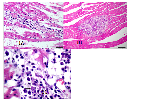
Mononuclear inflammatory infiltrate was observed in the histopathology of 77.8% (n=28) of VLG dogs. The infiltrate was predominantly lymphohistioplasmacytic and characterized by mild intensity (Fig. 1A, Table 4). Of the 28 affected animals, 85.7% (n=24) had myocardial inflammatory infiltrate, with 39.3% (n=11) showing infiltrate in the epicardium and 14.3% (n=4) showing infiltrate in the endocardium. The distribution of these lesions ranged from focal (25%, n=7) to focally extensive (10.7%, n=3) and multifocal (64.3%, n=18). Focal myocardial fibrosis and mild necrotizing subepicardial vasculitis (Fig. 1B) were detected in addition to the inflammatory infiltrate in two and four dogs, respectively. The presence of intracytoplasmic amastigotes of Leishmania sp. was confirmed in only one animal (Fig. 1C). No correlation was found between the intensity of histological lesions and the serum concentration of CK-MB (r= 0.2321, slope=80.77±66.40).
Myocardium of a dog from the Visceral Leishmaniasis group (VLG). Figure 1A. Discrete, focal lymphocytic inflammatory infiltration. HE, Bar = 50µm. Figure 1B. Artery with marked lymphohistioplasmacytic and neutrophilic vasculitis in the myocardium. The blood vessel is obliterated by intense inflammatory infiltrate. Bar = 200µm. Figure 1C. Presence of basophilic spherical structures in a macrophage cytoplasm, compatible with amastigotes of Leishmania sp. (arrow). HE, Bar = 20µm.
Percentage of occurrence of histopathological lesions in the myocardium of GLV dogs, classified according to the characteristic of the inflammatory infiltrate and its intensity
DISCUSSION
In the present study there was a higher prevalence of polysymptomatic dogs and mildly symptomatic dogs in the VLG. This outcome diverges from information reported byBarcelos (2009BARCELOS, D.S. Aspectos clínicos e parasitários de cães infectados naturalmente por Leishmania spp em duas áreas de transmissão intensa com diferentes características ambientais e sociais 2009. 91f. Dissertação (Mestrado em Ciências Clínicas) - Universidade Federal Rural do Rio de Janeiro, Seropédica, RJ. ), in which 57,1% (n=16) of VL dogs from the Rio de Janeiro region were classified as asymptomatic. However, mildly symptomatic and polysymptomatic dogs were observed at a frequency of 17.1% (n=7) and 73.2% (n=30), respectively, in a serological survey in the region of the state of Mato Grosso (Barcelos, 2009). In addition, mildly symptomatic and polysymptomatic dogs were seen at a rate of 50% and 47.2%, respectively, in the cases treated at a São Paulo State University Veterinary Hospital (Sonoda, 2007SONODA, M.C. Leishmaniose visceral canina: aspectos clinico-epidemiológicos de casos atendidos no período de 1997 a 2007, no Hospital Veterinário da Faculdade de Medicina Veterinária e Zootecnia da Universidade de São Paulo 2007. 115f. Dissertação (Mestrado), Universidade de São Paulo, São Paulo. ). It is believed that such variations may be related to the different species of Leishmaniainvolved, the immunity of the host and host-parasite interaction (Mancianti et al., 1988MANCIANTI, F.; GRAMICCIA, M.; GRADONI, L.; PIERI, S. Studies on canine leishmaniasis control. 1. Evolution of infection of different clinical forms of canine leishmaniasis following antimonial treatment. Trans. R. Soc. Trop. Med. Hyg. v.82, p.566-567, 1988.). Nevertheless, it is possible that the canine population evaluated in the present study influenced the results to the extent that it is believed that asymptomatic dogs are underrepresented in the hospital environment, as was similarly observed bySonoda (2007)SONODA, M.C. Leishmaniose visceral canina: aspectos clinico-epidemiológicos de casos atendidos no período de 1997 a 2007, no Hospital Veterinário da Faculdade de Medicina Veterinária e Zootecnia da Universidade de São Paulo 2007. 115f. Dissertação (Mestrado), Universidade de São Paulo, São Paulo. . Enlarged lymph nodes and integumentary signs were the most prevalent clinical findings for VLG dogs in this study, as also reported by Costa-Valet.al. (2007)COSTA-VAL, A.P.; CAVALCANTI, R.R.; FIGUEIREDO GONTIJO, N. et al Canine visceral leishmaniasis: Relationships between clinical status, humoral immune response, haematology and Lutzomyia longipalpis infectivity. Vet. J. v.174, p.636-643, 2007. and Alves et.al. (2010ALVES, G.B.B.; PINHO, F.A.; SILVA, S.M. et al Cardiac and pulmonary alterations in symptomatic and asymptomatic dogs infected naturally withLeishmania (Leishmania) chagasi Braz. J. Med. Biol. Res, v.43, p.310-315, 2010.).
Although heart murmur is a nonspecific clinical sign and has not been identified previously in VL dogs, a study of children and adolescents with the disease reported the detection of a systolic murmur in 35.9% (n=28) of patients (Diamantino, 2010DIAMANTINO, T.C.C. Leishmaniose visceral: avaliação das repercussões cardiovasculares secundárias à doença e ao tratamento em crianças e adolescentes tratadas com três esquemas terapêuticos. 2010. 221f. Tese (Doutorado) - Faculdade de Medicina, Universidade Federal de Minas Gerais, Belo Horizonte, MG. ). Neither of the two dogs in the present study that demonstrated a heart murmur showed any macroscopic or histological evidence of endocarditis, myxomatous valvular degeneration or congenital malformation of the mitral valve apparatus that would justify this clinical sign, which suggests a murmur that is hemodynamic in origin, caused by decreased blood viscosity secondary to the observed hematological changes. However, further studies must be conducted to confirm the prevalence of this clinical change and to determine its source.
The normocytic normochromic anemia observed in VLG dogs seems to be a common clinical finding in both dogs (López-Peña et al., 2009LÓPEZ-PEÑA, M.; ALEMAÑ, N.; MUÑOZ, F. et al Visceral leishmaniasis with cardiac involvement in a dog: a case report. Acta Vet. Scand v.51, p.1-3, 2009.; Alves et al., 2010ALVES, G.B.B.; PINHO, F.A.; SILVA, S.M. et al Cardiac and pulmonary alterations in symptomatic and asymptomatic dogs infected naturally withLeishmania (Leishmania) chagasi Braz. J. Med. Biol. Res, v.43, p.310-315, 2010.) and human beings with VL (Diamantino, 2010DIAMANTINO, T.C.C. Leishmaniose visceral: avaliação das repercussões cardiovasculares secundárias à doença e ao tratamento em crianças e adolescentes tratadas com três esquemas terapêuticos. 2010. 221f. Tese (Doutorado) - Faculdade de Medicina, Universidade Federal de Minas Gerais, Belo Horizonte, MG. ). Although the cellular mechanism of anemia has not been investigated in this study, evidence of the suppression of erythrocyte production in the bone marrow (Costa-Val et al., 2007COSTA-VAL, A.P.; CAVALCANTI, R.R.; FIGUEIREDO GONTIJO, N. et al Canine visceral leishmaniasis: Relationships between clinical status, humoral immune response, haematology and Lutzomyia longipalpis infectivity. Vet. J. v.174, p.636-643, 2007.) and splenic sequestration of erythrocytes (Luna et al. 2000LUNA, R.; FERRANTE, M.; SEVERINO, L. et al Decreased lipid fluidity of the erythrocyte membrane in dogs with leishmaniais associated anaemia. J. Comp. Pathol. v.122, p.213-216, 2000.) has been observed in dogs with VL.
However, contrary to the information obtained in the present study, neutrophilic leukocytosis was observed in only 21.5% of dogs with VL, while 9.5% of dogs with VL demonstrated leukopenia and 60% of dogs with VL showed no change in neutrophil count (Costa-Val et al., 2007COSTA-VAL, A.P.; CAVALCANTI, R.R.; FIGUEIREDO GONTIJO, N. et al Canine visceral leishmaniasis: Relationships between clinical status, humoral immune response, haematology and Lutzomyia longipalpis infectivity. Vet. J. v.174, p.636-643, 2007.). In children and adolescents naturally infected with VL, Diamantino (2010DIAMANTINO, T.C.C. Leishmaniose visceral: avaliação das repercussões cardiovasculares secundárias à doença e ao tratamento em crianças e adolescentes tratadas com três esquemas terapêuticos. 2010. 221f. Tese (Doutorado) - Faculdade de Medicina, Universidade Federal de Minas Gerais, Belo Horizonte, MG. ) verified leukopenia in 70.5% of cases, which was accompanied by neutropenia in 6.9% of cases. According to Mancianti et al. (1988MANCIANTI, F.; GRAMICCIA, M.; GRADONI, L.; PIERI, S. Studies on canine leishmaniasis control. 1. Evolution of infection of different clinical forms of canine leishmaniasis following antimonial treatment. Trans. R. Soc. Trop. Med. Hyg. v.82, p.566-567, 1988.), the host-parasite interaction may result in different clinical responses, which would explain the different responses observed. However, it is believed that hyperproteinemia in VLG dogs, as observed by Torrent et al.(2005TORRENT, E.; LEIVA, M.; SEGALÉS, J. et al Myocarditis and generalised vasculitis associated with leishmaniosis in a dog. J. Small Anim. Pract. v.46, p.549-552, 2005.) and Silvestrini et al. (2012SILVESTRINI, P.; PIVIANI, M.; ALBEROLA, J. et al Serum cardiac troponin I concentrations in dogs with leishmaniasis: correlation with age and clinicopathologic abnormalities. Vet. Clin. Pathol. v.41, p.568-574, 2012.), is due to hypergammaglobulinemia (Freitas et al., 2012FREITAS, J. C. C.; NUNES-PINHEIRO, D.C.S.; LOPES NETO, B.E. et al Clinical and laboratory alterations in dogs naturally infected byLeishmania chagasiRev. Soc. Bras. Med. Trop. v.45, p.24-29, 2012.), which in turn is due to a polyclonal lymphocyte response caused by the infection (Reis et al., 2009REIS, A.B.; MARTINS-FILHO, O.A.; TEIXEIRA-CARVALHO, A. et al Systemic and compartmentalized immune response in canine visceral Leishmaniasis.Vet. Immunol. Immunopathol., v.128, p.87-95, 2009.).
The higher serum activity of CK and MB-CK in VLG compared to CG suggests the occurrence of hypoxia and or cardiomyocyte necrosis (Yonezawa et al., 2009YONEZAWA, L.A., SILVEIRA, V.F., MACHADO, L.P.; KOHAYAGAWA A. Marcadores cardíacos na medicina veterinária. Cienc. Rural v.33, p.45-47, 2009.) in dogs infected with VL, as already observed by other authors (Medeiroset al., 2011MEDEIROS, A.A.; SOARES, N.P.; MUNDIM, A.V. et al Níveis de CK e CK-MB em cães naturalmente infectados por Leishmania. In: CONGRESSO BRASILEIRO DE MEDICINA VETERINÁRIA, 38., 2011, Florianópolis, Anais... Florianópolis: SBMV/Somevesc, 2011.. (Resumo)). However, there was no histopathological evidence of myocardial necrosis in the histological regions evaluated in the present study, which suggests that neither hypoxia nor myocardial necrosis in the areas assessed was responsible for the elevated serum activity of these biomarkers. Contrary to expectations, the activity of individual and mean serum activity of the CK-MB was higher than the total CK enzyme activity in both evaluated groups. One of the limitations of this analysis was that the measurements of serum activities of these enzymes (CK and MB-CK) were performed in different laboratories, which suggests that the differences observed may arise from the different techniques used, such as protein denaturation performed for different amounts of time, which may have influenced the outcomes.
In addition, cTnI is a useful and specific tool in the detection of myocardial necrosis in dogs (Yonezawa et al., 2009YONEZAWA, L.A., SILVEIRA, V.F., MACHADO, L.P.; KOHAYAGAWA A. Marcadores cardíacos na medicina veterinária. Cienc. Rural v.33, p.45-47, 2009.) when in concentrations greater than the normal limit of 0.02 ng/mL (Sleeper et al., 2001SLEEPER, M.M.; CLIFFORD, C.A.; LASTER, L.L. Cardiac troponin I in the normal dog and cat. J. Vet. Intern. Med.v.15, p.501-503, 2001.) or 0.03 ng/mL (Oyama & Solter, 2004OYAMA, M.A.; SOLTER, P.F. Validation of in immunoassay for measurement of canine cardiac troponin-I. J. Vet. Cardiol. v.6, p.17-24, 2004.). Although the elevation of this variable has been observed in dogs with VL (Silvestrini et al., 2012SILVESTRINI, P.; PIVIANI, M.; ALBEROLA, J. et al Serum cardiac troponin I concentrations in dogs with leishmaniasis: correlation with age and clinicopathologic abnormalities. Vet. Clin. Pathol. v.41, p.568-574, 2012.), it was not possible to verify this information in the present study because the technique used can only read values above 0.2ng/mL, which resulted in the detection of cTnI in only four VLG dogs (0.2474±0.1524 ng/mL). Although myocardial necrosis is suggested for these four dogs, the wide margin between the normal range and the minimum concentration measured by this technique may have hidden elevated cTnI in dogs from both the CG and the VLG, thus preventing a comparison between these two groups or the suggestion that the occurrence of myocardial necrosis is caused by infection with VL.
Regarding radiographic evaluation, no differences were observed in the qualitative or quantitative cardiac silhouettes of dogs from CG or VLG, which suggests that VL has no influence on the cardiac silhouette that can be detected with this diagnostic imaging tool. Although qualitative analysis can be influenced by the experience of the evaluator, the anatomic conformation of the thorax and the size of the animal (Ware 2006WARE, W.A. Radiografia torácica. In: NELSON, R.W.; COUTO, C.G. (Eds). Medicina interna de pequenos animais. Rio de Janeiro: Guanabara Koogan, 2006. p.30-34.), as well as the use of the quantitative method in this study provided greater reliability regarding the comparison of the two groups. Similar studies in dogs that might have allowed for a fuller discussion of these observations were not found during the time period in which this study was conducted. However, similar results have been reported in children and adolescents with VL (Diamantino, 2010DIAMANTINO, T.C.C. Leishmaniose visceral: avaliação das repercussões cardiovasculares secundárias à doença e ao tratamento em crianças e adolescentes tratadas com três esquemas terapêuticos. 2010. 221f. Tese (Doutorado) - Faculdade de Medicina, Universidade Federal de Minas Gerais, Belo Horizonte, MG. ), which reinforces the hypothesis of non-occurrence of cardiomegaly of any cardiac chamber that is secondary to this infection.
The electrocardiographic evaluation detected a higher occurrence of isolated events of sinus arrest associated with RSA in VLG dogs (8.3%). This phenomenon was also observed in 14.3% of the 105 dogs with VL that were evaluated bySousa et al. (2013SOUSA, M.G.; CARARETO R.; SILVA J.G.; OLIVEIRA J. Assesment of the electrocardiogram in dogs with visceral leishmaniasis. Pesqui. Vet. Bras. v.33, p.643-647, 2013.). First-degree atrioventricular block (Torrentet al., 2005TORRENT, E.; LEIVA, M.; SEGALÉS, J. et al Myocarditis and generalised vasculitis associated with leishmaniosis in a dog. J. Small Anim. Pract. v.46, p.549-552, 2005.), right bundle branch block and premature atrial contractions (Sousa et al., 2013SOUSA, M.G.; CARARETO R.; SILVA J.G.; OLIVEIRA J. Assesment of the electrocardiogram in dogs with visceral leishmaniasis. Pesqui. Vet. Bras. v.33, p.643-647, 2013.) were also detected in dogs with VL, which diverges from the findings of the present study. Although sinus arrest is considered a physiological rhythm in some dogs, it may be associated with increased vagal tone, electrolyte imbalance, cervical neoplasia and pathologies involving the region of the atrial sinus node (Tilley, 1992TILLEY, L.P. (Ed) Essentials of canine and feline electrocardiography. Philadelphia: Lea & Febiger, 1992. 470p.). In the present study, no correlation was found between the intensity of the inflammation of the myocardial regions and the occurrence of episodes of sinus arrest or RSA. However, atrial myocarditis was already associated with sinus arrest (Woolley, 2007WOOLLEY, R.R.; BLUNDELL, R.; ELSE, R. et al Atrial myocarditis as a cause of sinus arrest in a dog. J. Small Anim. Pract. v.48, p.455-457, 2007.) and right atrium predisposal to more severe lymphocytic infiltration of dogs with VL was reported (Rosa et al., 2013ROSA, F.A.; LEITE, J.H.A.C.; BRAGA, E.T. et al Cardiac lesions in 30 dogs naturally infected with Leishmania infantum chagasi. Vet. Patholv.51, p.503-606; 2013.). In this study, the regions used for histological evaluation of the atria of dogs from the VLG did not permit analysis of the sinus node. Accordingly, the possibility of myocarditis being involved in the electrocardiographic differences detected between the groups was not ruled out.
Likewise, atrial myocarditis may explain the lower average amplitude of the P wave found in VLG dogs because that would result in a decrease in the total number of atrial cells and a consequent reduction in the depolarizing millivoltage captured in the body surface by means of an electrocardiogram (Tilley, 1992TILLEY, L.P. (Ed) Essentials of canine and feline electrocardiography. Philadelphia: Lea & Febiger, 1992. 470p.). However, lower P wave amplitude is not a constant finding in studies involving dogs with VL, which reported suppression of R wave millivoltage (López-Peña et al., 2009LÓPEZ-PEÑA, M.; ALEMAÑ, N.; MUÑOZ, F. et al Visceral leishmaniasis with cardiac involvement in a dog: a case report. Acta Vet. Scand v.51, p.1-3, 2009.) and longer duration of the P wave and QRS complex (Sousa et al., 2013SOUSA, M.G.; CARARETO R.; SILVA J.G.; OLIVEIRA J. Assesment of the electrocardiogram in dogs with visceral leishmaniasis. Pesqui. Vet. Bras. v.33, p.643-647, 2013.). In the present study, all electrocardiographic variables remained within normal range for the species, but the differences between the CG and VLG groups suggest that atrial lesions may result in a decrease in P wave amplitude.
Microscopic changes were present in at least one of the regions subject to histological analysis in 77.8% of the dogs studied, while other authors have described this finding in 100% of dogs with VL (Rosaet al., 2013ROSA, F.A.; LEITE, J.H.A.C.; BRAGA, E.T. et al Cardiac lesions in 30 dogs naturally infected with Leishmania infantum chagasi. Vet. Patholv.51, p.503-606; 2013.). The inflammatory infiltrate was predominantly lymphohistioplasmacytic, similar to the findings described by Alves et al. (2010ALVES, G.B.B.; PINHO, F.A.; SILVA, S.M. et al Cardiac and pulmonary alterations in symptomatic and asymptomatic dogs infected naturally withLeishmania (Leishmania) chagasi Braz. J. Med. Biol. Res, v.43, p.310-315, 2010.), López-Peña et al. (2009)LÓPEZ-PEÑA, M.; ALEMAÑ, N.; MUÑOZ, F. et al Visceral leishmaniasis with cardiac involvement in a dog: a case report. Acta Vet. Scand v.51, p.1-3, 2009. andSilva et al. (2009SILVA, B.C.; RACHID, M.A.; VIEIRA, F.G. et al Chronic pericarditis in a naturally Leishmania chagasi infected dog. Braz. J.. Vet. Pathol v.2, p.107-10, 2009.). Lymphocytic infiltration was observed in 22.2% of dogs in VLG, although other researchers have reported a higher prevalence of lymphocytic infiltration (Rosa et al., 2013ROSA, F.A.; LEITE, J.H.A.C.; BRAGA, E.T. et al Cardiac lesions in 30 dogs naturally infected with Leishmania infantum chagasi. Vet. Patholv.51, p.503-606; 2013.). Although mild infiltrate is the most consistent finding, mild to moderate intensities were observed in dogs tested in the present study while other studies have reported intensities ranging from mild to moderate (Alveset al., 2010ALVES, G.B.B.; PINHO, F.A.; SILVA, S.M. et al Cardiac and pulmonary alterations in symptomatic and asymptomatic dogs infected naturally withLeishmania (Leishmania) chagasi Braz. J. Med. Biol. Res, v.43, p.310-315, 2010.), moderate to severe (Silva et al., 2009SILVA, B.C.; RACHID, M.A.; VIEIRA, F.G. et al Chronic pericarditis in a naturally Leishmania chagasi infected dog. Braz. J.. Vet. Pathol v.2, p.107-10, 2009.), intense (López-Peña et al., 2009LÓPEZ-PEÑA, M.; ALEMAÑ, N.; MUÑOZ, F. et al Visceral leishmaniasis with cardiac involvement in a dog: a case report. Acta Vet. Scand v.51, p.1-3, 2009.) and mild to intense (Rosa et al., 2013ROSA, F.A.; LEITE, J.H.A.C.; BRAGA, E.T. et al Cardiac lesions in 30 dogs naturally infected with Leishmania infantum chagasi. Vet. Patholv.51, p.503-606; 2013.). Likewise, there does not seem to be a consensus in the literature regarding the predominant location of the infiltrate. Specifically, the myocardium and pericardium regions were the predominant locations of infiltrate in dogs in the VLG, as well as those studied by Alves et al. (2010)ALVES, G.B.B.; PINHO, F.A.; SILVA, S.M. et al Cardiac and pulmonary alterations in symptomatic and asymptomatic dogs infected naturally withLeishmania (Leishmania) chagasi Braz. J. Med. Biol. Res, v.43, p.310-315, 2010. and Silvaet al. (2009)SILVA, B.C.; RACHID, M.A.; VIEIRA, F.G. et al Chronic pericarditis in a naturally Leishmania chagasi infected dog. Braz. J.. Vet. Pathol v.2, p.107-10, 2009., although the endocardial and subepicardial regions have also been noted (Rosaet al., 2013ROSA, F.A.; LEITE, J.H.A.C.; BRAGA, E.T. et al Cardiac lesions in 30 dogs naturally infected with Leishmania infantum chagasi. Vet. Patholv.51, p.503-606; 2013.).
In the present study, it was not possible to relate myocarditis to direct injury by the parasite, because the amastigote form was found in the histopathology of the myocardium in only one dog. However, Rosa et al. (2013ROSA, F.A.; LEITE, J.H.A.C.; BRAGA, E.T. et al Cardiac lesions in 30 dogs naturally infected with Leishmania infantum chagasi. Vet. Patholv.51, p.503-606; 2013.) detected Leishmania sp. in 20 of 30 dogs studied by immunohistochemistry and positively correlated this finding with the degree of the severity of parasitic inflammation. In addition, the vascular lesions observed in the present study, which were also mentioned by other authors (Torrent et al., 2005TORRENT, E.; LEIVA, M.; SEGALÉS, J. et al Myocarditis and generalised vasculitis associated with leishmaniosis in a dog. J. Small Anim. Pract. v.46, p.549-552, 2005.;Rosa et al., 2013ROSA, F.A.; LEITE, J.H.A.C.; BRAGA, E.T. et al Cardiac lesions in 30 dogs naturally infected with Leishmania infantum chagasi. Vet. Patholv.51, p.503-606; 2013.), may suggest an indirect mechanism of myocardial injury exacerbated by the formation and deposition of immune complexes (hypersensitivity type III) in the vascular endothelium, which results in the activation of the inflammatory cell response (hypersensitivity type IV) and tissue damage (Torrent et al., 2005TORRENT, E.; LEIVA, M.; SEGALÉS, J. et al Myocarditis and generalised vasculitis associated with leishmaniosis in a dog. J. Small Anim. Pract. v.46, p.549-552, 2005.; Rosa et al., 2013ROSA, F.A.; LEITE, J.H.A.C.; BRAGA, E.T. et al Cardiac lesions in 30 dogs naturally infected with Leishmania infantum chagasi. Vet. Patholv.51, p.503-606; 2013.). However, further studies should be conducted to confirm the hypothesis of direct and indirect sources of myocarditis associated with VL in dogs.
Other histological lesions not identified in the present study that were described in VL dogs include hyaline degeneration of myocardial fibers (Alves et.al., 2010ALVES, G.B.B.; PINHO, F.A.; SILVA, S.M. et al Cardiac and pulmonary alterations in symptomatic and asymptomatic dogs infected naturally withLeishmania (Leishmania) chagasi Braz. J. Med. Biol. Res, v.43, p.310-315, 2010.), cardiomyocyte necrosis (López-Peña et.al., 2009LÓPEZ-PEÑA, M.; ALEMAÑ, N.; MUÑOZ, F. et al Visceral leishmaniasis with cardiac involvement in a dog: a case report. Acta Vet. Scand v.51, p.1-3, 2009.;Alves et.al., 2010ALVES, G.B.B.; PINHO, F.A.; SILVA, S.M. et al Cardiac and pulmonary alterations in symptomatic and asymptomatic dogs infected naturally withLeishmania (Leishmania) chagasi Braz. J. Med. Biol. Res, v.43, p.310-315, 2010.; Rosa et al., 2013ROSA, F.A.; LEITE, J.H.A.C.; BRAGA, E.T. et al Cardiac lesions in 30 dogs naturally infected with Leishmania infantum chagasi. Vet. Patholv.51, p.503-606; 2013.), increased interstitial collagen and coagulation necrosis in the right atrium and ventricle (Rosa et al., 2013ALVES, G.B.B.; PINHO, F.A.; SILVA, S.M. et al Cardiac and pulmonary alterations in symptomatic and asymptomatic dogs infected naturally withLeishmania (Leishmania) chagasi Braz. J. Med. Biol. Res, v.43, p.310-315, 2010.) and cardiac muscle fiber atrophy (López-Peñaet.al., 2009LÓPEZ-PEÑA, M.; ALEMAÑ, N.; MUÑOZ, F. et al Visceral leishmaniasis with cardiac involvement in a dog: a case report. Acta Vet. Scand v.51, p.1-3, 2009.). These findings highlight both the complexity of the injuries promoted by this infection and the need for further research in this area. Possibly, the use of long term electrocardiographic (Holter) and immunohistochemical analysis could elucidate the cardiovascular effects of this infection.
CONCLUSIONS
Nonetheless, under the conditions in which the present study was conducted, it was determined that, when compared to the CG, dogs from the VLG showed polysymptomatic clinical features (characterized by a predominance of enlarged lymph nodes and skin changes) that were associated with (1) normocytic normochromic anemia; (2) leukocytosis with neutrophilia; (3) elevated serum activity of CK and MB-CK; and (4) a higher incidence of respiratory sinus arrhythmia and episodes of migratory pacemaker and sinus arrest. All of these findings were accompanied by primarily mild, multifocal myocarditis and inflammatory lymphohistioplasmacytic infiltrate.
REFERENCES
- ALVES, G.B.B.; PINHO, F.A.; SILVA, S.M. et al Cardiac and pulmonary alterations in symptomatic and asymptomatic dogs infected naturally withLeishmania (Leishmania) chagasi Braz. J. Med. Biol. Res, v.43, p.310-315, 2010.
- BARCELOS, D.S. Aspectos clínicos e parasitários de cães infectados naturalmente por Leishmania spp em duas áreas de transmissão intensa com diferentes características ambientais e sociais 2009. 91f. Dissertação (Mestrado em Ciências Clínicas) - Universidade Federal Rural do Rio de Janeiro, Seropédica, RJ.
- BUCHANAN, J.W.; BÜCHELER, J. Vertebral scale system to measure canine heart size in radiographs. J. Am. Vet. Med. Assoc v.206, p.194-199, 1995.
- COSTA-VAL, A.P.; CAVALCANTI, R.R.; FIGUEIREDO GONTIJO, N. et al Canine visceral leishmaniasis: Relationships between clinical status, humoral immune response, haematology and Lutzomyia longipalpis infectivity. Vet. J. v.174, p.636-643, 2007.
- DIAMANTINO, T.C.C. Leishmaniose visceral: avaliação das repercussões cardiovasculares secundárias à doença e ao tratamento em crianças e adolescentes tratadas com três esquemas terapêuticos. 2010. 221f. Tese (Doutorado) - Faculdade de Medicina, Universidade Federal de Minas Gerais, Belo Horizonte, MG.
- FREITAS, J. C. C.; NUNES-PINHEIRO, D.C.S.; LOPES NETO, B.E. et al Clinical and laboratory alterations in dogs naturally infected byLeishmania chagasiRev. Soc. Bras. Med. Trop. v.45, p.24-29, 2012.
- LÓPEZ-PEÑA, M.; ALEMAÑ, N.; MUÑOZ, F. et al Visceral leishmaniasis with cardiac involvement in a dog: a case report. Acta Vet. Scand v.51, p.1-3, 2009.
- LUNA, R.; FERRANTE, M.; SEVERINO, L. et al Decreased lipid fluidity of the erythrocyte membrane in dogs with leishmaniais associated anaemia. J. Comp. Pathol. v.122, p.213-216, 2000.
- MANCIANTI, F.; GRAMICCIA, M.; GRADONI, L.; PIERI, S. Studies on canine leishmaniasis control. 1. Evolution of infection of different clinical forms of canine leishmaniasis following antimonial treatment. Trans. R. Soc. Trop. Med. Hyg. v.82, p.566-567, 1988.
- MANUAL de vigilância e controle da leishmaniose visceral. Secretaria de Vigilância em Saúde Brasília: Ministério da Saúde. 2006.
- MEDEIROS, A.A.; SOARES, N.P.; MUNDIM, A.V. et al Níveis de CK e CK-MB em cães naturalmente infectados por Leishmania In: CONGRESSO BRASILEIRO DE MEDICINA VETERINÁRIA, 38., 2011, Florianópolis, Anais... Florianópolis: SBMV/Somevesc, 2011.. (Resumo)
- MONTEIRO, E.M.; SILVA, J.C.F.; COSTA, R.T. et al Leishmaniose visceral: estudo de flebotomíneos e infecção canina em Montes Claros, Minas Gerais., Rev. Soc. Bras. Med. Trop. v.38, p.147-152, 2005.
- OYAMA, M.A.; SOLTER, P.F. Validation of in immunoassay for measurement of canine cardiac troponin-I. J Vet. Cardiol. v.6, p.17-24, 2004.
- REBAR, A. H.; MAC WILLIANS, P.S.; FELDMAN, B.F. et al (Eds) Guia de hematologia para cães e gatos São Paulo: Roca, 2003. 285p.
- REIS, A.B.; MARTINS-FILHO, O.A.; TEIXEIRA-CARVALHO, A. et al Systemic and compartmentalized immune response in canine visceral Leishmaniasis.Vet. Immunol. Immunopathol, v.128, p.87-95, 2009.
- ROSA, F.A.; LEITE, J.H.A.C.; BRAGA, E.T. et al Cardiac lesions in 30 dogs naturally infected with Leishmania infantum chagasi Vet. Patholv.51, p.503-606; 2013.
- SILVA, B.C.; RACHID, M.A.; VIEIRA, F.G. et al Chronic pericarditis in a naturally Leishmania chagasi infected dog. Braz. J.. Vet. Pathol v.2, p.107-10, 2009.
- SILVESTRINI, P.; PIVIANI, M.; ALBEROLA, J. et al Serum cardiac troponin I concentrations in dogs with leishmaniasis: correlation with age and clinicopathologic abnormalities. Vet. Clin. Pathol. v.41, p.568-574, 2012.
- SLEEPER, M.M.; CLIFFORD, C.A.; LASTER, L.L. Cardiac troponin I in the normal dog and cat. J Vet. Intern. Med.v.15, p.501-503, 2001.
- SONODA, M.C. Leishmaniose visceral canina: aspectos clinico-epidemiológicos de casos atendidos no período de 1997 a 2007, no Hospital Veterinário da Faculdade de Medicina Veterinária e Zootecnia da Universidade de São Paulo 2007. 115f. Dissertação (Mestrado), Universidade de São Paulo, São Paulo.
- SOUSA, M.G.; CARARETO R.; SILVA J.G.; OLIVEIRA J. Assesment of the electrocardiogram in dogs with visceral leishmaniasis. Pesqui. Vet. Bras. v.33, p.643-647, 2013.
- TILLEY, L.P. (Ed) Essentials of canine and feline electrocardiography. Philadelphia: Lea & Febiger, 1992. 470p.
- TORRENT, E.; LEIVA, M.; SEGALÉS, J. et al Myocarditis and generalised vasculitis associated with leishmaniosis in a dog. J. Small Anim. Pract. v.46, p.549-552, 2005.
- YONEZAWA, L.A., SILVEIRA, V.F., MACHADO, L.P.; KOHAYAGAWA A. Marcadores cardíacos na medicina veterinária. Cienc. Rural v.33, p.45-47, 2009.
- XAVIER, S.C.; CHIARELLI, I.M.; LIMA, W.G. et al Canine visceral leishmaniasis: a remarkable histopathological picture of one asymptomatic animal reported from Belo Horizonte, Minas Gerais, Brasil. Arq. Bras. Med. Vet. Zootec. v.58, p.994-1000, 2006.
- WARE, W.A. Radiografia torácica. In: NELSON, R.W.; COUTO, C.G. (Eds). Medicina interna de pequenos animais. Rio de Janeiro: Guanabara Koogan, 2006. p.30-34.
- WOOLLEY, R.R.; BLUNDELL, R.; ELSE, R. et al Atrial myocarditis as a cause of sinus arrest in a dog. J. Small Anim. Pract. v.48, p.455-457, 2007.
Publication Dates
-
Publication in this collection
Nov-Dec 2015
History
-
Received
23 July 2014 -
Accepted
06 May 2015