ABSTRACT
This paper reports a case of a rare variant of the cervical spinal cord astrocytoma diagnosed in a dog with progressive neurological signs, initially asymmetrical, not ambulatory tetraparesis, segmental reflexes and normal muscle tone in all four limbs and absence of pain upon palpation of the cervical spine. Myelography revealed attenuation of the ventral and dorsal contrast line in the third region of the fifth cervical vertebra. At necropsy intramedullary cylindrical mass that stretched from the third to the sixth cervical vertebra, which replaced all the gray matter of the spinal cord was observed. In the histological study, there was the replacement of the substance by neoplastic cells mantle arranged loosely. The cells were large and slightly rounded. The eosinophilic cytoplasm was well defined, sometimes forming processes interconnecting cells. The nucleus was eccentric, round, oval or kidney-shaped, and the nucleolus was evident. Thus, the microscopic changes observed in the cervical spinal cord were consistent with gemistocytic astrocytoma.
Keywords:
canine; neoplasm; glioma; spinal cord
RESUMO
Relata-se um caso de uma variante rara de astrocitoma na medula cervical, diagnosticado em cadela com sinais neurológicos progressivos, inicialmente assimétricos, de tetraparesia não ambulatória, com reflexos segmentares e tônus muscular normais nos quatro membros e ausência de dor à palpação da coluna cervical. A mielografia revelou atenuação da linha de contraste ventral e dorsal na região da terceira à quinta vértebra cervical. À necropsia, foi observada massa cilíndrica intramedular que se estendia da terceira à sexta vértebra cervical, a qual substituía toda a substância cinzenta da medula espinhal. No estudo histológico, observou-se substituição da substância por manto de células neoplásicas arranjadas frouxamente. As células eram grandes e levemente arredondadas. O citoplasma eosinofílico, bem delineado, por vezes formava processos interligando as células. O núcleo era excêntrico, redondo, oval ou reniforme, e o nucléolo evidente. Logo, as alterações microscópicas observadas na medula espinhal cervical foram compatíveis com astrocitoma gemistocítico.
Palavras-chave:
canino; neoplasma; glioma; medula espinhal
INTRODUCTION
The spinal neoplasms is an important cause of locomotor neurological signs in dogs, affecting mainly large breed and middle-aged to elderly animals (Da Costa, 2009COSTA, R.C. Neoplasias do sistema nervoso. In: DALECK, R.C.; DE NARDI, A.B.; RODASKI, S. (Eds.). Oncologia em cães e gatos. São Paulo: ROCA, 2009. p.431-455.; Bagley, 2010BAGLEY, R.S. Spinal neoplasms in small animals. Vet. Clin. North Am. Small Anim. Pract., v.40, p.915-927, 2010.). Anatomically, the spinal neoplasms can be classified into extradural (localized tumors external to the dura mater), intradural-extramedullary (internal to the dura mater but external to the spinal cord) and intramedullary (inside the spinal cord), the latter being the least common (Da Costa, 2009; Bagley, 2010; Lorenz et al., 2011LORENZ, M.D.; COATES, J.R.; KENT, M. Handbook of veterinary neurology. 5.ed. Philadelphia: W.B. Saunders, 2011. 545p.).
The clinical neurological signs depend on the location, size and growth rate of the neoplasm (Da Costa, 2009COSTA, R.C. Neoplasias do sistema nervoso. In: DALECK, R.C.; DE NARDI, A.B.; RODASKI, S. (Eds.). Oncologia em cães e gatos. São Paulo: ROCA, 2009. p.431-455.), and are the result of compression, direct invasion of tissues, edema, inflammation and necrosis (Da Costa, 2009; Bagley, 2010BAGLEY, R.S. Spinal neoplasms in small animals. Vet. Clin. North Am. Small Anim. Pract., v.40, p.915-927, 2010.). The definitive diagnosis can be established only by histological analysis, performed by biopsy or necropsy (Da Costa, 2009; Bagley, 2010; Pancotto et al., 2013PANCOTTO, T.E.; ROSSMEISL JR, J.H.; ZIMMERMAN, K. et al. Intramedullary spinal cord neoplasia in 53 dogs (1990-2010): distribution, clinicopathologic characteristics, and clinical behavior. J. Vet. Intern. Med., v.27, p.1500-1508, 2013.).
Astrocytomas are the most common glial tumors and can be classified according to the cell subtypes involved: low grade (fibrillary, protoplasmic, pilocytic and gemistocytic), medium grade (anaplastic) and high grade (glioblastoma multiforme) (Koestner et al., 1999KOESTNER, A.; BILZER, T.; FATZER, R.; et al. Histological classification of tumors of the nervous system of domestic animals. [s.l.]: World Health Organization, 1999. p.17-21.; Maxie and Youssef, 2007MAXIE, M.G.; YOUSSEF, S. Nervous system. In: MAXIE, M.G. Jubb, Kennedy, and Palmer's pathology of domestic animals. 5.ed. Philadelphia: Elsevier Saunders, 2007. p.281-457.). The gemistocytic astrocytoma is the least common variant, classified as low grade. The main histological features of this variant are: large, round cells, eosinophilic cytoplasm, and eccentric and oval nucleus (Kostner et al., 1999; Maxie and Youssef, 2007). In addition, it occurs most often in the brain, and is rarely found in the spinal cord (Maxie and Youssef, 2007; Coelho et al., 2013COELHO, M.P.R.C.; MARTINS, B.C.; MOREIRA, M.V.L. et al. Astrocitoma fibrilar medular multifocal em cão - relato de caso. Acta Vet. Brasilica., v.7, p.378-379, 2013.; Pancotto et al., 2013PANCOTTO, T.E.; ROSSMEISL JR, J.H.; ZIMMERMAN, K. et al. Intramedullary spinal cord neoplasia in 53 dogs (1990-2010): distribution, clinicopathologic characteristics, and clinical behavior. J. Vet. Intern. Med., v.27, p.1500-1508, 2013.). Therefore, the aim here is to report a case of gemistocytic astrocytoma in the cervical cord in a dog.
CASE DESCRIPTION
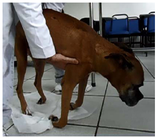
At the University Veterinary Hospital of the Federal University of Santa Maria, a Boxer bitch, 11 years old, weighing 18kg, with monoparesis history of the left forelimb for three months was assisted. After 30 days of the onset of clinical signs she presented difficulty walking in the four members, with progressive worsening until the patient could no longer walk. Upon neurological examination, the dog was alert. Non ambulatory tetraparesis, postural reactions (proprioception and hopping) absent in the four limbs (Figure 1), segmental spinal reflexes in the hind limbs (patellar, flexor and perineal) and thoracic (flexor) and normal muscle tone and absence of pain cervical epaxial palpation were observed. The neurological signs found characterized a lesion between segments C1-C5 of the spinal cord. Spinal neoplasms, intervertebral disc disease and meningomyelitis (infectious and non-infectious) were differential diagnoses.
The complementary exams performed included: hemogram, serum biochemistry (creatinine, blood urea nitrogen, ALT, AST, serum alkaline phosphatase, glucose, total protein, and albumin), urinalysis, abdominal ultrasound, analysis of cerebrospinal fluid and plain radiography of thorax and cervical spine, which showed no changes. In myelography the attenuation of ventral and dorsal line of contrast in the lateral projection and attenuation of the lateral line of contrast in the ventrodorsal projection the third of the region's fifth cervical vertebra (Figure 2A and 2B) were observed.
A. Myelography of the cervical spine. A. In the lateral projection, the attenuation of ventral and dorsal contrast lines in the third region of the fifth cervical vertebra is observed. B. In the ventrodorsal projection, there is attenuation in lateral contrast lines in the third region of the fifth cervical vertebra.
According to the historical, clinical, neurological and laboratory test findings, the presumptive diagnosis was intramedullary neoplasm. After the presumptive diagnosis, the owner chose to conduct euthanasia on the patient. The owner did not authorize a necropsy, so only a fragment of the cervical spinal cord, of 5.5 cm long was removed. Macroscopically, intramedullary cylindrical mass was observed which extended from C3 to C6, which replaced the entire gray matter of the spinal cord (Figure 3).
In the histological study, a replacement of the gray matter by loosely arranged neoplastic cell mantle was observed. The cells were large and slightly rounded. The eosinophilic cytoplasm was well delineated, and sometimes formed processes interconnecting cells. The nucleus was eccentric, round, oval to kidney-shaped, whit evident nucleolus (Fig. 4). We also observed multiple cells with a pyknotic nucleus distributed by the tumor, indicating apoptosis. The neoplastic cells infiltrated the white matter. A focal area of hemorrhage and necrosis was observed. Thus, the microscopic changes observed in the cervical spinal cord of this dog were consistent with gemistocytic astrocytoma.
Spinal cord, gemistocytic astrocytoma. A. Proliferated cells arranged in dense sheets that completely obliterate the neuropil. Hematoxylin and eosin. Obj.10x. B. Higher magnification of the previous figure (Fig. A). The cells are highly pleomorphic, but predominantly stellate to spindle shaped. Hematoxylin and eosin. Obj.20x. C. Multiple round to polygonal cells with eccentric, round to reniform nuclei. A markedly anaplastic astrocyte is seen in the center of the figure. Hematoxylin and eosin. Obj.40x. D. Multiple astrocytes (cell body and cytoplasmic processes) strongly immunostained. Streptavidin-biotin method (GFAP). Obj.20x.
DISCUSSION
Primary or secondary, the intramedullary neoplasms are not common and account for approximately 15% of the spinal neoplasms in dogs (Da Costa, 2009COSTA, R.C. Neoplasias do sistema nervoso. In: DALECK, R.C.; DE NARDI, A.B.; RODASKI, S. (Eds.). Oncologia em cães e gatos. São Paulo: ROCA, 2009. p.431-455.; Pancotto et al., 2013PANCOTTO, T.E.; ROSSMEISL JR, J.H.; ZIMMERMAN, K. et al. Intramedullary spinal cord neoplasia in 53 dogs (1990-2010): distribution, clinicopathologic characteristics, and clinical behavior. J. Vet. Intern. Med., v.27, p.1500-1508, 2013.). In the study by Pancotto et al. (2013), of 331 dogs with spinal neoplasms, 53 (16%) were diagnosed with intramedullary neoplasms, of which 35 (66%) were of primary origin and 18 (34%) of secondary origin. Of the 35 primary origin neoplasms, ependymoma was the most common (n=13), followed by astrocytoma (n=7) and nephroblastoma (n=4). Primarily affecting dogs older than five years (Da Costa, 2009; Santos et al., 2012SANTOS, R.P.; FIGHERA, R.A.; BECKMANN, D.V. et al. Neoplasmas envolvendo o sistema nervoso central de cães: 26 casos (2003-2011). Pesqui. Vet. Bras., v.32, p.153-158, 2012.; Pancotto et al., 2013), of brachycephalic breeds such as Boxers and Boston Terriers (Maxie and Youssef, 2007MAXIE, M.G.; YOUSSEF, S. Nervous system. In: MAXIE, M.G. Jubb, Kennedy, and Palmer's pathology of domestic animals. 5.ed. Philadelphia: Elsevier Saunders, 2007. p.281-457.), females appear to be more prevalent (Pancotto et al., 2013). The dog in this report was a Boxer breed, and age and sex were found according to the literature as the most prevalent.
Signs of spinal neoplasms depend on the location of the tumor, and as in this case and in most cases, the signals are compatible with the focal myelopathy. Less common clinical signs may also be multifocal depending on the number and location of neoplasms and local alterations caused (Da Costa, 2009COSTA, R.C. Neoplasias do sistema nervoso. In: DALECK, R.C.; DE NARDI, A.B.; RODASKI, S. (Eds.). Oncologia em cães e gatos. São Paulo: ROCA, 2009. p.431-455.; Coelho et al., 2013COELHO, M.P.R.C.; MARTINS, B.C.; MOREIRA, M.V.L. et al. Astrocitoma fibrilar medular multifocal em cão - relato de caso. Acta Vet. Brasilica., v.7, p.378-379, 2013.; Pancotto et al., 2013PANCOTTO, T.E.; ROSSMEISL JR, J.H.; ZIMMERMAN, K. et al. Intramedullary spinal cord neoplasia in 53 dogs (1990-2010): distribution, clinicopathologic characteristics, and clinical behavior. J. Vet. Intern. Med., v.27, p.1500-1508, 2013.). The site most commonly affected by spinal neoplasms is the spinal region between T3-L3 and C1-C5 (Da Costa, 2009; Santos et al., 2012SANTOS, R.P.; FIGHERA, R.A.; BECKMANN, D.V. et al. Neoplasmas envolvendo o sistema nervoso central de cães: 26 casos (2003-2011). Pesqui. Vet. Bras., v.32, p.153-158, 2012.; Pancotto et al., 2013). However, for the intramedullary neoplasms, a possible predisposition to the primary origin, such as astrocytoma, occurring in the cervical spinal cord was identified (Luttgen et al., 1980LUTTGEN, P.J.; BRAUND, K.G.; BRAWNER, W.R JR.; VANDEVELDE, M. A retrospective study of twenty-nine spinal tumours in the dog and cat. J. Small Anim. Pract., v.21, p.213-226, 1980.; Pancotto et al., 2013).
The upper motor neurons, whose nuclei are predominantly in the brain stem, are responsible for sending stimuli to initiate voluntary movements and maintain muscle tone and posture during movement (Lorenz et al., 2011LORENZ, M.D.; COATES, J.R.; KENT, M. Handbook of veterinary neurology. 5.ed. Philadelphia: W.B. Saunders, 2011. 545p.). Therefore, a lesion between C1-C5 segments of the spinal cord can cause tetraparesis and ataxia of the four members (which may, in severe cases, reach tetraplegia), muscle tone and normal or increased spinal reflexes in the fore and hindlimbs (Da Costa, 2009COSTA, R.C. Neoplasias do sistema nervoso. In: DALECK, R.C.; DE NARDI, A.B.; RODASKI, S. (Eds.). Oncologia em cães e gatos. São Paulo: ROCA, 2009. p.431-455.). Therefore, the neurological signs observed in the patient in question, such as non-ambulatory tetraparesis and segmental reflexes and muscle tone loss in the four normal members characterized injury between C1-C5 segment of the spinal cord.
The presence of spinal pain varies with the location of the neoplasm, and are commonly observed in extradural neoplasms, occasionally in intradural-extramedullary, and rarely in the intramedullary (Da Costa, 2009COSTA, R.C. Neoplasias do sistema nervoso. In: DALECK, R.C.; DE NARDI, A.B.; RODASKI, S. (Eds.). Oncologia em cães e gatos. São Paulo: ROCA, 2009. p.431-455.). On the other hand, in the study by Pancotto et al. (2013PANCOTTO, T.E.; ROSSMEISL JR, J.H.; ZIMMERMAN, K. et al. Intramedullary spinal cord neoplasia in 53 dogs (1990-2010): distribution, clinicopathologic characteristics, and clinical behavior. J. Vet. Intern. Med., v.27, p.1500-1508, 2013.), 89% of neoplasm intramedullary dogs showed signs of pain when receiving palpation of the vertebral column. In the dog in this report, this signal was not observed during the neurological examination. One hypothesis for the development of pain associated with intramedullary lesions is changing the modulation in neurotransmitters due to remodeling or destruction of the dorsal horn of the spinal cord (Rusbridge and Jeffery, 2008RUSBRIDGE, C.; JEFFERY, N.D. Pathophysiology and treatment of neuropathic pain associated with syringomyelia. Vet. J., v.175, p.164-172, 2008.). Another probable cause is the mass effect, which could contribute to the stretching of the meninges, dorsal nerve roots, or both, causing discomfort upon palpation (Pancotto et al., 2013).
The presumptive diagnosis of intramedullary neoplasm was based on history, breed, age, neurological signs, the evolution of the signs and the results of complementary tests (chest radiograph, abdominal ultrasound, analysis of LCE, radiography and myelography of the vertebral column). Advanced image examinations such as computed tomography (CT) and magnetic resonance imaging (MRI), can also aid in the diagnosis (Da Costa, 2009COSTA, R.C. Neoplasias do sistema nervoso. In: DALECK, R.C.; DE NARDI, A.B.; RODASKI, S. (Eds.). Oncologia em cães e gatos. São Paulo: ROCA, 2009. p.431-455.). However, due to unavailability of this equipment in the region, these procedures were not performed in this case.
The images used for exams to aid diagnosis were plain radiography of the spine, in which there was no change, myelography, which revealed attenuation of ventral and dorsal line of contrast in the lateral projection and attenuation of lateral line of contrast in the ventrodorsal projection the third of the region's fifth cervical vertebra, chest radiography and abdominal ultrasound. There was no evidence of metastasis in radiographic images of the chest and the abdominal ultrasound. Myelography is an important diagnostic modality, especially in Brazil, where the possibility of obtaining a CT or MRI is not yet widely available (Da Costa, 2009COSTA, R.C. Neoplasias do sistema nervoso. In: DALECK, R.C.; DE NARDI, A.B.; RODASKI, S. (Eds.). Oncologia em cães e gatos. São Paulo: ROCA, 2009. p.431-455.). As observed in the bitch in this report, the intramedullary neoplasms have myelography image characterized by the divergence of both contrast columns from the spinal cord. However, it should be noted that the standard intramedullary is not specific to neoplasia and can be viewed on any injury that causes spinal cord tumefaction (Da Costa, 2009).
As in the present case, the clinical signs of spinal neoplasms tend to evolve slowly and progressively. However, clinical signs of acute or subacute (Da Costa, 2009COSTA, R.C. Neoplasias do sistema nervoso. In: DALECK, R.C.; DE NARDI, A.B.; RODASKI, S. (Eds.). Oncologia em cães e gatos. São Paulo: ROCA, 2009. p.431-455.; Pancotto et al., 2013PANCOTTO, T.E.; ROSSMEISL JR, J.H.; ZIMMERMAN, K. et al. Intramedullary spinal cord neoplasia in 53 dogs (1990-2010): distribution, clinicopathologic characteristics, and clinical behavior. J. Vet. Intern. Med., v.27, p.1500-1508, 2013.) neoplasms can also be observed. This relatively rapid evolution probably occurs in the sudden decompensation of the secondary spinal cord compression, pathological fractures in cases of vertebral neoplasms or the tumor can cause sudden vascular interference, causing ischemic or hemorrhagic areas (Da Costa, 2009; Bagley, 2010BAGLEY, R.S. Spinal neoplasms in small animals. Vet. Clin. North Am. Small Anim. Pract., v.40, p.915-927, 2010.).
Surgical treatment is usually recommended for patients with spinal neoplasms to be more effective and result in longer survival (Bagley, 2010BAGLEY, R.S. Spinal neoplasms in small animals. Vet. Clin. North Am. Small Anim. Pract., v.40, p.915-927, 2010.), which was not indicated in this case, due to the time course of clinical signs and extent of lesions observed in myelography. In addition, in cases of intramedullary tumors, the ideal is to make a preoperative planning based on some method of advanced neuroimaging, such as CT and MRI, to know the exact extent and characteristics of the neoplasm (Da Costa, 2009COSTA, R.C. Neoplasias do sistema nervoso. In: DALECK, R.C.; DE NARDI, A.B.; RODASKI, S. (Eds.). Oncologia em cães e gatos. São Paulo: ROCA, 2009. p.431-455.). There are few reports of surgical procedures in dogs with intramedullary neoplasms (Jeffery and Phillips, 1995; Ueno et al., 2006UENO, H.; MORIMOTO, M.; KOBAYASHI, Y. et al. Surgical and radiotherapy treatment of a spinal cord ependymoma in a dog. Aust. Vet. J., v.84, p.36-39, 2006.; Brewer et al., 2011BREWER, D.M.; CERDA-GONZALEZ, S.; DEWEY, C.W. et al. Spinal cord nephroblastoma in dogs: 11 cases (1985-2007). J. Am. Vet. Med. Assoc., v.238, p.618-624, 2011.; Liebel et al., 2011LIEBEL, F.X.; ROSSMEISL JR, J.H.; LANZ, O.I.; ROBERTSON, J.L. Canine spinal nephroblastoma: long-term outcomes associated with treatment of 10 cases (1996-2009). Vet. Surg., v.40, p.244-252, 2011.; Coelho et al., 2013COELHO, M.P.R.C.; MARTINS, B.C.; MOREIRA, M.V.L. et al. Astrocitoma fibrilar medular multifocal em cão - relato de caso. Acta Vet. Brasilica., v.7, p.378-379, 2013.). Knowing that, early diagnosis increases the chances of success in therapy and the combination of therapeutic modalities may offer a cure or prolonged survival (Da Costa, 2009; Bagley, 2010), as already reported in some cases, and ependymoma and nephroblastoma intramedullary treated with surgery associated with radiotherapy, which made it possible to increase the survival of these patients (Jeffery and Phillips, 1995; Ueno et al., 2006; Brewer et al., 2011; Liebel et al., 2011). Therefore, it is essential to consider spinal neoplasm in the differential diagnosis in dogs with chronic and progressive spinal cord signs (Da Costa, 2009).
The case brings the clinically relevant progressive clinical signs, initially asymmetrical neurological findings, the images found in myelography, the histological description of a rare variant astrocytoma, and that although infrequent, this should be considered a differential diagnosis in dogs with neurological signs of injury in the cervical spinal cord.
REFERENCES
- BAGLEY, R.S. Spinal neoplasms in small animals. Vet. Clin. North Am. Small Anim. Pract., v.40, p.915-927, 2010.
- BREWER, D.M.; CERDA-GONZALEZ, S.; DEWEY, C.W. et al. Spinal cord nephroblastoma in dogs: 11 cases (1985-2007). J. Am. Vet. Med. Assoc., v.238, p.618-624, 2011.
- COELHO, M.P.R.C.; MARTINS, B.C.; MOREIRA, M.V.L. et al. Astrocitoma fibrilar medular multifocal em cão - relato de caso. Acta Vet. Brasilica., v.7, p.378-379, 2013.
- COSTA, R.C. Neoplasias do sistema nervoso. In: DALECK, R.C.; DE NARDI, A.B.; RODASKI, S. (Eds.). Oncologia em cães e gatos. São Paulo: ROCA, 2009. p.431-455.
- JEFFERY, N.D.; PHILLIPS, S.M. Surgical treatment of intramedullary spinal cord neoplasia in two dogs. J. Small Anim. Pract., v.36, p.553-557, 1995.
- KOESTNER, A.; BILZER, T.; FATZER, R.; et al. Histological classification of tumors of the nervous system of domestic animals. [s.l.]: World Health Organization, 1999. p.17-21.
- LIEBEL, F.X.; ROSSMEISL JR, J.H.; LANZ, O.I.; ROBERTSON, J.L. Canine spinal nephroblastoma: long-term outcomes associated with treatment of 10 cases (1996-2009). Vet. Surg., v.40, p.244-252, 2011.
- LORENZ, M.D.; COATES, J.R.; KENT, M. Handbook of veterinary neurology. 5.ed. Philadelphia: W.B. Saunders, 2011. 545p.
- LUTTGEN, P.J.; BRAUND, K.G.; BRAWNER, W.R JR.; VANDEVELDE, M. A retrospective study of twenty-nine spinal tumours in the dog and cat. J. Small Anim. Pract., v.21, p.213-226, 1980.
- MAXIE, M.G.; YOUSSEF, S. Nervous system. In: MAXIE, M.G. Jubb, Kennedy, and Palmer's pathology of domestic animals. 5.ed. Philadelphia: Elsevier Saunders, 2007. p.281-457.
- PANCOTTO, T.E.; ROSSMEISL JR, J.H.; ZIMMERMAN, K. et al. Intramedullary spinal cord neoplasia in 53 dogs (1990-2010): distribution, clinicopathologic characteristics, and clinical behavior. J. Vet. Intern. Med., v.27, p.1500-1508, 2013.
- RUSBRIDGE, C.; JEFFERY, N.D. Pathophysiology and treatment of neuropathic pain associated with syringomyelia. Vet. J., v.175, p.164-172, 2008.
- SANTOS, R.P.; FIGHERA, R.A.; BECKMANN, D.V. et al. Neoplasmas envolvendo o sistema nervoso central de cães: 26 casos (2003-2011). Pesqui. Vet. Bras., v.32, p.153-158, 2012.
- UENO, H.; MORIMOTO, M.; KOBAYASHI, Y. et al. Surgical and radiotherapy treatment of a spinal cord ependymoma in a dog. Aust. Vet. J., v.84, p.36-39, 2006.
Publication Dates
-
Publication in this collection
Jul-Aug 2016
History
-
Received
12 July 2015 -
Accepted
10 Dec 2015