ABSTRACT
Trauma or disease inflicted by tissue injuries may cause tissue degeneration. The use of biomaterials for direct or indirect repair has emerged as a promising alternative, and has become an important research topic. The pequi fruit (Caryocar brasiliense Camb.) has shown antifungal, antibacterial, anti-inflammatory, healing, antitumor, and antioxidant properties. The objective of this study was to develop a new biomaterial using a combination of collagen, gelatin, and pulp pequi oil, and to evaluate its biocompatibility in comparison with that of biomaterials produced without pulp pequi oil. Membranes were prepared from a mixture of bovine tendon collagen, commercial gelatin, and pulp pequi oil. The inflammatory and cicatricial processes were assessed via histopathology of the tissue interface/implants in the subcutaneous tissues and quantitative evaluation of leukocyte and collagen production in Wistar rats. It was observed that the presence of pequi oil reduced the amount of foreign-body giant cells and favored the recruitment of fibroblasts (P< 0.01), thereby promoting greater production of collagen membrane than that in the membranes of control samples. Therefore, it can be concluded that the addition of pequi oil improved the biocompatibility of collagen and accelerated the healing process.
Keywords:
pequi oil; membranes collagen; tissue injuries
RESUMO
Trauma ou lesões causadas por doenças podem enfraquecer e degenerar os tecidos humanos e animais. O uso de biomateriais para reparação direta ou indireta surgiu como uma alternativa promissora e tornou-se um importante tema de pesquisa. O óleo de pequi (Caryocar brasiliense Camb.) mostrou propriedades antifúngicas, antibacterianas, anti-inflamatórias, curativas, antitumorais e antioxidantes. O objetivo deste estudo foi obter um novo biomaterial, produzido pela combinação de óleo de pequi, colágeno e gelatina, para avaliar sua biocompatibilidade em comparação às membranas produzidas sem o óleo. As membranas foram preparadas por meio da mistura de colágeno de tendão bovino, gelatina comercial e óleo de pequi. Os processos inflamatórios e cicatriciais foram avaliados por histopatologia da interface / implantes de tecido subcutâneo de ratos Wistar para avaliação quantitativa da produção de leucócitos e colágeno. Observou-se que a presença de óleo de pequi reduziu a quantidade de células gigantes de corpo estranho e favoreceu o recrutamento de fibroblastos (P<0,01), promovendo, assim, maior produção da membrana de colágeno em comparação com a membrana de controle. Portanto, pode-se concluir que a adição de óleo de pequi melhorou a biocompatibilidade do colágeno e acelerou o processo de cicatrização.
Palavras-chave:
colágeno de membranas; lesões de tecidos; óleo de pequi
INTRODUCTION
Trauma or disease inflicted by tissue injuries may lead to degeneration of human and animal tissues and require specific treatments for tissue repair, replacement, or regeneration. The use of biomaterials for direct or indirect repair has emerged as a promising alternative, and has become an important research topic (O’Brein, 2011; Pires et al., 2015PIRES, A.L.R.; BIERHALZ, A.C.K.; MORAES, A.M. Biomateriais: tipos, aplicações e mercado, Quim. Nova, v.38, p.957-971, 2015.).
Biomaterials are applied at the interface of biological systems to treat, augment, or replace any tissue, organ, or organic function. An appropriate interaction of a material with receptor tissues can stimulate cell growth and influence physical-chemical phenomena that alleviate inflammation and stimulate tissue healing (Anderson et al., 2008ANDERSON, J.M.; RODRIGUEZ, A.; CHANG, D.T. Foreign body reaction to biomaterials. Semin. Immunol., v.20, p.86-100, 2008.).
Biomaterials can be classified according to their molecular composition into metal, ceramics, polymers, and composites. Natural polymeric materials such as collagen have received special attention owing to their versatility, good biocompatibility, flexibility, processability, and low cost (Oréfice et al., 2006ORÉFICE, R.L.; PEREIRA, M.M.; MANSUR, M.S. Biomateriais: fundamentos e aplicações. Rio de Janeiro: Guanabara Koogan, 2006. 552p; Shoulders and Raines, 2009SHOULDERS, M.D.; RAINES, R.T. Collagen structure and stability. Annu. Rev. Biochem., v.78, p.929-958, 2009.).
Collagen can be used for biomedical purposes in its native form; however, it can be chemically modified either by esterification, acylation, and deamination of the (-amino lysine groups or through protection of the guanidine group of arginine residues to produce biocompatible materials with physical properties that are quite different from those of the native collagen. The resulting materials are collagen matrices that are either positively or negatively charged (Bauer et al., 2014BAUER, J.P.; LIU, J.; WINDSOR, L.J. et al. Current development of collagen-based biomaterials for tissue repair and regeneration. Soft Materials, v.12, p.359-370, 2014.; Chattopadhyay and Raines, 2014CHATTOPADHYAY, S.; RAINES, R.T. Review collagen-based biomaterials for wound healing. Biopolymers, v.101, p.821-833, 2014.).
One of the chemical modifications that can be performed with collagen is the selective hydrolysis of carboxyamides from asparagine and glutamine amino acid residues, leading to an increase of up to 134 negative charges per molecule. Through this modification, it is possible to obtain collagen matrices that are negatively charged at pH 7.4 with high biocompatibility (Rodrigues et al., 2010RODRIGUES, F.T.; MARTINS, V.C.A.; PLEPIS, A.M.G. Porcine skin as a source of biodegradable matrices: alkaline treatment and glutaraldehyde crosslinking. Polímeros, v.20, p.92-97, 2010.).
Gelatin is a heterogeneous biopolymer obtained from a mixture of collagen-derived peptides. It is a translucent material and is biodegradable, biocompatible, and soluble at room temperature at pH 7.0. Gelatin has plastic and adhesive properties, and promotes cell growth (Huang et al., 2012HUANG, C.; FU, X.; LIU, J. et al. The involvement of integrin β1 signaling in the migration and myofibroblastic differentiation of skin fibroblasts on anisotropic collagen-containing nanofibers, Biomaterials, v.33, p.1791-1800, 2012.). It can be obtained by acidic or basic hydrolysis, or by thermal processing of collagen in a moist atmosphere (Gómez-Guillén et al., 2011).
Pequi (Caryocar brasiliense Camb.) is a very common fruit in the midwest region of Brazil. Pequi oil, obtained from the pulp, has been used by popular medicine in the treatment of burns, flu, and lung diseases (Batista et al., 2010BATISTA, J.S.; SILVA, A.E.; RODRIGUES, C.M.F. et al. Avaliação da atividade cicatrizante do óleo de pequi (Caryocar coriaceum Wittm) em feridas cutâneas produzidas experimentalmente em ratos. Arq. Inst. Biol., v.77, p.441-447, 2010.). Studies have shown that pequi oil has antifungal, antibacterial, anti-inflammatory, healing, antitumor, and antioxidant properties (Faria-Machado et al., 2015; Lima et al., 2007LIMA, A.; SILVA, A.M.O.; TRINDADE, R.A. et al. Composição química e compostos bioativos presentes na polpa e na amêndoa de pequi (Caryocar brasiliense Camb). Rev. Bras. Fruticult., v.29, p.695-698, 2007. ; Rogério et al., 2012ROGÉRIO, J.B.; SANTOS, M.C.S.; ANTONIASSI, R. et al. Variação da composição dos ácidos graxos dos óleos de polpa e amêndoa de pequi. In: CONGRESSO BRASILEIRO DE PLANTAS OLEAGINOSAS: GORDURAS E BIODISEL, 8., 2012, Salvador. Anais... Salvador: BDPA, 2012. p.275-276. (Resumo).). Although pequi oil is used in several applications, there is no information on the in vivo biocompatibility of membranes containing pequi oil associated with collagen and gelatin.
Therefore, the objective of this study was to evaluate the in vivo biocompatibility of a new biomaterial produced from the combination of collagen, gelatin, and pequi oil in comparison with membranes produced without the oil.
MATERIALS AND METHODS
Collagen was obtained from adult bovine tendons, washed with 0.9% saline solution and distilled water, and placed in an alkaline solution containing salts, alkaline, and alkaline-earth metal hydroxides for 96h at 25°C. The bovine tendons were placed in a solution containing sulfates and chlorides of sodium, potassium, and calcium for 6 hours, washed in a 3% boric acid solution, then washed in a 0.3% EDTA and deionized water solution. Collagen was extracted with an acetic acid solution (pH 3.5) (Lima et al., 2007LIMA, A.; SILVA, A.M.O.; TRINDADE, R.A. et al. Composição química e compostos bioativos presentes na polpa e na amêndoa de pequi (Caryocar brasiliense Camb). Rev. Bras. Fruticult., v.29, p.695-698, 2007. ; Rogério et al., 2012ROGÉRIO, J.B.; SANTOS, M.C.S.; ANTONIASSI, R. et al. Variação da composição dos ácidos graxos dos óleos de polpa e amêndoa de pequi. In: CONGRESSO BRASILEIRO DE PLANTAS OLEAGINOSAS: GORDURAS E BIODISEL, 8., 2012, Salvador. Anais... Salvador: BDPA, 2012. p.275-276. (Resumo).). A concentration of 1.1% was determined by lyophilization (Dryer Modulyo, Edwards, lyophilizer). The oil from the pequi pulp was obtained from the Comunidades da Central do Cerrado de Minas Gerais, Brazil and used without any form of processing.
To improve the homogenization of the pequi oil mixture and the fibrillar structure of collagen, 1% SIGMA® type A gelatin was solubilized in acetic acid (pH 3.5) and stirred for 30min at 60°C. Membranes were prepared using the 1:1 collagen/gelatin mixture added with 0.5mL of the X solution for each 10g of the mixture. Solution X was prepared in two ways: X1 - 1.5mL of acetic acid solution pH 3.5 + 1.5mL of acetone (control membrane - CM, containing collagen and gelatin); and X2 - 1.5mL of pequi oil + 1.5mL acetone (experimental membrane - EM, containing collagen, gelatin and pequi oil). After deaeration and casting, the membranes were neutralized in ammonia vapor, and kept under airflow for ten days. The membranes were cut (1.5cm2 and 2mm thick), placed in individual containers, and sterilized with ethylene oxide.
In this study, we used 30 adult male rats (Rattus norvegicus Wistar Albinus) that were healthy at clinical examination, weighed between 200 and 300g, and were approximately 90 days old. The animals were kept in cage-boxes at 23°C, with a 12-hour light/dark cycle in the vivarium of the Federal University of Goiás, Brazil. All procedures used for the in vivo experiments were previously approved by the Ethics Committee on Animal Use of the Dean of Research and Innovation of the Federal University of Goiás (CEUA-PRPI-UFG), Protocol 058/2014. The animals were fed a rat-specific solid diet and water ad libitum, according to the ethics recommendation of the National Council of Animal Experimentation Control (Brasil, 2013). After a seven-day period of adaptation the animals were randomly divided into six equal groups (n= 5) to receive the CM e EM implants, which were extracted at 3, 7, 14, 21, 28, and 35 days after implantation (Biological…, 10993-6, 2007; Subramanian et al., 2013SUBRAMANIAN, A.; KRISHNAN, U.M.; SETHURAMAN, S. In vivo biocompatibility of PLGA-Polyhexylthiophene nanofiber scaffolds in a rat model. BioMed Res. Int., v.2013, p.1-8, 2013. ).
For surgical procedures, the animals were subjected to intraperitoneal injectable dissociative general anesthetics with 8mg/kg of 2% xylazine hydrochloride, in association with 60mg/kg of 5% ketamine hydrochloride. After trichotomy and antisepsis of the dorsal region, two 1cm-incisions were made 1cm apart in the craniocaudal direction. A divulsion in the subcutaneous tissue was performed using blunt scissors, creating a tunnel for the insertion of the membrane implant. Each animal was implanted with one experimental membrane (EM), in the upper incision and one control membrane (CM) in the lower incision (Biological…, 10993-6, 2007). A simple suture was performed using 3-0 polyamide suture and the stitches were removed seven days after implantation.
A subcutaneous (SID) antibiotic therapy was used postoperatively with 10% enrofloxacin (5mg/kg) for five days. SID dipyrone (100mg/kg) was used for analgesia immediately after the surgical procedure for three consecutive days. Local surgical dressings were applied daily with chlorhexidine solution until clinical cicatrization of the wound occurred. The animals were evaluated daily, with feed and water consumption being monitored, as well as possible behavioral changes.
For implant removal, the animals were euthanized via inhalational anesthesia with isoflurane at a high concentration using an induction box and handmade vaporizer, as described by Miranda et al. (2011MIRANDA, E.G.; NASCIMENTO, V.P.; WAISBERG, D.R. et al. Inhalation anestesia equipment for rats with provision of simultaneous anesthetic and oxygen. Acta. Circ. Bras., v.26, p.140-143, 2011.). After a safe anesthetic plan was established, the animals were subjected to hypovolemia through cardiac puncture to achieve exsanguination and cervical dislocation (Brasil, 2013).
During the removal of the biomaterial, the difficulty in dissection was analyzed based on tissue adhesion to the implant according to the following classification: minimal adherence (1), when the sample was released from adjacent tissues by gentle blunt dissection; moderate adherence (2), when the sample was released from adjacent tissues by gross blunt dissection; maximum adherence (3), when it was impossible to release the implant by blunt dissection (Vulcani et al., 2008).
Fragments containing the biomaterial and adjacent tissue were fixed in a 1% formalin solution in buffered phosphate (pH 7.2) and processed via staining with hematoxylin and eosin (HE) and picrosirius. Analysis of the histological sections was performed under a common light, optical microscope Leica DM 750, 103, with digital camera ICC 50 (HD-521 420 221). For the capture and standardization of the histological analysis of the HE sections, five areas of each sample were selected as criteria, with a 400× total magnification. A single observer performed the inflammatory cell count in the interface region using the ImageJ Software. Every cell found within the field was counted. The following types of cells were counted separately: neutrophils, eosinophils, macrophages, foam cells, giant cells, fibroblasts, lymphocytes, and new blood vessels. To measure the total area of collagen produced, the images were captured from the slides stained with picrosirius in polarized light microscope, coupled with a digital camera. Using the ImageJ software, the area was measured by selecting the corresponding birefringent shades of collagen bands, where types I and II were not differentiated. The results from both tests were treated in the 3.5 ( SigmaStat software using the Dunnett's test (P< 0.01), and the Holm-Sidak test (P= 0.5).
RESULTS
No animals died during the experimental period and no alterations in behavior, feed, and water consumption were observed. Upon removal of the implants, there was no difficulty in the dissection of adjacent tissues for both EM and CM membranes, and minimal adherence (1) was predominant in all periods. The presence of new vessels in the direction of the implant region, hemorrhagic areas, and severe swelling for EM and CM were noted after 3 days, but with regression over time (Figure 1).
Macroscopic appearance of EM and CM (outlined arrows) in the subcutaneous rats on days 3, 7, 14, 21, 28, and 35 after implantation. In A, there is a hemorrhagic, swollen appearance and the presence of blood vessels around EM and CM (white arrow). In B to F, there is reduced hemorrhage and edema, increased scar tissue, and persistence of blood vessels around MS and MC (white arrow in B, C, and D). The dimensions of EM and CM decreased over time. In F, CM is not observed in the subcutaneous tissue (dashed circle). EM - experimental membrane; CM - control membrane.
There was no significant difference (P> 0.01) in relation to cellular infiltration of neutrophils, macrophages, foam cells, and angiogenesis during all removal periods for both CM and EM membranes. Giant cells (foreign-body type) showed significant differences (P< 0.01) at 28 days, with a higher mean for CM (Table 1). Fibroblast count was higher for EM membranes than for CM membranes, with significant differences (P< 0.01) at 7, 14, 28, and 35 days (Table 1).
For every analyzed variable, mean values followed by the same capital letters in the rows and mean values followed by same small letter in the columns are not significantly different according to the Dunnett’s test (P> 0.01).
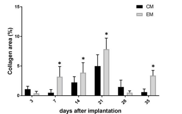
The same trend was observed for collagen production in the subcutaneous tissue of animals that received the CM and EM membranes, with a gradual increase and peak at 21 days. There was higher collagen production with a significant difference at 7, 14, 21, and 35 days in animals receiving the EM (P˂ 0.01) (Figure 2). Collagen deposition was organized around the membranes, forming a capsule and englobing the inflammatory cells together with the implanted materials (Figure 3). The capsules formed around EM were thicker when compared to the capsules formed around CM, with significant differences at 14 and 28 days. The values for the EM membrane were: 91.71(m±10.46 at 14 days and 80.00±5.00 at 28 days, and the values for CM were: 56.22(m±2.63 at 14 days and 60.00±11.5 at 28 days (P˂ 0.01).
Collagen area (%) in the subcutaneous tissue of rats implanted with CM and EM membranes. Removal at 3, 7, 14, 21, 28, and 35 days after implantation. * Significant difference (P˂ 0.01).
Organization of collagen produced in the subcutaneous tissue of rats implanted with EM. In A, the EM (highlighted by asterisk) is surrounded by a fibrous capsule (black quadrangle). The implant was withdrawn at 14 days, HE, 100X. In B, the fibrous capsule is shown. Fibroblasts (black arrow), collagen fibers (white arrow), and blood vessels (arrowhead) are observed. The implant was withdrawn at 14 days, HE, 400X.
DISCUSSION
Biocompatibility is an essential aspect in the development of new materials with potential for clinical applications. This study demonstrated that the EM membrane was effective in reducing the recruitment of foreign-body type cells and accelerating the healing process. This study used the conventional membrane CM and the new EM membrane containing pequi oil as implants in the subcutaneous tissue of rats to compare their biocompatibility in vivo.
Macroscopic observation demonstrated swelling around the implants because of increased inflammation and an accumulation of exudate stemming from surgical trauma, especially within the first three days (Oréfice et al., 2006ORÉFICE, R.L.; PEREIRA, M.M.; MANSUR, M.S. Biomateriais: fundamentos e aplicações. Rio de Janeiro: Guanabara Koogan, 2006. 552p; Cheville, 2009CHEVILLE, N.F. Introdução à patologia veterinária. In: COELHO, H.E. Patologia veterinária. 3.ed. São Paulo: Manole, 2009. p.556-580.).
The microscopic analysis showed a reduction in the recruitment of foreign-body giant cells in the EM compared to the CM at 28 days, which can be attributed to the anti-inflammatory activity of the oil on the tissues. Studies related to the chemical composition of pequi oil have shown high contents of β-carotene, lycopene, and total carotenoids with antioxidant activities, which prevent lipid peroxidation and thereby reduce the release of inflammation mediators and the recruitment of inflammatory cells.
Some in vitro studies have shown that, although the foreign-body response cannot be completely avoided, the adhesion of inflammatory cells is influenced by the chemical structure of the material surface (Anderson et al., 2008ANDERSON, J.M.; RODRIGUEZ, A.; CHANG, D.T. Foreign body reaction to biomaterials. Semin. Immunol., v.20, p.86-100, 2008.; Rolfe et al., 2011ROLFE, B.; MOONEY, J.; ZHANG, B. et al. The fibrotic response to implanted biomaterials: implications for tissue engineering. In: ROLFE, B.; MOONEY, J.; ZHANG, B. et al. Regenerative medicine and tissue engineering - cells and biomaterials. Rijeka: Tech Europe, 2011. p.551-568.). In line with this, several materials have been used to cover implantable devices and modulate the foreign-body giant cell response. Among these are synthetic materials, such as the poly-vinyl alcohol, polylactic acid, and poly (co-glycolic) acid, as well as biopolymers such as chitosan, collagen, and alginate (Mitchell et al., 2010MITCHELL, E.A.; CHAFFEY, B.T.; MCCASKIE, A.W. et al. Controlled spatial and conformational display of immobilised bone morphogenetic protein-2 and osteopontin signalling motifs regulates osteoblast adhesion and differentiation in vitro. BMC Biol., v.8, p.57-69, 2010.).
Notably, the antimicrobial activity of pequi oil may contribute to reducing the inflammatory process in rats implanted with EM. Inflammatory processes present an excessive amount of exudate, which likely favors dehiscence, bacterial growth, and consequently inhibits fibroblast proliferation and collagen deposition (Cotran et al., 2000COTRAN, R.S.; KUMAR, V.; COLLINS, T. Reparo dos tecidos: crescimento celular, fibrose e cicatrização de feridas. In: ROBBINS, S.L et al. Patologia estrutural e funcional. 6.ed. Rio de Janeiro: Guanabara Koogan, 2000. p.79-100.). Some authors have demonstrated the antimicrobial activity of the hydroethanol extract from the leaf and bark of the pequi plant over Enterococcus faecalis, Escherichia coli, Pseudomonas aeruginosa, and Staphylococcus aureus (Paula-Júnior et al., 2006; Pinho et al., 2012PINHO, L.; SOUZA, P.N.S.; SOBRINHO, E.M. et al. Atividade antimicrobiana de extratos hidroalcoólicos das folhas de alecrim-pimenta, aroeira, barbatimão, erva baleeira e do farelo da casca de pequi. Ciênc. Rural, v.42, p.326-331, 2012.).
One study found that inflammation and ear edema in rats were reduced upon treatment with pequi oil, either directly or in ointment form. The authors found a significant reduction in the wound area and an optimized healing process in the treatment group. The anti-inflammatory activity of the pulp pequi oil was found to be similar to those of drugs that modulate the production of metabolites of arachidonic acid in rats (Batista et al., 2010BATISTA, J.S.; SILVA, A.E.; RODRIGUES, C.M.F. et al. Avaliação da atividade cicatrizante do óleo de pequi (Caryocar coriaceum Wittm) em feridas cutâneas produzidas experimentalmente em ratos. Arq. Inst. Biol., v.77, p.441-447, 2010.). The oil had a positive influence on the healing process of experimental skin injuries on rats, with reduced intensity of the inflammatory reaction and faster wound healing than that of the control group (Huang et al., 2012HUANG, C.; FU, X.; LIU, J. et al. The involvement of integrin β1 signaling in the migration and myofibroblastic differentiation of skin fibroblasts on anisotropic collagen-containing nanofibers, Biomaterials, v.33, p.1791-1800, 2012.).
In the rats implanted with EM, an increase in the levels of fibroblasts and collagen production was also observed during the different assessed periods. Similar results were observed with the use of pequi oil on induced skin injuries in rats, with the effects attributed to the presence of fatty acids in the oil. The authors also reported a gastroprotective effect of pequi oil on ethanol-induced gastric lesions in rats, highlighting its protective effect on the gastric mucosa. The increased intake of fatty acids is known to be associated with a decreased incidence of peptic ulcers, possibly due to an increased synthesis of prostaglandins. The healing and gastroprotective effects of pequi oil were attributed to its phytoconstituents that may act individually or in an additive manner to facilitate the healing process (Saraiva et al., 2011SARAIVA, R.A.; ARARUNA, M.K.; OLIVEIRA, R.C. et al. Topical anti-inflammatory effect of Caryocar coriaceum Wittm. (Caryocaraceae) fruit pulp fixed oil on mice ear edema induced by different irritant agents. J. Ethnopharmacol., v.136, p.504-510, 2011.).
Regarding collagen production, the various collagen subtypes were not measured in our study. However, microscopic visualization of the slides stained with picrosirius highlighted the predominance of type I collagen in the group of animals implanted with EM. Type I collagen is most often synthesized by fibroblasts, predominantly in bone and tendon, whereas type III is most commonly found in soft tissues, such as blood vessels and dermis. The dermis contains approximately 80% of type I collagen and 20% type III collagen. During the healing process, type III collagen is formed first, and is subsequently replaced by fibers of collagen I (Ribeiro et al., 2015RIBEIRO, M.; MORAES, M.A.; BEPPU, M.M. et al. Development of silk fibroin/nano hydroxyapatite composite hydrogels for bone tissue engineering. Eur. Polym. J., v.67, p.66-77, 2015.).
Over the last 20 years, a great variety of biomaterials have been obtained and synthesized for applications in soft tissue to promote tissue repair and wound healing. However, this is the first study to report an association between collagen, gelatin, and pequi oil, and to provide evidence supporting the use of pequi oil as a biomaterial in membrane form. It has been shown that the presence pequi oil reduced inflammation in the subcutaneous tissue of rats and stimulated the recruitment of fibroblasts and collagen production. However, the next step in this research will be to evaluate the components of pequi oil and the molecular mechanisms involved in regulating inflammation, recruitment of fibroblasts, and collagen production via cell culture and molecular biology techniques.
CONCLUSION
The presence of pulp pequi oil in the collagen and gelatin membrane reduced the recruitment of inflammatory cells and increased collagen production with deposition centralized around the membranes, thus improving biocompatibility and accelerating the healing process in rats.
ACKNOWLEDGMENTS
The Vulcani V.A.S. laboratory was supported by funding from FAPEG-Fundo de Amparo à pesquisa do estado de Goiás, Brazil. Edital Universal n. 05/2012. Author Rabbers A.S. has received research grants from Coordenação de Aperfeiçoamento Pessoal de Nível Superior (CAPES) scholarship from Brazil.
REFERENCES
- ANDERSON, J.M.; RODRIGUEZ, A.; CHANG, D.T. Foreign body reaction to biomaterials. Semin. Immunol., v.20, p.86-100, 2008.
- BATISTA, J.S.; SILVA, A.E.; RODRIGUES, C.M.F. et al. Avaliação da atividade cicatrizante do óleo de pequi (Caryocar coriaceum Wittm) em feridas cutâneas produzidas experimentalmente em ratos. Arq. Inst. Biol., v.77, p.441-447, 2010.
- BAUER, J.P.; LIU, J.; WINDSOR, L.J. et al. Current development of collagen-based biomaterials for tissue repair and regeneration. Soft Materials, v.12, p.359-370, 2014.
- BIOLOGICAL evaluation of medical devices - part 6: tests for local effects after implantation. Vernie: International Organization for Standardization, 2007. (ISO 10993-6). Available in: <https://www.iso.org>. Accessed in: 01 Dec. 2016.
» https://www.iso.org - BRASIL. Conselho Nacional de Controle de Experimentação Animal. Resolução Normativa n.12, de 20 de setembro de 2013. Dispõe sobre o cuidado e a utilização de animais para fins científicos e didáticos. Diário Oficial da União. Brasília, 25 de setembro 2013. Disponível em: <http://www.mct.gov.br/updblob/0228/228352.pdf>. Acessado em: 20 out. 2016
» http://www.mct.gov.br/updblob/0228/228352.pdf - CHATTOPADHYAY, S.; RAINES, R.T. Review collagen-based biomaterials for wound healing. Biopolymers, v.101, p.821-833, 2014.
- CHEVILLE, N.F. Introdução à patologia veterinária. In: COELHO, H.E. Patologia veterinária. 3.ed. São Paulo: Manole, 2009. p.556-580.
- COTRAN, R.S.; KUMAR, V.; COLLINS, T. Reparo dos tecidos: crescimento celular, fibrose e cicatrização de feridas. In: ROBBINS, S.L et al. Patologia estrutural e funcional. 6.ed. Rio de Janeiro: Guanabara Koogan, 2000. p.79-100.
- FARIA-MACHADO, F.; TRES, A.; VAN RUTH, S. M. et al. Discrimination of pulp oil and kernel oil from pequi (Caryocar brasiliense) by fatty acid methyl esters fingerprinting, using GC-FID and multivariate analysis. J. Agric. Food Chem., v.63, p.10064-10069, 2015.
- GÓMEZ-GUILLÉN, M.; GIMÉNEZ, B.; LOPEZ-CABALLERO, M.A.; MONTERO, M. Functional and bioactive properties of collagen and gelatin from alternative sources: a review. Food Hydrocolloids, v.25, p.1813-1827, 2011.
- HUANG, C.; FU, X.; LIU, J. et al. The involvement of integrin β1 signaling in the migration and myofibroblastic differentiation of skin fibroblasts on anisotropic collagen-containing nanofibers, Biomaterials, v.33, p.1791-1800, 2012.
- LIMA, A.; SILVA, A.M.O.; TRINDADE, R.A. et al. Composição química e compostos bioativos presentes na polpa e na amêndoa de pequi (Caryocar brasiliense Camb). Rev. Bras. Fruticult., v.29, p.695-698, 2007.
- MIRANDA, E.G.; NASCIMENTO, V.P.; WAISBERG, D.R. et al. Inhalation anestesia equipment for rats with provision of simultaneous anesthetic and oxygen. Acta. Circ. Bras., v.26, p.140-143, 2011.
- MITCHELL, E.A.; CHAFFEY, B.T.; MCCASKIE, A.W. et al. Controlled spatial and conformational display of immobilised bone morphogenetic protein-2 and osteopontin signalling motifs regulates osteoblast adhesion and differentiation in vitro. BMC Biol., v.8, p.57-69, 2010.
- O’BREIN, F.J. Biomaterials & scaffolds for tissue engineering. Materialstoday, v.14, p.88-95, 2011.
- ORÉFICE, R.L.; PEREIRA, M.M.; MANSUR, M.S. Biomateriais: fundamentos e aplicações. Rio de Janeiro: Guanabara Koogan, 2006. 552p
- PAULA-JÚNIOR, W.; ROCHA, F.H.; DONATTI, L. Leishmanicidal, antibacterial, and antioxidant activities of Caryocar brasiliense Cambess leaves hydroethanolic extract. Rev. Bras Farmacogn., v.14, p.625-630, 2006.
- PINHO, L.; SOUZA, P.N.S.; SOBRINHO, E.M. et al. Atividade antimicrobiana de extratos hidroalcoólicos das folhas de alecrim-pimenta, aroeira, barbatimão, erva baleeira e do farelo da casca de pequi. Ciênc. Rural, v.42, p.326-331, 2012.
- PIRES, A.L.R.; BIERHALZ, A.C.K.; MORAES, A.M. Biomateriais: tipos, aplicações e mercado, Quim. Nova, v.38, p.957-971, 2015.
- RIBEIRO, M.; MORAES, M.A.; BEPPU, M.M. et al. Development of silk fibroin/nano hydroxyapatite composite hydrogels for bone tissue engineering. Eur. Polym. J., v.67, p.66-77, 2015.
- RODRIGUES, F.T.; MARTINS, V.C.A.; PLEPIS, A.M.G. Porcine skin as a source of biodegradable matrices: alkaline treatment and glutaraldehyde crosslinking. Polímeros, v.20, p.92-97, 2010.
- ROGÉRIO, J.B.; SANTOS, M.C.S.; ANTONIASSI, R. et al. Variação da composição dos ácidos graxos dos óleos de polpa e amêndoa de pequi. In: CONGRESSO BRASILEIRO DE PLANTAS OLEAGINOSAS: GORDURAS E BIODISEL, 8., 2012, Salvador. Anais... Salvador: BDPA, 2012. p.275-276. (Resumo).
- ROLFE, B.; MOONEY, J.; ZHANG, B. et al. The fibrotic response to implanted biomaterials: implications for tissue engineering. In: ROLFE, B.; MOONEY, J.; ZHANG, B. et al. Regenerative medicine and tissue engineering - cells and biomaterials. Rijeka: Tech Europe, 2011. p.551-568.
- SARAIVA, R.A.; ARARUNA, M.K.; OLIVEIRA, R.C. et al. Topical anti-inflammatory effect of Caryocar coriaceum Wittm. (Caryocaraceae) fruit pulp fixed oil on mice ear edema induced by different irritant agents. J. Ethnopharmacol., v.136, p.504-510, 2011.
- SHOULDERS, M.D.; RAINES, R.T. Collagen structure and stability. Annu. Rev. Biochem., v.78, p.929-958, 2009.
- SUBRAMANIAN, A.; KRISHNAN, U.M.; SETHURAMAN, S. In vivo biocompatibility of PLGA-Polyhexylthiophene nanofiber scaffolds in a rat model. BioMed Res. Int., v.2013, p.1-8, 2013.
- VULCANI; V.A.S.; MACORIS, D.G.; PLEPIS, A.M.G. et al. Obtenção, caracterização e aplicação cirúrgica de matrizes de colágeno na parede abdominal de equinos. Ciênc. Anim. Bras., v.9, p.778-785, 2008.
-
FUNDING:
This study was funded by FAPEG-Fundo de Amparo à pesquisa do estado de Goiás, Brazil. Edital Universal n. 05/2012. Author Rabbers, A.S. has received research grants from Company Coordenação de Aperfeiçoamento Pessoal de Nível Superior (CAPES) scholarship from Brazil. There was no direct involvement of the funding source.
Publication Dates
-
Publication in this collection
14 June 2019 -
Date of issue
May-Jun 2019
History
-
Received
23 Nov 2017 -
Accepted
09 Oct 2018